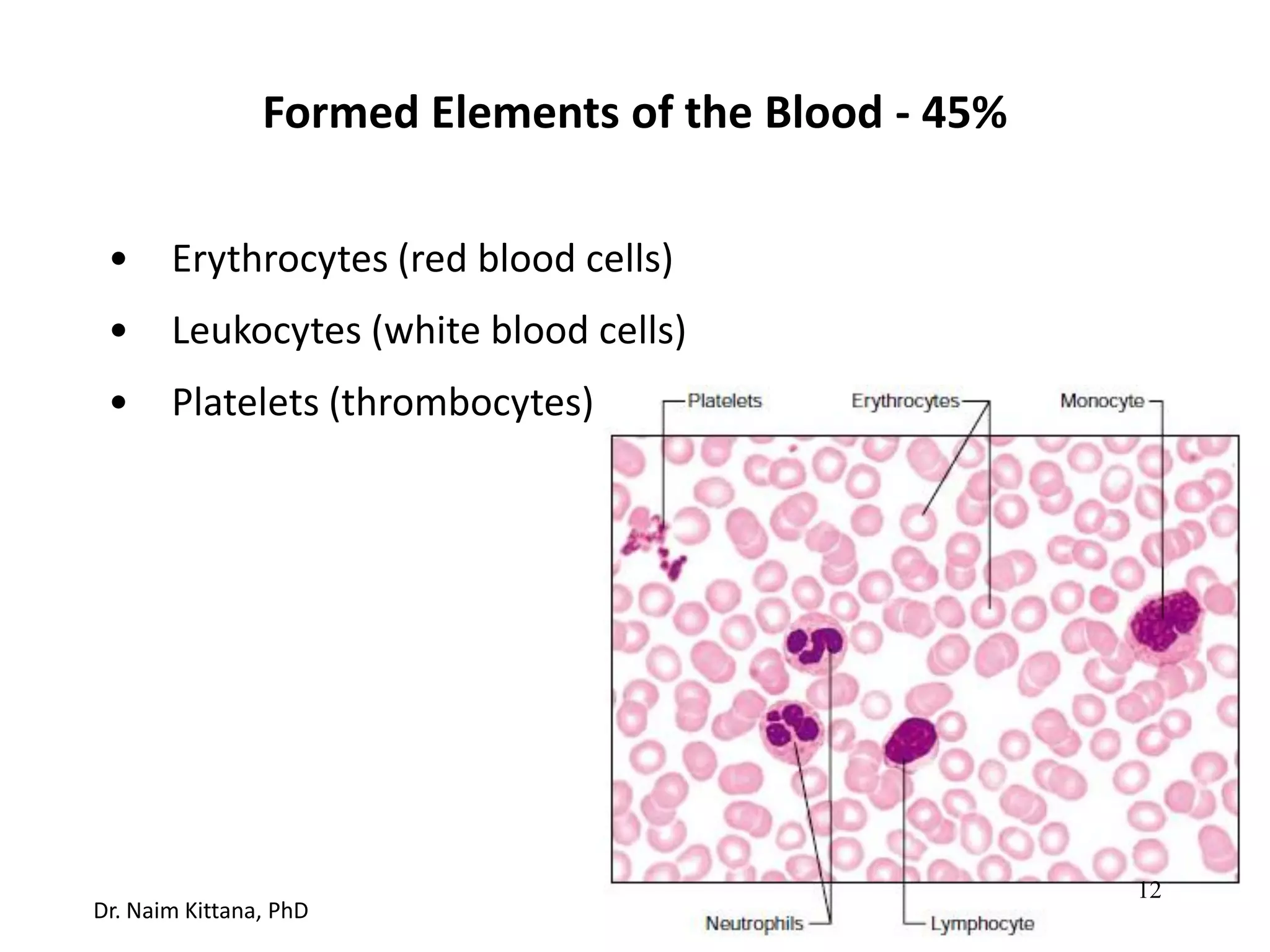
SlideShare a Scribd company logo
1 of 106
Download to read offline
Physiology and Anatomy of
Blood
Prepared by
Dr. Naim Kittana, PhD
An-Najah National University
Faculty of Medicine and Health Sciences
Department of Biomedical Sciences
Disclosure
• The material and the illustrations are adopted from the
textbook “Human Anatomy and Physiology / Ninth
edition/ Eliane N. Marieb 2013”
Dr. Naim Kittana, PhD 2
White blood cells
Platelets
Red blood cells
Artery
Blood
3
Dr. Naim Kittana, PhD
• Deliver O2
• Remove metabolic wastes
• Maintain temperature, pH, and fluid volume
• Protection from blood loss- platelets
• Prevent infection- antibodies and WBC
• Transport hormones
Functions of Blood
4
Dr. Naim Kittana, PhD
Blood Composition
• Hematocrit: % of erythrocytes volume to the total blood volume.
• Normal hematocrit values in healthy males is 47% ± 5% and in females
it is 42% ± 5%
• Normal pH between 7.35 and 7.45
5
Dr. Naim Kittana, PhD
Blood Composition
• Approximately 8% of body weight
• Average volume in healthy adult males is 5-6 L, but 4-5 L in healthy
adult females
6
Dr. Naim Kittana, PhD
Blood Plasma Components (55%)
• 90% Water
• Proteins 8% w/v
– Albumin (60 %):
Produced by the liver
Maintain osmotic pressure
Transport hormones and enzymes
7
Dr. Naim Kittana, PhD
Blood Plasma Components (55%)
• Proteins 8% w/v
– Globulins (36%) :
Alpha and Beta Globulins: produced by the liver,
transport lipids, metals and fat soluble vitamins
Gamma Globulins: Antibodies released by plasma
cells in response to immune response
– Fibrinogens (4%):
Produced by the liver, form fibrin fibers of blood
clots
8
Dr. Naim Kittana, PhD
Blood Plasma Components (55%)
• Gas
• Electrolytes:
 Na+, K+, Ca2+, Mg2+, Cl-, SO4-, HCO3
 Maintain plasma osmotic pressure and pH
9
Dr. Naim Kittana, PhD
Blood Plasma Components (55%)
• Organic Nutrients
– Carbohydrates
– Amino Acids
– Lipids
– Vitamins
• Hormones: Steroid and thyroid hormones are carried by
plasma proteins
• Metabolic waste
– CO2, urea, uric acid, creatinin, ammonium salts
10
Dr. Naim Kittana, PhD
• Platelets
• Leukocytes
Buffy Coat < 1%
11
Dr. Naim Kittana, PhD
• Erythrocytes (red blood cells)
• Leukocytes (white blood cells)
• Platelets (thrombocytes)
Formed Elements of the Blood - 45%
12
Dr. Naim Kittana, PhD
Erythrocytes
13
• Diameter :7.5 m
• Morphology: biconcave discs—flattened discs
with depressed centers
• Anucleate and have essentially no organelles
• Contain 97% hemoglobin
• Contain antioxidant enzymes that rid the
body of harmful oxygen radicals
• Function- transport respiratory gases
Erythrocytes
14
Dr. Naim Kittana, PhD
• Hemoglobin- quaternary structure, 2  chains and 2  chains
• Lack mitochondria, because they generate ATP by anaerobic
mechanisms, therefore they do not consume any of the
oxygen they carry
• An RBC contains 280 million hemoglobin molecules
• Life span 100-120 days and then are destroyed in spleen
(RBC graveyard)
Erythrocytes
15
Dr. Naim Kittana, PhD
• Men: 4.7–6.1 million cells/ microliter
• Women: 4.2- 5.4 million cells/ microliter
• Erythrocytes are the major factor contributing to blood
viscosity.
• When the number of Erythrocytes increases beyond the
normal range, blood becomes more viscous and flows more
slowly.
• When the number of Erythrocytes drops below the lower
end of the range, the blood thins and flows more rapidly
Erythrocytes count
16
Dr. Naim Kittana, PhD
Hemoglobin
17
Dr. Naim Kittana, PhD
Hemoglobin
• Normal values for hemoglobin are 13–18 g/100 ml in adult
males, and 12–16 g/100 ml in adult females
• Hemoglobin is made up of the red heme pigment bound
to the protein globin.
• Globin consists of four polypeptide chains: two alpha (α)
and two beta (β)—each binding a ring-like heme
• Each heme group bears an atom of iron in its center
18
Dr. Naim Kittana, PhD
Hemoglobin
• A hemoglobin molecule can transport four molecules of
oxygen because each iron atom can combine reversibly
with one molecule of oxygen
• A single RBC contains about 250 million hemoglobin
molecules, so each RBC can carry about 1 billion
molecules of oxygen
19
Dr. Naim Kittana, PhD
Why Hemoglobin does not exist free in the plasma?
1) To protect hemoglobin from breaking into fragments that
would leak out of the bloodstream (through porous
capillary walls)
2) To prevent hemoglobin from making blood more viscous
and raising osmotic pressure
20
Dr. Naim Kittana, PhD
Hematopoiesis
• Hematopoiesis (hemopoiesis): blood cell formation
• Occurs in red bone marrow of axial skeleton, girdles and
proximal epiphyses of humerus and femur
• On average, the marrow turns out an 29.5 ml of new
blood containing 100 billion new cells every day
21
Dr. Naim Kittana, PhD
Hematopoiesis
• Hemocytoblasts (hematopoietic stem cells)
• Give rise to all formed elements
• Hormones and growth factors push the cell toward a specific
pathway of blood cell development
22
Dr. Naim Kittana, PhD
Erythropoiesis: formation of red blood cells
Reticulocytes
are released
into the
bloodstream
23
Dr. Naim Kittana, PhD
Erythropoiesis
• Erythropoiesis: red blood cell production
• A hematopoietic stem cell descendant called a myeloid
stem cell transforms into a proerythroblast
• Proerythroblasts develop into basophilic erythroblasts
24
Dr. Naim Kittana, PhD
Erythropoiesis: formation of red blood cells
Reticulocytes
are released
into the
bloodstream
25
Dr. Naim Kittana, PhD
Erythropoiesis
• Hemoglobin is synthesized and iron accumulates as the
basophilic erythroblast transforms into a polychromatic
erythroblast and then an orthochromatic erythroblast
• Reticulocytes (account for 1–2% of all erythrocytes) are
formed after ejection all of the organelles and the nuclei
• Reticulocytes fully mature to erythrocytes within two days
of release as their ribosomes are degraded by intracellular
enzymes
26
Dr. Naim Kittana, PhD
Erythropoiesis: formation of red blood cells
Reticulocytes
are released
into the
bloodstream
27
Dr. Naim Kittana, PhD
Regulation of Erythropoiesis
• Too few RBCs leads to tissue hypoxia
• Too many RBCs increases blood viscosity
• Balance between RBC production and destruction depends
on
– Hormonal controls: Erythropoietin (EPO)
– Adequate supplies of iron, amino acids, and B vitamins
28
Dr. Naim Kittana, PhD
Hormonal Control of Erythropoiesis
• Erythropoietin (EPO)
– Direct stimulus for erythropoiesis
– Released by the kidneys in response to hypoxia
29
Hormonal Control of Erythropoiesis
• Causes of hypoxia
– Hemorrhage or increased RBC destruction reduces RBC
numbers
– Insufficient hemoglobin per RBC (e.g., iron deficiency)
– Reduced availability of O2 (e.g., high altitudes) or during
pneumonia
30
Dr. Naim Kittana, PhD
Hormonal Control of Erythropoiesis
• Effects of EPO
– More rapid maturation of committed bone marrow
cells
• Increased circulating reticulocyte count in 1–2 days after
erythropoietin levels rise in the blood
• hypoxia does not activate the bone marrow directly.
Instead it stimulates the kidneys to provide EPO
• Testosterone also enhances EPO production, resulting in
higher RBC counts in males
31
Dr. Naim Kittana, PhD
Imbalance of EPO
• Kidneys of renal dialysis patients fail to produce enough EPO
to support normal erythropoiesis.
• Consequently, their RBC counts becomes less than half
those of healthy individuals.
• Treatment: Genetically engineered (recombinant) EPO is
frequently administered
32
Dr. Naim Kittana, PhD
Dietary Requirements
• Iron is essential for hemoglobin synthesis.
• Iron is available from the diet, and it is absorbed by the
intestinal cells.
• Approximately 65% of the body’s iron supply (about 4000
mg) is in hemoglobin
33
Dr. Naim Kittana, PhD
Dietary Requirements
• Free iron ions (Fe2+, Fe3+) are toxic, so iron is stored inside
cells as protein-iron complexes such as ferritin and
hemosiderin
• In blood, iron is transported loosely bound to a transport
protein called transferrin
• Developing erythrocytes take up iron as needed to form
hemoglobin
34
Dr. Naim Kittana, PhD
Dietary Requirements
• Small amounts of iron are lost each day in feces, urine, and
perspiration.
• The average daily loss of iron is 1.7 mg in women and 0.9 mg
in men.
• In women, the menstrual flow accounts for the additional
losses.
35
Dr. Naim Kittana, PhD
Dietary Requirements
• Vitamin B12 and folic acid are necessary for normal DNA
synthesis.
• Even slight deficits endanger rapidly dividing cell
populations, such as developing erythrocytes.
36
Dr. Naim Kittana, PhD
Fate and Destruction of Erythrocytes
• Life span of RBC ranges from 100 to 120 days
• Erythrocytes become “old” as they lose their flexibility,
become increasingly rigid and fragile, and their hemoglobin
begins to degenerate.
• They are trapped and fragmented by the spleen “red blood
cell graveyard.”
37
Dr. Naim Kittana, PhD
Fate and Destruction of Erythrocytes
• Macrophages engulf and destroy dying erythrocytes.
• The heme is seperated from globin.
• Its core of iron is salvaged, bound to protein (as ferritin or
hemosiderin), and stored for reuse.
38
Dr. Naim Kittana, PhD
Fate
and
Destruction
of
Erythrocytes
39
Formation & Destruction of RBCs
40
Fate and Destruction of Erythrocytes
• Most of this degraded pigment leaves the body in feces, as a
brown pigment called stercobilin.
• Some of urobilinogen (coloreless) is reabsorbed from the
intestine, and converted into urobilin (yellow color) that is
excreted by the kidneys.
• The protein (globin) part of hemoglobin is metabolized or
broken down to amino acids, which are released to the
circulation.
41
Dr. Naim Kittana, PhD
Erythrocyte Disorders
42
Dr. Naim Kittana, PhD
Blood Cell Production
43
Anemia
• It is a condition in which the blood’s oxygen-carrying
capacity is too low to support normal metabolism.
• It is a sign of some disorder rather than a disease in itself.
• Anemic individuals are fatigued, often pale, short of breath,
and chilled
• The causes of anemia can be divided into three groups:
 Blood loss
 Not enough red blood cells produced
 or too many of them destroyed
44
Dr. Naim Kittana, PhD
Blood loss (Hemorrhagic anemia)
• Acute hemorrhagic anemia: blood loss is rapid
• Persistent blood loss: due to hemorrhoids, an undiagnosed
bleeding ulcer or malignancies
45
Dr. Naim Kittana, PhD
Iron-deficiency anemia
• Cause:
 A secondary result of hemorrhagic anemia
 Inadequate intake of iron-containing foods and
impaired iron absorption.
• Erythrocytes produced, called microcytes, are small and
pale because they cannot synthesize their normal
complement of hemoglobin
46
Dr. Naim Kittana, PhD
Pernicious anemia
• Autoimmune disease most often affects the elderly.
• The immune system destroys cells in the stomach mucosa
that produce a substance called intrinsic factor
(important for vitamin B12 absorption) by intestinal cells.
• Without vitamin B12, the developing erythrocytes grow
but cannot divide, and large, pale cells called macrocytes
result (megaloblastic anemia).
• Treatment involves regular intramuscular injections of
vitamin B12 or as sublingual tablets
• Lack of vitamin B12 in the diet also leads to anemia.
47
Renal anemia
• Frequently accompanies renal disease because damaged
or diseased kidneys cannot produce enough EPO.
• It can be treated with synthetic EPO.
48
Dr. Naim Kittana, PhD
Aplastic anemia
• Results from destruction or inhibition of the red marrow
by certain drugs and chemicals, ionizing radiation, or
viruses.
• In most cases, though, the cause is unknown.
• Because marrow destruction impairs formation of all
formed elements, anemia is just one of its signs
49
Dr. Naim Kittana, PhD
Aplastic anemia
• Defects in blood clotting and immunity are also present.
• Blood transfusions provide a temporal treatment until
stem cells harvested from a donor’s blood, bone marrow,
or umbilical cord blood can be transplanted.
50
Dr. Naim Kittana, PhD
Hemolytic anemias
• Erythrocytes rupture, or lyse, prematurely and with high
rate
• Common causes:
 Hemoglobin abnormalities: thalassemia and sickle-cell
anemia
 Transfusion of mismatched blood
 Certain bacterial and parasitic infections
51
Dr. Naim Kittana, PhD
Thalassemias
• Occur in people of Mediterranean ancestry
• One of the globin chains is absent or faulty, and the
erythrocytes are thin, delicate, and deficient in
hemoglobin
• Severity of the diseased varies according to the associated
mutation
52
Dr. Naim Kittana, PhD
Sickle-cell anemia
• Caused by the abnormal hemoglobin, hemoglobin S (HbS)
• Results from a change in just one of the 146 amino acids
in a beta chain of the globin molecule!
• This alteration causes the beta chains to link together
under low-oxygen conditions, forming stiff rods so that
hemoglobin S becomes spiky and sharp
53
Genetics of Sickle Cell Anemia
54
Dr. Naim Kittana, PhD
Sickle-cell anemia
• This, causes the red blood cells to become crescent
shaped when they unload oxygen molecules or when the
oxygen content of the blood is lower than normal, as
during vigorous exercise and other activities that increase
metabolic rate.
• The stiff, deformed erythrocytes rupture easily and tend
to occlude small blood vessels
55
Sickle-cell anemia
• This causes organ ischemia, leaving the victims gasping for
air and in extreme pain.
56
Dr. Naim Kittana, PhD
Polycythemia
• Abnormal excess of erythrocytes that increases blood
viscosity
• Risk of stroke and heart failure due to high hematocrit and
high blood viscosity
• Types:
 Polycythemia vera
 Secondary polycythemia
57
Polycythemia vera
• Bone marrow cancer, characterized by dizziness and an
exceptionally high RBC count (8–11 million cells/μl).
• The hematocrit may be as high as 80% and blood volume
may double, causing the vascular system to become
engorged with blood and severely impairing circulation
58
Dr. Naim Kittana, PhD
Secondary polycythemias
• Due to high level of EPO secretion
• Detected in smokers and individuals living at high altitudes
59
Dr. Naim Kittana, PhD
Leukocytes (White Blood Cells)
60
Dr. Naim Kittana, PhD
Leukocytes (White Blood Cells)
• On average, there are 4800–10,800 WBCs/μl of blood
• Leukocytes are grouped into two major categories on the
basis of structural and chemical characteristics.
 Granulocytes: contain obvious membrane-bound
cytoplasmic granules, and
 Agranulocytes: lack obvious granules
61
Dr. Naim Kittana, PhD
Granulocytes
• Include: neutrophils, eosinophils, and basophils
• They have lobed nuclei
• Functionally, all granulocytes are phagocytes to some
degree
62
Neutrophils
• Also called Polymorphonuclear leukocytes (PMNs)
• Account for 50–70% of the WBC population
• About twice as large as erythrocytes
• Their numbers increase explosively during acute bacterial
infections
63
Dr. Naim Kittana, PhD
Neutrophils
• Some of their granules contain hydrolytic enzymes, and
are regarded as lysosomes.
• Others, especially the smaller granules, contain a potent
“brew” of antimicrobial proteins, called defensins that
pierce holes in the membrane of the ingested pathogen.
• They also kill pathogens by respiratory burst (the cells
metabolize oxygen to produce potent germ-killer oxidizing
substances such as bleach and hydrogen peroxide.
• Neutrophils are chemically attracted to sites of
inflammation and are active phagocytes 64
Eosinophils
• Account for 2–4% of all leukocytes
• Their granules are lysosome-like and filled with a unique
variety of digestive enzymes.
• They lack enzymes that specifically digest bacteria.
65
Dr. Naim Kittana, PhD
Eosinophils
• The most important role is to lead the counterattack
against parasitic worms, such as flatworms (tapeworms
and flukes) and roundworms (pinworms and hookworms)
that are too large to be phagocytized
• They release the enzymes from their cytoplasmic granules
onto the parasite’s surface to digesting it
• Eosinophils have complex roles in many other diseases
including allergies and asthma
66
Dr. Naim Kittana, PhD
Basophils
• The rarest white blood cells, accounting for only 0.5–1% of
the leukocyte population
• Their cytoplasm contains large, coarse, histamine-
containing granules
• Histamine is an inflammatory chemical that acts as a
vasodilator (makes blood vessels dilate) and attracts other
white blood cells to the inflamed site
• Granulated cells similar to basophils, called mast cells, are
found in connective tissues. They also release histamine
67
Dr. Naim Kittana, PhD
Agranulocytes
• Include lymphocytes and monocytes
• Lack visible cytoplasmic granules
68
Lymphocytes
• Accounting for 25% of the WBC population
• Few are found in the bloodstream, as they are closely
associated with lymphoid tissues (lymph nodes, spleen,
• etc.), where they play a crucial role in immunity.
• T lymphocytes (T cells) function in the immune response
by acting directly against virus-infected cells and tumor
cells.
• B lymphocytes (B cells) give rise to plasma cells, which
produce antibodies
69
Dr. Naim Kittana, PhD
Monocytes
• Account for 3–8% of WBCs
• When circulating monocytes leave the bloodstream and
enter the tissues, they differentiate into highly mobile
macrophages.
• Macrophages are actively phagocytic, and they are crucial
in the body’s defense against viruses, certain intracellular
bacterial parasites, and chronic infections such as
tuberculosis
70
Dr. Naim Kittana, PhD
Leukocyte Disorders
• Leukemia and infectious mononucleosis: overproduction
of abnormal leukocytes
• Leukopenia: is an abnormally low white blood cell count,
commonly induced by drugs, particularly glucocorticoids
and anticancer agents.
71
Dr. Naim Kittana, PhD
Leukemia
• a group of cancerous conditions involving overproduction
of abnormal white blood cells
• The diseased leukocytes are members of a single clone
(descendants of a single cell) that remain unspecialized
and proliferate out of control, impairing normal red bone
marrow function.
72
Dr. Naim Kittana, PhD
Leukemia
• The leukemias are named according to the cell type
primarily involved.
• For example, myeloid leukemia involves myeloblast
descendants, whereas lymphocytic leukemia involves the
lymphocytes.
• Leukemia is acute (quickly advancing) if it derives from
stem cells, and chronic (slowly advancing) if it involves
proliferation of later cell stages.
73
Dr. Naim Kittana, PhD
Leukemia
• The more serious acute forms primarily affect children.
• Chronic leukemia occurs more often in elderly people.
• Without therapy all leukemias are fatal
• Cancerous leukocytes fill the red bone marrow and
immature WBCs flood into the bloodstream.
74
Dr. Naim Kittana, PhD
Leukemia
• The other blood cell lines are crowded out, so severe
anemia and bleeding problems result
• Other symptoms include fever, weight loss, and bone pain.
• Although tremendous numbers of leukocytes are
produced, they are nonfunctional and cannot defend the
body in the usual way.
• The most common causes of death are internal
hemorrhage and overwhelming infections.
75
Dr. Naim Kittana, PhD
Infectious Mononucleosis
• Highly contagious viral disease
• Caused by the Epstein- Barr virus
• Its hallmark is excessive numbers of agranulocytes
• The affected individual complains of being tired and achy,
and has a chronic sore throat and a low-grade fever.
• There is no cure, but with rest the condition typically runs
its course to recovery in a few weeks.
76
Dr. Naim Kittana, PhD
Platelets
• They are not cells in the strict sense
• they are cytoplasmic fragments of extraordinarily large
cells called megakaryocytes
• Their granules contain lots of chemicals that act in the
clotting process, (including serotonin, Ca2+, a variety of
enzymes, ADP, and platelet derived growth factor (PDGF)).
77
Dr. Naim Kittana, PhD
Stem cell Developmental pathway
Hemocyto-
blast Megakaryoblast
Promegakaryocyte
Megakaryocyte Platelets
78
Dr. Naim Kittana, PhD
Hemostasis
Step 1: Vascular spasm
Smooth muscle contracts
causing vasoconstriction
Step 2:
• Platelet plug formation
Injury to lining of vessel exposing
collagen fibers, platelets adhere.
• Platelets release chemicals
that make nearby platelets
sticky, platelet plug forms
Step 3: Coagulation
• Fibrin forms a mesh that
traps RBCs & platelets,
forming the clot.
direct injury to vascular smooth muscle,
chemicals released by endothelial cells and
platelets, and reflexes initiated by local pain
receptors
79
Platelets
• As a rule, platelets do not stick to each other or to the smooth
endothelial linings of blood vessels.
• Intact endothelial cells release nitric oxide and a prostaglandin
called prostacyclin (or PGI2).
• Both chemicals prevent platelet aggregation in undamaged
tissue and restrict aggregation to the site of injury.
80
Dr. Naim Kittana, PhD
Platelets
• However, when the endothelium is damaged and the
underlying collagen fibers are exposed, platelets adhere
tenaciously to the collagen fibers.
• A large plasma protein called von Willebrand factor
stabilizes bound platelets by forming a bridge between
collagen and platelets.
81
Dr. Naim Kittana, PhD
Platelets
• Platelets swell, form spiked processes, become stickier,
and release chemical messengers including the following:
 Adenosine diphosphate (ADP)—a potent aggregating
agent that causes more platelets to stick to the area
and release their contents
 Serotonin and Thromboxane A2 messengers that
enhance vascular spasm and platelet aggregation
82
Dr. Naim Kittana, PhD
Coagulation (blood clotting)
• Reinforces the platelet plug with fibrin threads that act as
a “molecular glue” for the aggregated platelets
• Blood is transformed from a liquid to a gel in a multistep
process hat involves a series of substances called clotting
factors (procoagulants)
• Most clotting factors are plasma proteins synthesized by
the liver. They are numbered I to XIII according to the
order of their discovery
83
Dr. Naim Kittana, PhD
Coagulation (blood clotting)
• Vitamin K (fat-soluble vitamin) is required for synthesizing
four of the clotting factors
• In most cases, activation turns clotting factors into
enzymes, except factor IV (Ca2+) and I (Fibrinogen)
84
Dr. Naim Kittana, PhD
Two Pathways to Prothrombin Activator
• Coagulation may be initiated by either the intrinsic or the
extrinsic pathway
• In the body, the same tissue-damaging events usually
trigger both pathways.
• Outside the body (such as in a test tube), only the intrinsic
pathway initiates blood clotting.
85
Dr. Naim Kittana, PhD
Blood coagulation
XII XIIa
XI XIa
Prothrombin (II)
IX IXa
X Xa
VIIa VII
X
Thrombin (IIa)
Fibrinogen (I) Fibrin
Vascular injury
Expose Tissue Factor (TF)
(Thromboplastin)
Vascular injury Expose collagen
to blood
Intrinsic pathway Extrensic pathway
86
Blood coagulation
87
Blood Clot
Fibrin thread
Platelet
RBC
88
Disorders of Hemostasis
• Thromboembolytic disorders: undesirable clot formation
• Bleeding disorders: abnormalities that prevent normal clot
formation
89
Dr. Naim Kittana, PhD
Thromboembolytic Conditions
• Thrombus: clot that develops and persists in an unbroken
blood vessel
– May block circulation, leading to tissue death
• Embolus: a thrombus freely floating in the blood stream
– Pulmonary emboli impair the ability of the body to
obtain oxygen
– Cerebral emboli can cause strokes
90
Dr. Naim Kittana, PhD
Thromboembolytic Conditions
Prevented by
– Aspirin
• Antiprostaglandin that inhibits thromboxane A2
– Heparin
• Anticoagulant used clinically for pre- and
postoperative cardiac care
– Warfarin
• Used for those prone to atrial fibrillation
91
Dr. Naim Kittana, PhD
Thrombocytosis- too many platelets due to inflammation,
infection or cancer
Thrombocytopenia- too few platelets
• causes spontaneous bleeding
• due to suppression or destruction of bone marrow (e.g.,
malignancy, radiation)
– Platelet count <50,000/mm3 is diagnostic
– Treated with transfusion of concentrated platelets
Bleeding disorders
92
Dr. Naim Kittana, PhD
• Impaired liver function
– Inability to synthesize procoagulants
– Causes include vitamin K deficiency, hepatitis, and
cirrhosis
– Liver disease can also prevent the liver from producing
bile, impairing fat and vitamin K absorption
Bleeding disorders
93
Dr. Naim Kittana, PhD
• Hemophilias include several similar hereditary bleeding
disorders
• Symptoms include prolonged bleeding, especially into
joint cavities
• Treated with plasma transfusions and injection of missing
factors
Bleeding disorders
94
Dr. Naim Kittana, PhD
Blood types
• Type A
• Type B
• Type AB
• Type O
95
Dr. Naim Kittana, PhD
Blood type is based on the presence of 2 major antigens in RBC
membranes-- A and B
Blood type Antigen Antibody
A A Anti-B
B B Anti-A
A & B AB no anti body
Neither A or B O anti-A and anti-B
Antigen- protein on the surface of a RBC membrane
Antibody- proteins made by lymphocytes in plasma which are made
in response to the presence of antigens.
They attack foreign antigens, which result in clumping (agglutination)
Blood types
96
Dr. Naim Kittana, PhD
Type A
b
b
b
b
b
b
b
97
Dr. Naim Kittana, PhD
Type B
a
a
a
a
a
a
a
98
Dr. Naim Kittana, PhD
Type O
a
a
a
b
a
a
a
b
b
b
99
Dr. Naim Kittana, PhD
Type AB
100
Dr. Naim Kittana, PhD
101
Rh Factor and Pregnancy
RH- indicates no protein
RH+ indicates protein
102
Rh Factor and
Pregnancy
Rh+ mother w/Rh- baby– no problem
Rh- mother w/Rh+ baby– problem
Rh- mother w/Rh- father– no problem
Rh- mother w/Rh- baby-- no problem
103
Rho(D) immune globulin (anti-RhD)
• IgG anti-D antibodies
• Given at the 28th week of pregnancy as IM for pregnant
women how are Rho-negative if the father is Rho-positive
• Target fetal RBCs that leak to the maternal blood
circulation before the maternal immune system reacts to it
• Prevents Rh disease (hemolytic disease of new born) in the
subsequent pregnancies
104
Dr. Naim Kittana, PhD
Type AB- universal recipients
Type O- universal donor
Rh factor:
Rh+ 85% dominant in pop
Rh- 15% recessive
Blood Type Clumping Antibody
A antigen A anti-A serum antibody anti-b
B antigen B anti-B serum antibody anti-a
AB antigen A & B anti A & B serum -
O neither A or B no clumping w/ either anti A or B anti-a, anti-b
105
Dr. Naim Kittana, PhD
Serum
Anti-A
RBCs
Anti-B
Type AB (contains
agglutinogens A and B;
agglutinates with both
sera)
Blood test
Type A (contains
agglutinogen A;
agglutinates with anti-A)
Type B (contains
agglutinogen B;
agglutinates with anti-B)
Type O (contains no
agglutinogens; does not
agglutinate with either
serum) 106

More Related Content

What's hot

Posterior pituitary by m. pandian
Posterior pituitary by m. pandianPosterior pituitary by m. pandian
Posterior pituitary by m. pandianPandian M
 
M-2- General Reactions of amino acids.pptx
M-2- General Reactions of amino acids.pptxM-2- General Reactions of amino acids.pptx
M-2- General Reactions of amino acids.pptxDr. Santhosh Kumar. N
 
Liver function test (LFT)
Liver function test (LFT)Liver function test (LFT)
Liver function test (LFT)Maniz Joshi
 
ISOENZYMES & CLINICAL ENZYMOLOGY
ISOENZYMES & CLINICAL ENZYMOLOGYISOENZYMES & CLINICAL ENZYMOLOGY
ISOENZYMES & CLINICAL ENZYMOLOGYYESANNA
 
Enzymes in diagnostics
Enzymes in diagnosticsEnzymes in diagnostics
Enzymes in diagnosticsNiKita Q
 
Disorders of purine and pyrimidine metabolism
Disorders of purine and pyrimidine metabolismDisorders of purine and pyrimidine metabolism
Disorders of purine and pyrimidine metabolismAzeem Aslam
 
Kidney function tests by moustafa rizk
Kidney function tests by moustafa rizkKidney function tests by moustafa rizk
Kidney function tests by moustafa rizkMoustafa Rezk
 
A quick revision of Carbohydrate metabolism with case- based discussions and ...
A quick revision of Carbohydrate metabolism with case- based discussions and ...A quick revision of Carbohydrate metabolism with case- based discussions and ...
A quick revision of Carbohydrate metabolism with case- based discussions and ...Namrata Chhabra
 
Digestion & absorption of lipids by Dr. Santhosh Kumar N.pptx
Digestion & absorption of lipids by Dr. Santhosh Kumar N.pptxDigestion & absorption of lipids by Dr. Santhosh Kumar N.pptx
Digestion & absorption of lipids by Dr. Santhosh Kumar N.pptxDr. Santhosh Kumar. N
 
Clinical Biochemistry Laboratory
Clinical Biochemistry LaboratoryClinical Biochemistry Laboratory
Clinical Biochemistry LaboratoryTapeshwar Yadav
 
DISORDERS OF PHENYLALANINE METABOLISM
DISORDERS OF PHENYLALANINE METABOLISMDISORDERS OF PHENYLALANINE METABOLISM
DISORDERS OF PHENYLALANINE METABOLISMYESANNA
 

What's hot (20)

Posterior pituitary by m. pandian
Posterior pituitary by m. pandianPosterior pituitary by m. pandian
Posterior pituitary by m. pandian
 
M-2- General Reactions of amino acids.pptx
M-2- General Reactions of amino acids.pptxM-2- General Reactions of amino acids.pptx
M-2- General Reactions of amino acids.pptx
 
Liver function test (LFT)
Liver function test (LFT)Liver function test (LFT)
Liver function test (LFT)
 
ISOENZYMES & CLINICAL ENZYMOLOGY
ISOENZYMES & CLINICAL ENZYMOLOGYISOENZYMES & CLINICAL ENZYMOLOGY
ISOENZYMES & CLINICAL ENZYMOLOGY
 
Oxytocin and vasopressin
Oxytocin and vasopressinOxytocin and vasopressin
Oxytocin and vasopressin
 
Enzymes in diagnostics
Enzymes in diagnosticsEnzymes in diagnostics
Enzymes in diagnostics
 
Liver fxn
Liver fxnLiver fxn
Liver fxn
 
Blood glucose Regulation Dr veerendra
Blood glucose Regulation Dr veerendraBlood glucose Regulation Dr veerendra
Blood glucose Regulation Dr veerendra
 
Liver Function Test
Liver Function TestLiver Function Test
Liver Function Test
 
Disorders of purine and pyrimidine metabolism
Disorders of purine and pyrimidine metabolismDisorders of purine and pyrimidine metabolism
Disorders of purine and pyrimidine metabolism
 
Kidney function tests by moustafa rizk
Kidney function tests by moustafa rizkKidney function tests by moustafa rizk
Kidney function tests by moustafa rizk
 
LIVER FUNCTIONS TESTS -1-
LIVER FUNCTIONS TESTS -1-LIVER FUNCTIONS TESTS -1-
LIVER FUNCTIONS TESTS -1-
 
Growth hormone
Growth hormoneGrowth hormone
Growth hormone
 
A quick revision of Carbohydrate metabolism with case- based discussions and ...
A quick revision of Carbohydrate metabolism with case- based discussions and ...A quick revision of Carbohydrate metabolism with case- based discussions and ...
A quick revision of Carbohydrate metabolism with case- based discussions and ...
 
Uric acid metabolism and Gout
Uric acid metabolism and GoutUric acid metabolism and Gout
Uric acid metabolism and Gout
 
Hb A1c by Dr Sarma RVSN
Hb A1c by Dr Sarma RVSNHb A1c by Dr Sarma RVSN
Hb A1c by Dr Sarma RVSN
 
Digestion & absorption of lipids by Dr. Santhosh Kumar N.pptx
Digestion & absorption of lipids by Dr. Santhosh Kumar N.pptxDigestion & absorption of lipids by Dr. Santhosh Kumar N.pptx
Digestion & absorption of lipids by Dr. Santhosh Kumar N.pptx
 
ENDOCRINE PANCREAS, INSULIN, GLUCAGON
ENDOCRINE PANCREAS, INSULIN, GLUCAGONENDOCRINE PANCREAS, INSULIN, GLUCAGON
ENDOCRINE PANCREAS, INSULIN, GLUCAGON
 
Clinical Biochemistry Laboratory
Clinical Biochemistry LaboratoryClinical Biochemistry Laboratory
Clinical Biochemistry Laboratory
 
DISORDERS OF PHENYLALANINE METABOLISM
DISORDERS OF PHENYLALANINE METABOLISMDISORDERS OF PHENYLALANINE METABOLISM
DISORDERS OF PHENYLALANINE METABOLISM
 

Similar to Blood physiology.pdf

Anemias interpretation of cbc 2010rev
Anemias   interpretation of cbc 2010revAnemias   interpretation of cbc 2010rev
Anemias interpretation of cbc 2010revkaycase
 
Blood and its components
Blood and its componentsBlood and its components
Blood and its componentsjunni86
 
Blood physiology: Part I
Blood physiology: Part IBlood physiology: Part I
Blood physiology: Part IFawaz A.M.
 
Circulatory system -The blood
Circulatory system -The bloodCirculatory system -The blood
Circulatory system -The bloodLivson Thomas
 
Blood, It's constituents and Functions
Blood, It's constituents and FunctionsBlood, It's constituents and Functions
Blood, It's constituents and FunctionsMaryangela Ohanu
 
Blood (RBC, Anemia, Polycythemia)
Blood (RBC, Anemia, Polycythemia)Blood (RBC, Anemia, Polycythemia)
Blood (RBC, Anemia, Polycythemia)Pharmacy Universe
 
Hematologic disorders
Hematologic disordersHematologic disorders
Hematologic disordersKiran
 
theblood-211019111147 (1).pdf...............
theblood-211019111147 (1).pdf...............theblood-211019111147 (1).pdf...............
theblood-211019111147 (1).pdf...............chetanmeti13
 
Introduction To Hematology
Introduction To HematologyIntroduction To Hematology
Introduction To HematologyMiami Dade
 

Similar to Blood physiology.pdf (20)

2 blood rev12
2 blood rev122 blood rev12
2 blood rev12
 
Hematology_Comprehensive Blood Physiology Review
 Hematology_Comprehensive Blood Physiology Review Hematology_Comprehensive Blood Physiology Review
Hematology_Comprehensive Blood Physiology Review
 
Anemias interpretation of cbc 2010rev
Anemias   interpretation of cbc 2010revAnemias   interpretation of cbc 2010rev
Anemias interpretation of cbc 2010rev
 
Blood
BloodBlood
Blood
 
Blood and its components
Blood and its componentsBlood and its components
Blood and its components
 
1. BLOOD Part 2.pptx
1. BLOOD Part 2.pptx1. BLOOD Part 2.pptx
1. BLOOD Part 2.pptx
 
Blood physiology: Part I
Blood physiology: Part IBlood physiology: Part I
Blood physiology: Part I
 
Blood and its components
Blood and its componentsBlood and its components
Blood and its components
 
Circulatory system -The blood
Circulatory system -The bloodCirculatory system -The blood
Circulatory system -The blood
 
Blood, It's constituents and Functions
Blood, It's constituents and FunctionsBlood, It's constituents and Functions
Blood, It's constituents and Functions
 
Blood (RBC, Anemia, Polycythemia)
Blood (RBC, Anemia, Polycythemia)Blood (RBC, Anemia, Polycythemia)
Blood (RBC, Anemia, Polycythemia)
 
Hematologic disorders
Hematologic disordersHematologic disorders
Hematologic disorders
 
Red blood cell &amp; bleeding disorders
Red blood cell &amp; bleeding disordersRed blood cell &amp; bleeding disorders
Red blood cell &amp; bleeding disorders
 
The blood
The bloodThe blood
The blood
 
theblood-211019111147 (1).pdf...............
theblood-211019111147 (1).pdf...............theblood-211019111147 (1).pdf...............
theblood-211019111147 (1).pdf...............
 
Blood composition_ Physiology
Blood composition_ PhysiologyBlood composition_ Physiology
Blood composition_ Physiology
 
Introduction To Hematology
Introduction To HematologyIntroduction To Hematology
Introduction To Hematology
 
BLOOD ITS STRUCTURE.pptx
BLOOD ITS STRUCTURE.pptxBLOOD ITS STRUCTURE.pptx
BLOOD ITS STRUCTURE.pptx
 
Lecture 9 CVS.pptx
Lecture 9 CVS.pptxLecture 9 CVS.pptx
Lecture 9 CVS.pptx
 
Lecture 9 CVS.pptx
Lecture 9 CVS.pptxLecture 9 CVS.pptx
Lecture 9 CVS.pptx
 

More from sathishvsathish1

typhoid fever public health care .pptx
typhoid fever public health care   .pptxtyphoid fever public health care   .pptx
typhoid fever public health care .pptxsathishvsathish1
 
Diabetic retinopathy from pathophysiology.pptx
Diabetic retinopathy from pathophysiology.pptxDiabetic retinopathy from pathophysiology.pptx
Diabetic retinopathy from pathophysiology.pptxsathishvsathish1
 
Lecture 12. Antiepileptic drugs pharmacology.pptx
Lecture 12. Antiepileptic drugs pharmacology.pptxLecture 12. Antiepileptic drugs pharmacology.pptx
Lecture 12. Antiepileptic drugs pharmacology.pptxsathishvsathish1
 
protozoans kd sync dud six ch vii goo .pptx
protozoans kd sync dud six ch vii goo .pptxprotozoans kd sync dud six ch vii goo .pptx
protozoans kd sync dud six ch vii goo .pptxsathishvsathish1
 
Pathology_Stom_Eng_2021_(1).pdf
Pathology_Stom_Eng_2021_(1).pdfPathology_Stom_Eng_2021_(1).pdf
Pathology_Stom_Eng_2021_(1).pdfsathishvsathish1
 
internationalredcross-171220093004.pdf
internationalredcross-171220093004.pdfinternationalredcross-171220093004.pdf
internationalredcross-171220093004.pdfsathishvsathish1
 
presentationofstreptococcus-180731145601.pdf
presentationofstreptococcus-180731145601.pdfpresentationofstreptococcus-180731145601.pdf
presentationofstreptococcus-180731145601.pdfsathishvsathish1
 
qualitycontrolinbiochemistry-190731013722.pdf
qualitycontrolinbiochemistry-190731013722.pdfqualitycontrolinbiochemistry-190731013722.pdf
qualitycontrolinbiochemistry-190731013722.pdfsathishvsathish1
 
b-150112001834-conversion-gate02.pptx
b-150112001834-conversion-gate02.pptxb-150112001834-conversion-gate02.pptx
b-150112001834-conversion-gate02.pptxsathishvsathish1
 
streptococcus-160204030310.pdf
streptococcus-160204030310.pdfstreptococcus-160204030310.pdf
streptococcus-160204030310.pdfsathishvsathish1
 

More from sathishvsathish1 (18)

typhoid fever public health care .pptx
typhoid fever public health care   .pptxtyphoid fever public health care   .pptx
typhoid fever public health care .pptx
 
Diabetic retinopathy from pathophysiology.pptx
Diabetic retinopathy from pathophysiology.pptxDiabetic retinopathy from pathophysiology.pptx
Diabetic retinopathy from pathophysiology.pptx
 
Lecture 12. Antiepileptic drugs pharmacology.pptx
Lecture 12. Antiepileptic drugs pharmacology.pptxLecture 12. Antiepileptic drugs pharmacology.pptx
Lecture 12. Antiepileptic drugs pharmacology.pptx
 
protozoans kd sync dud six ch vii goo .pptx
protozoans kd sync dud six ch vii goo .pptxprotozoans kd sync dud six ch vii goo .pptx
protozoans kd sync dud six ch vii goo .pptx
 
Pathology_Stom_Eng_2021_(1).pdf
Pathology_Stom_Eng_2021_(1).pdfPathology_Stom_Eng_2021_(1).pdf
Pathology_Stom_Eng_2021_(1).pdf
 
lower limb.pdf
 lower limb.pdf lower limb.pdf
lower limb.pdf
 
DEFENCE MECHANISMS.pptx
DEFENCE MECHANISMS.pptxDEFENCE MECHANISMS.pptx
DEFENCE MECHANISMS.pptx
 
TESTIS.pptx
TESTIS.pptxTESTIS.pptx
TESTIS.pptx
 
Health insurance 2.pdf
Health insurance 2.pdfHealth insurance 2.pdf
Health insurance 2.pdf
 
internationalredcross-171220093004.pdf
internationalredcross-171220093004.pdfinternationalredcross-171220093004.pdf
internationalredcross-171220093004.pdf
 
presentationofstreptococcus-180731145601.pdf
presentationofstreptococcus-180731145601.pdfpresentationofstreptococcus-180731145601.pdf
presentationofstreptococcus-180731145601.pdf
 
qualitycontrolinbiochemistry-190731013722.pdf
qualitycontrolinbiochemistry-190731013722.pdfqualitycontrolinbiochemistry-190731013722.pdf
qualitycontrolinbiochemistry-190731013722.pdf
 
b-150112001834-conversion-gate02.pptx
b-150112001834-conversion-gate02.pptxb-150112001834-conversion-gate02.pptx
b-150112001834-conversion-gate02.pptx
 
Human anatomy.pdf
Human anatomy.pdfHuman anatomy.pdf
Human anatomy.pdf
 
09 - Carbon
09 - Carbon09 - Carbon
09 - Carbon
 
04 - Peptides.pptx
04 - Peptides.pptx04 - Peptides.pptx
04 - Peptides.pptx
 
streptococcus-160204030310.pdf
streptococcus-160204030310.pdfstreptococcus-160204030310.pdf
streptococcus-160204030310.pdf
 
07 - Vitamins.ppt
07 - Vitamins.ppt07 - Vitamins.ppt
07 - Vitamins.ppt
 

Recently uploaded

Types of Journalistic Writing Grade 8.pptx
Types of Journalistic Writing Grade 8.pptxTypes of Journalistic Writing Grade 8.pptx
Types of Journalistic Writing Grade 8.pptxEyham Joco
 
Roles & Responsibilities in Pharmacovigilance
Roles & Responsibilities in PharmacovigilanceRoles & Responsibilities in Pharmacovigilance
Roles & Responsibilities in PharmacovigilanceSamikshaHamane
 
Like-prefer-love -hate+verb+ing & silent letters & citizenship text.pdf
Like-prefer-love -hate+verb+ing & silent letters & citizenship text.pdfLike-prefer-love -hate+verb+ing & silent letters & citizenship text.pdf
Like-prefer-love -hate+verb+ing & silent letters & citizenship text.pdfMr Bounab Samir
 
Earth Day Presentation wow hello nice great
Earth Day Presentation wow hello nice greatEarth Day Presentation wow hello nice great
Earth Day Presentation wow hello nice greatYousafMalik24
 
Alper Gobel In Media Res Media Component
Alper Gobel In Media Res Media ComponentAlper Gobel In Media Res Media Component
Alper Gobel In Media Res Media ComponentInMediaRes1
 
What is Model Inheritance in Odoo 17 ERP
What is Model Inheritance in Odoo 17 ERPWhat is Model Inheritance in Odoo 17 ERP
What is Model Inheritance in Odoo 17 ERPCeline George
 
ENGLISH 7_Q4_LESSON 2_ Employing a Variety of Strategies for Effective Interp...
ENGLISH 7_Q4_LESSON 2_ Employing a Variety of Strategies for Effective Interp...ENGLISH 7_Q4_LESSON 2_ Employing a Variety of Strategies for Effective Interp...
ENGLISH 7_Q4_LESSON 2_ Employing a Variety of Strategies for Effective Interp...JhezDiaz1
 
Computed Fields and api Depends in the Odoo 17
Computed Fields and api Depends in the Odoo 17Computed Fields and api Depends in the Odoo 17
Computed Fields and api Depends in the Odoo 17Celine George
 
Introduction to AI in Higher Education_draft.pptx
Introduction to AI in Higher Education_draft.pptxIntroduction to AI in Higher Education_draft.pptx
Introduction to AI in Higher Education_draft.pptxpboyjonauth
 
Hierarchy of management that covers different levels of management
Hierarchy of management that covers different levels of managementHierarchy of management that covers different levels of management
Hierarchy of management that covers different levels of managementmkooblal
 
MARGINALIZATION (Different learners in Marginalized Group
MARGINALIZATION (Different learners in Marginalized GroupMARGINALIZATION (Different learners in Marginalized Group
MARGINALIZATION (Different learners in Marginalized GroupJonathanParaisoCruz
 
Final demo Grade 9 for demo Plan dessert.pptx
Final demo Grade 9 for demo Plan dessert.pptxFinal demo Grade 9 for demo Plan dessert.pptx
Final demo Grade 9 for demo Plan dessert.pptxAvyJaneVismanos
 
Proudly South Africa powerpoint Thorisha.pptx
Proudly South Africa powerpoint Thorisha.pptxProudly South Africa powerpoint Thorisha.pptx
Proudly South Africa powerpoint Thorisha.pptxthorishapillay1
 
Gas measurement O2,Co2,& ph) 04/2024.pptx
Gas measurement O2,Co2,& ph) 04/2024.pptxGas measurement O2,Co2,& ph) 04/2024.pptx
Gas measurement O2,Co2,& ph) 04/2024.pptxDr.Ibrahim Hassaan
 
Presiding Officer Training module 2024 lok sabha elections
Presiding Officer Training module 2024 lok sabha electionsPresiding Officer Training module 2024 lok sabha elections
Presiding Officer Training module 2024 lok sabha electionsanshu789521
 
Employee wellbeing at the workplace.pptx
Employee wellbeing at the workplace.pptxEmployee wellbeing at the workplace.pptx
Employee wellbeing at the workplace.pptxNirmalaLoungPoorunde1
 
call girls in Kamla Market (DELHI) 🔝 >༒9953330565🔝 genuine Escort Service 🔝✔️✔️
call girls in Kamla Market (DELHI) 🔝 >༒9953330565🔝 genuine Escort Service 🔝✔️✔️call girls in Kamla Market (DELHI) 🔝 >༒9953330565🔝 genuine Escort Service 🔝✔️✔️
call girls in Kamla Market (DELHI) 🔝 >༒9953330565🔝 genuine Escort Service 🔝✔️✔️9953056974 Low Rate Call Girls In Saket, Delhi NCR
 
Capitol Tech U Doctoral Presentation - April 2024.pptx
Capitol Tech U Doctoral Presentation - April 2024.pptxCapitol Tech U Doctoral Presentation - April 2024.pptx
Capitol Tech U Doctoral Presentation - April 2024.pptxCapitolTechU
 

Recently uploaded (20)

Types of Journalistic Writing Grade 8.pptx
Types of Journalistic Writing Grade 8.pptxTypes of Journalistic Writing Grade 8.pptx
Types of Journalistic Writing Grade 8.pptx
 
TataKelola dan KamSiber Kecerdasan Buatan v022.pdf
TataKelola dan KamSiber Kecerdasan Buatan v022.pdfTataKelola dan KamSiber Kecerdasan Buatan v022.pdf
TataKelola dan KamSiber Kecerdasan Buatan v022.pdf
 
Roles & Responsibilities in Pharmacovigilance
Roles & Responsibilities in PharmacovigilanceRoles & Responsibilities in Pharmacovigilance
Roles & Responsibilities in Pharmacovigilance
 
Like-prefer-love -hate+verb+ing & silent letters & citizenship text.pdf
Like-prefer-love -hate+verb+ing & silent letters & citizenship text.pdfLike-prefer-love -hate+verb+ing & silent letters & citizenship text.pdf
Like-prefer-love -hate+verb+ing & silent letters & citizenship text.pdf
 
Earth Day Presentation wow hello nice great
Earth Day Presentation wow hello nice greatEarth Day Presentation wow hello nice great
Earth Day Presentation wow hello nice great
 
Alper Gobel In Media Res Media Component
Alper Gobel In Media Res Media ComponentAlper Gobel In Media Res Media Component
Alper Gobel In Media Res Media Component
 
What is Model Inheritance in Odoo 17 ERP
What is Model Inheritance in Odoo 17 ERPWhat is Model Inheritance in Odoo 17 ERP
What is Model Inheritance in Odoo 17 ERP
 
ENGLISH 7_Q4_LESSON 2_ Employing a Variety of Strategies for Effective Interp...
ENGLISH 7_Q4_LESSON 2_ Employing a Variety of Strategies for Effective Interp...ENGLISH 7_Q4_LESSON 2_ Employing a Variety of Strategies for Effective Interp...
ENGLISH 7_Q4_LESSON 2_ Employing a Variety of Strategies for Effective Interp...
 
Computed Fields and api Depends in the Odoo 17
Computed Fields and api Depends in the Odoo 17Computed Fields and api Depends in the Odoo 17
Computed Fields and api Depends in the Odoo 17
 
Model Call Girl in Tilak Nagar Delhi reach out to us at 🔝9953056974🔝
Model Call Girl in Tilak Nagar Delhi reach out to us at 🔝9953056974🔝Model Call Girl in Tilak Nagar Delhi reach out to us at 🔝9953056974🔝
Model Call Girl in Tilak Nagar Delhi reach out to us at 🔝9953056974🔝
 
Introduction to AI in Higher Education_draft.pptx
Introduction to AI in Higher Education_draft.pptxIntroduction to AI in Higher Education_draft.pptx
Introduction to AI in Higher Education_draft.pptx
 
Hierarchy of management that covers different levels of management
Hierarchy of management that covers different levels of managementHierarchy of management that covers different levels of management
Hierarchy of management that covers different levels of management
 
MARGINALIZATION (Different learners in Marginalized Group
MARGINALIZATION (Different learners in Marginalized GroupMARGINALIZATION (Different learners in Marginalized Group
MARGINALIZATION (Different learners in Marginalized Group
 
Final demo Grade 9 for demo Plan dessert.pptx
Final demo Grade 9 for demo Plan dessert.pptxFinal demo Grade 9 for demo Plan dessert.pptx
Final demo Grade 9 for demo Plan dessert.pptx
 
Proudly South Africa powerpoint Thorisha.pptx
Proudly South Africa powerpoint Thorisha.pptxProudly South Africa powerpoint Thorisha.pptx
Proudly South Africa powerpoint Thorisha.pptx
 
Gas measurement O2,Co2,& ph) 04/2024.pptx
Gas measurement O2,Co2,& ph) 04/2024.pptxGas measurement O2,Co2,& ph) 04/2024.pptx
Gas measurement O2,Co2,& ph) 04/2024.pptx
 
Presiding Officer Training module 2024 lok sabha elections
Presiding Officer Training module 2024 lok sabha electionsPresiding Officer Training module 2024 lok sabha elections
Presiding Officer Training module 2024 lok sabha elections
 
Employee wellbeing at the workplace.pptx
Employee wellbeing at the workplace.pptxEmployee wellbeing at the workplace.pptx
Employee wellbeing at the workplace.pptx
 
call girls in Kamla Market (DELHI) 🔝 >༒9953330565🔝 genuine Escort Service 🔝✔️✔️
call girls in Kamla Market (DELHI) 🔝 >༒9953330565🔝 genuine Escort Service 🔝✔️✔️call girls in Kamla Market (DELHI) 🔝 >༒9953330565🔝 genuine Escort Service 🔝✔️✔️
call girls in Kamla Market (DELHI) 🔝 >༒9953330565🔝 genuine Escort Service 🔝✔️✔️
 
Capitol Tech U Doctoral Presentation - April 2024.pptx
Capitol Tech U Doctoral Presentation - April 2024.pptxCapitol Tech U Doctoral Presentation - April 2024.pptx
Capitol Tech U Doctoral Presentation - April 2024.pptx
 

Blood physiology.pdf

  • 1. Physiology and Anatomy of Blood Prepared by Dr. Naim Kittana, PhD An-Najah National University Faculty of Medicine and Health Sciences Department of Biomedical Sciences
  • 2. Disclosure • The material and the illustrations are adopted from the textbook “Human Anatomy and Physiology / Ninth edition/ Eliane N. Marieb 2013” Dr. Naim Kittana, PhD 2
  • 3. White blood cells Platelets Red blood cells Artery Blood 3 Dr. Naim Kittana, PhD
  • 4. • Deliver O2 • Remove metabolic wastes • Maintain temperature, pH, and fluid volume • Protection from blood loss- platelets • Prevent infection- antibodies and WBC • Transport hormones Functions of Blood 4 Dr. Naim Kittana, PhD
  • 5. Blood Composition • Hematocrit: % of erythrocytes volume to the total blood volume. • Normal hematocrit values in healthy males is 47% ± 5% and in females it is 42% ± 5% • Normal pH between 7.35 and 7.45 5 Dr. Naim Kittana, PhD
  • 6. Blood Composition • Approximately 8% of body weight • Average volume in healthy adult males is 5-6 L, but 4-5 L in healthy adult females 6 Dr. Naim Kittana, PhD
  • 7. Blood Plasma Components (55%) • 90% Water • Proteins 8% w/v – Albumin (60 %): Produced by the liver Maintain osmotic pressure Transport hormones and enzymes 7 Dr. Naim Kittana, PhD
  • 8. Blood Plasma Components (55%) • Proteins 8% w/v – Globulins (36%) : Alpha and Beta Globulins: produced by the liver, transport lipids, metals and fat soluble vitamins Gamma Globulins: Antibodies released by plasma cells in response to immune response – Fibrinogens (4%): Produced by the liver, form fibrin fibers of blood clots 8 Dr. Naim Kittana, PhD
  • 9. Blood Plasma Components (55%) • Gas • Electrolytes:  Na+, K+, Ca2+, Mg2+, Cl-, SO4-, HCO3  Maintain plasma osmotic pressure and pH 9 Dr. Naim Kittana, PhD
  • 10. Blood Plasma Components (55%) • Organic Nutrients – Carbohydrates – Amino Acids – Lipids – Vitamins • Hormones: Steroid and thyroid hormones are carried by plasma proteins • Metabolic waste – CO2, urea, uric acid, creatinin, ammonium salts 10 Dr. Naim Kittana, PhD
  • 11. • Platelets • Leukocytes Buffy Coat < 1% 11 Dr. Naim Kittana, PhD
  • 12. • Erythrocytes (red blood cells) • Leukocytes (white blood cells) • Platelets (thrombocytes) Formed Elements of the Blood - 45% 12 Dr. Naim Kittana, PhD
  • 14. • Diameter :7.5 m • Morphology: biconcave discs—flattened discs with depressed centers • Anucleate and have essentially no organelles • Contain 97% hemoglobin • Contain antioxidant enzymes that rid the body of harmful oxygen radicals • Function- transport respiratory gases Erythrocytes 14 Dr. Naim Kittana, PhD
  • 15. • Hemoglobin- quaternary structure, 2  chains and 2  chains • Lack mitochondria, because they generate ATP by anaerobic mechanisms, therefore they do not consume any of the oxygen they carry • An RBC contains 280 million hemoglobin molecules • Life span 100-120 days and then are destroyed in spleen (RBC graveyard) Erythrocytes 15 Dr. Naim Kittana, PhD
  • 16. • Men: 4.7–6.1 million cells/ microliter • Women: 4.2- 5.4 million cells/ microliter • Erythrocytes are the major factor contributing to blood viscosity. • When the number of Erythrocytes increases beyond the normal range, blood becomes more viscous and flows more slowly. • When the number of Erythrocytes drops below the lower end of the range, the blood thins and flows more rapidly Erythrocytes count 16 Dr. Naim Kittana, PhD
  • 18. Hemoglobin • Normal values for hemoglobin are 13–18 g/100 ml in adult males, and 12–16 g/100 ml in adult females • Hemoglobin is made up of the red heme pigment bound to the protein globin. • Globin consists of four polypeptide chains: two alpha (α) and two beta (β)—each binding a ring-like heme • Each heme group bears an atom of iron in its center 18 Dr. Naim Kittana, PhD
  • 19. Hemoglobin • A hemoglobin molecule can transport four molecules of oxygen because each iron atom can combine reversibly with one molecule of oxygen • A single RBC contains about 250 million hemoglobin molecules, so each RBC can carry about 1 billion molecules of oxygen 19 Dr. Naim Kittana, PhD
  • 20. Why Hemoglobin does not exist free in the plasma? 1) To protect hemoglobin from breaking into fragments that would leak out of the bloodstream (through porous capillary walls) 2) To prevent hemoglobin from making blood more viscous and raising osmotic pressure 20 Dr. Naim Kittana, PhD
  • 21. Hematopoiesis • Hematopoiesis (hemopoiesis): blood cell formation • Occurs in red bone marrow of axial skeleton, girdles and proximal epiphyses of humerus and femur • On average, the marrow turns out an 29.5 ml of new blood containing 100 billion new cells every day 21 Dr. Naim Kittana, PhD
  • 22. Hematopoiesis • Hemocytoblasts (hematopoietic stem cells) • Give rise to all formed elements • Hormones and growth factors push the cell toward a specific pathway of blood cell development 22 Dr. Naim Kittana, PhD
  • 23. Erythropoiesis: formation of red blood cells Reticulocytes are released into the bloodstream 23 Dr. Naim Kittana, PhD
  • 24. Erythropoiesis • Erythropoiesis: red blood cell production • A hematopoietic stem cell descendant called a myeloid stem cell transforms into a proerythroblast • Proerythroblasts develop into basophilic erythroblasts 24 Dr. Naim Kittana, PhD
  • 25. Erythropoiesis: formation of red blood cells Reticulocytes are released into the bloodstream 25 Dr. Naim Kittana, PhD
  • 26. Erythropoiesis • Hemoglobin is synthesized and iron accumulates as the basophilic erythroblast transforms into a polychromatic erythroblast and then an orthochromatic erythroblast • Reticulocytes (account for 1–2% of all erythrocytes) are formed after ejection all of the organelles and the nuclei • Reticulocytes fully mature to erythrocytes within two days of release as their ribosomes are degraded by intracellular enzymes 26 Dr. Naim Kittana, PhD
  • 27. Erythropoiesis: formation of red blood cells Reticulocytes are released into the bloodstream 27 Dr. Naim Kittana, PhD
  • 28. Regulation of Erythropoiesis • Too few RBCs leads to tissue hypoxia • Too many RBCs increases blood viscosity • Balance between RBC production and destruction depends on – Hormonal controls: Erythropoietin (EPO) – Adequate supplies of iron, amino acids, and B vitamins 28 Dr. Naim Kittana, PhD
  • 29. Hormonal Control of Erythropoiesis • Erythropoietin (EPO) – Direct stimulus for erythropoiesis – Released by the kidneys in response to hypoxia 29
  • 30. Hormonal Control of Erythropoiesis • Causes of hypoxia – Hemorrhage or increased RBC destruction reduces RBC numbers – Insufficient hemoglobin per RBC (e.g., iron deficiency) – Reduced availability of O2 (e.g., high altitudes) or during pneumonia 30 Dr. Naim Kittana, PhD
  • 31. Hormonal Control of Erythropoiesis • Effects of EPO – More rapid maturation of committed bone marrow cells • Increased circulating reticulocyte count in 1–2 days after erythropoietin levels rise in the blood • hypoxia does not activate the bone marrow directly. Instead it stimulates the kidneys to provide EPO • Testosterone also enhances EPO production, resulting in higher RBC counts in males 31 Dr. Naim Kittana, PhD
  • 32. Imbalance of EPO • Kidneys of renal dialysis patients fail to produce enough EPO to support normal erythropoiesis. • Consequently, their RBC counts becomes less than half those of healthy individuals. • Treatment: Genetically engineered (recombinant) EPO is frequently administered 32 Dr. Naim Kittana, PhD
  • 33. Dietary Requirements • Iron is essential for hemoglobin synthesis. • Iron is available from the diet, and it is absorbed by the intestinal cells. • Approximately 65% of the body’s iron supply (about 4000 mg) is in hemoglobin 33 Dr. Naim Kittana, PhD
  • 34. Dietary Requirements • Free iron ions (Fe2+, Fe3+) are toxic, so iron is stored inside cells as protein-iron complexes such as ferritin and hemosiderin • In blood, iron is transported loosely bound to a transport protein called transferrin • Developing erythrocytes take up iron as needed to form hemoglobin 34 Dr. Naim Kittana, PhD
  • 35. Dietary Requirements • Small amounts of iron are lost each day in feces, urine, and perspiration. • The average daily loss of iron is 1.7 mg in women and 0.9 mg in men. • In women, the menstrual flow accounts for the additional losses. 35 Dr. Naim Kittana, PhD
  • 36. Dietary Requirements • Vitamin B12 and folic acid are necessary for normal DNA synthesis. • Even slight deficits endanger rapidly dividing cell populations, such as developing erythrocytes. 36 Dr. Naim Kittana, PhD
  • 37. Fate and Destruction of Erythrocytes • Life span of RBC ranges from 100 to 120 days • Erythrocytes become “old” as they lose their flexibility, become increasingly rigid and fragile, and their hemoglobin begins to degenerate. • They are trapped and fragmented by the spleen “red blood cell graveyard.” 37 Dr. Naim Kittana, PhD
  • 38. Fate and Destruction of Erythrocytes • Macrophages engulf and destroy dying erythrocytes. • The heme is seperated from globin. • Its core of iron is salvaged, bound to protein (as ferritin or hemosiderin), and stored for reuse. 38 Dr. Naim Kittana, PhD
  • 41. Fate and Destruction of Erythrocytes • Most of this degraded pigment leaves the body in feces, as a brown pigment called stercobilin. • Some of urobilinogen (coloreless) is reabsorbed from the intestine, and converted into urobilin (yellow color) that is excreted by the kidneys. • The protein (globin) part of hemoglobin is metabolized or broken down to amino acids, which are released to the circulation. 41 Dr. Naim Kittana, PhD
  • 44. Anemia • It is a condition in which the blood’s oxygen-carrying capacity is too low to support normal metabolism. • It is a sign of some disorder rather than a disease in itself. • Anemic individuals are fatigued, often pale, short of breath, and chilled • The causes of anemia can be divided into three groups:  Blood loss  Not enough red blood cells produced  or too many of them destroyed 44 Dr. Naim Kittana, PhD
  • 45. Blood loss (Hemorrhagic anemia) • Acute hemorrhagic anemia: blood loss is rapid • Persistent blood loss: due to hemorrhoids, an undiagnosed bleeding ulcer or malignancies 45 Dr. Naim Kittana, PhD
  • 46. Iron-deficiency anemia • Cause:  A secondary result of hemorrhagic anemia  Inadequate intake of iron-containing foods and impaired iron absorption. • Erythrocytes produced, called microcytes, are small and pale because they cannot synthesize their normal complement of hemoglobin 46 Dr. Naim Kittana, PhD
  • 47. Pernicious anemia • Autoimmune disease most often affects the elderly. • The immune system destroys cells in the stomach mucosa that produce a substance called intrinsic factor (important for vitamin B12 absorption) by intestinal cells. • Without vitamin B12, the developing erythrocytes grow but cannot divide, and large, pale cells called macrocytes result (megaloblastic anemia). • Treatment involves regular intramuscular injections of vitamin B12 or as sublingual tablets • Lack of vitamin B12 in the diet also leads to anemia. 47
  • 48. Renal anemia • Frequently accompanies renal disease because damaged or diseased kidneys cannot produce enough EPO. • It can be treated with synthetic EPO. 48 Dr. Naim Kittana, PhD
  • 49. Aplastic anemia • Results from destruction or inhibition of the red marrow by certain drugs and chemicals, ionizing radiation, or viruses. • In most cases, though, the cause is unknown. • Because marrow destruction impairs formation of all formed elements, anemia is just one of its signs 49 Dr. Naim Kittana, PhD
  • 50. Aplastic anemia • Defects in blood clotting and immunity are also present. • Blood transfusions provide a temporal treatment until stem cells harvested from a donor’s blood, bone marrow, or umbilical cord blood can be transplanted. 50 Dr. Naim Kittana, PhD
  • 51. Hemolytic anemias • Erythrocytes rupture, or lyse, prematurely and with high rate • Common causes:  Hemoglobin abnormalities: thalassemia and sickle-cell anemia  Transfusion of mismatched blood  Certain bacterial and parasitic infections 51 Dr. Naim Kittana, PhD
  • 52. Thalassemias • Occur in people of Mediterranean ancestry • One of the globin chains is absent or faulty, and the erythrocytes are thin, delicate, and deficient in hemoglobin • Severity of the diseased varies according to the associated mutation 52 Dr. Naim Kittana, PhD
  • 53. Sickle-cell anemia • Caused by the abnormal hemoglobin, hemoglobin S (HbS) • Results from a change in just one of the 146 amino acids in a beta chain of the globin molecule! • This alteration causes the beta chains to link together under low-oxygen conditions, forming stiff rods so that hemoglobin S becomes spiky and sharp 53
  • 54. Genetics of Sickle Cell Anemia 54 Dr. Naim Kittana, PhD
  • 55. Sickle-cell anemia • This, causes the red blood cells to become crescent shaped when they unload oxygen molecules or when the oxygen content of the blood is lower than normal, as during vigorous exercise and other activities that increase metabolic rate. • The stiff, deformed erythrocytes rupture easily and tend to occlude small blood vessels 55
  • 56. Sickle-cell anemia • This causes organ ischemia, leaving the victims gasping for air and in extreme pain. 56 Dr. Naim Kittana, PhD
  • 57. Polycythemia • Abnormal excess of erythrocytes that increases blood viscosity • Risk of stroke and heart failure due to high hematocrit and high blood viscosity • Types:  Polycythemia vera  Secondary polycythemia 57
  • 58. Polycythemia vera • Bone marrow cancer, characterized by dizziness and an exceptionally high RBC count (8–11 million cells/μl). • The hematocrit may be as high as 80% and blood volume may double, causing the vascular system to become engorged with blood and severely impairing circulation 58 Dr. Naim Kittana, PhD
  • 59. Secondary polycythemias • Due to high level of EPO secretion • Detected in smokers and individuals living at high altitudes 59 Dr. Naim Kittana, PhD
  • 60. Leukocytes (White Blood Cells) 60 Dr. Naim Kittana, PhD
  • 61. Leukocytes (White Blood Cells) • On average, there are 4800–10,800 WBCs/μl of blood • Leukocytes are grouped into two major categories on the basis of structural and chemical characteristics.  Granulocytes: contain obvious membrane-bound cytoplasmic granules, and  Agranulocytes: lack obvious granules 61 Dr. Naim Kittana, PhD
  • 62. Granulocytes • Include: neutrophils, eosinophils, and basophils • They have lobed nuclei • Functionally, all granulocytes are phagocytes to some degree 62
  • 63. Neutrophils • Also called Polymorphonuclear leukocytes (PMNs) • Account for 50–70% of the WBC population • About twice as large as erythrocytes • Their numbers increase explosively during acute bacterial infections 63 Dr. Naim Kittana, PhD
  • 64. Neutrophils • Some of their granules contain hydrolytic enzymes, and are regarded as lysosomes. • Others, especially the smaller granules, contain a potent “brew” of antimicrobial proteins, called defensins that pierce holes in the membrane of the ingested pathogen. • They also kill pathogens by respiratory burst (the cells metabolize oxygen to produce potent germ-killer oxidizing substances such as bleach and hydrogen peroxide. • Neutrophils are chemically attracted to sites of inflammation and are active phagocytes 64
  • 65. Eosinophils • Account for 2–4% of all leukocytes • Their granules are lysosome-like and filled with a unique variety of digestive enzymes. • They lack enzymes that specifically digest bacteria. 65 Dr. Naim Kittana, PhD
  • 66. Eosinophils • The most important role is to lead the counterattack against parasitic worms, such as flatworms (tapeworms and flukes) and roundworms (pinworms and hookworms) that are too large to be phagocytized • They release the enzymes from their cytoplasmic granules onto the parasite’s surface to digesting it • Eosinophils have complex roles in many other diseases including allergies and asthma 66 Dr. Naim Kittana, PhD
  • 67. Basophils • The rarest white blood cells, accounting for only 0.5–1% of the leukocyte population • Their cytoplasm contains large, coarse, histamine- containing granules • Histamine is an inflammatory chemical that acts as a vasodilator (makes blood vessels dilate) and attracts other white blood cells to the inflamed site • Granulated cells similar to basophils, called mast cells, are found in connective tissues. They also release histamine 67 Dr. Naim Kittana, PhD
  • 68. Agranulocytes • Include lymphocytes and monocytes • Lack visible cytoplasmic granules 68
  • 69. Lymphocytes • Accounting for 25% of the WBC population • Few are found in the bloodstream, as they are closely associated with lymphoid tissues (lymph nodes, spleen, • etc.), where they play a crucial role in immunity. • T lymphocytes (T cells) function in the immune response by acting directly against virus-infected cells and tumor cells. • B lymphocytes (B cells) give rise to plasma cells, which produce antibodies 69 Dr. Naim Kittana, PhD
  • 70. Monocytes • Account for 3–8% of WBCs • When circulating monocytes leave the bloodstream and enter the tissues, they differentiate into highly mobile macrophages. • Macrophages are actively phagocytic, and they are crucial in the body’s defense against viruses, certain intracellular bacterial parasites, and chronic infections such as tuberculosis 70 Dr. Naim Kittana, PhD
  • 71. Leukocyte Disorders • Leukemia and infectious mononucleosis: overproduction of abnormal leukocytes • Leukopenia: is an abnormally low white blood cell count, commonly induced by drugs, particularly glucocorticoids and anticancer agents. 71 Dr. Naim Kittana, PhD
  • 72. Leukemia • a group of cancerous conditions involving overproduction of abnormal white blood cells • The diseased leukocytes are members of a single clone (descendants of a single cell) that remain unspecialized and proliferate out of control, impairing normal red bone marrow function. 72 Dr. Naim Kittana, PhD
  • 73. Leukemia • The leukemias are named according to the cell type primarily involved. • For example, myeloid leukemia involves myeloblast descendants, whereas lymphocytic leukemia involves the lymphocytes. • Leukemia is acute (quickly advancing) if it derives from stem cells, and chronic (slowly advancing) if it involves proliferation of later cell stages. 73 Dr. Naim Kittana, PhD
  • 74. Leukemia • The more serious acute forms primarily affect children. • Chronic leukemia occurs more often in elderly people. • Without therapy all leukemias are fatal • Cancerous leukocytes fill the red bone marrow and immature WBCs flood into the bloodstream. 74 Dr. Naim Kittana, PhD
  • 75. Leukemia • The other blood cell lines are crowded out, so severe anemia and bleeding problems result • Other symptoms include fever, weight loss, and bone pain. • Although tremendous numbers of leukocytes are produced, they are nonfunctional and cannot defend the body in the usual way. • The most common causes of death are internal hemorrhage and overwhelming infections. 75 Dr. Naim Kittana, PhD
  • 76. Infectious Mononucleosis • Highly contagious viral disease • Caused by the Epstein- Barr virus • Its hallmark is excessive numbers of agranulocytes • The affected individual complains of being tired and achy, and has a chronic sore throat and a low-grade fever. • There is no cure, but with rest the condition typically runs its course to recovery in a few weeks. 76 Dr. Naim Kittana, PhD
  • 77. Platelets • They are not cells in the strict sense • they are cytoplasmic fragments of extraordinarily large cells called megakaryocytes • Their granules contain lots of chemicals that act in the clotting process, (including serotonin, Ca2+, a variety of enzymes, ADP, and platelet derived growth factor (PDGF)). 77 Dr. Naim Kittana, PhD
  • 78. Stem cell Developmental pathway Hemocyto- blast Megakaryoblast Promegakaryocyte Megakaryocyte Platelets 78 Dr. Naim Kittana, PhD
  • 79. Hemostasis Step 1: Vascular spasm Smooth muscle contracts causing vasoconstriction Step 2: • Platelet plug formation Injury to lining of vessel exposing collagen fibers, platelets adhere. • Platelets release chemicals that make nearby platelets sticky, platelet plug forms Step 3: Coagulation • Fibrin forms a mesh that traps RBCs & platelets, forming the clot. direct injury to vascular smooth muscle, chemicals released by endothelial cells and platelets, and reflexes initiated by local pain receptors 79
  • 80. Platelets • As a rule, platelets do not stick to each other or to the smooth endothelial linings of blood vessels. • Intact endothelial cells release nitric oxide and a prostaglandin called prostacyclin (or PGI2). • Both chemicals prevent platelet aggregation in undamaged tissue and restrict aggregation to the site of injury. 80 Dr. Naim Kittana, PhD
  • 81. Platelets • However, when the endothelium is damaged and the underlying collagen fibers are exposed, platelets adhere tenaciously to the collagen fibers. • A large plasma protein called von Willebrand factor stabilizes bound platelets by forming a bridge between collagen and platelets. 81 Dr. Naim Kittana, PhD
  • 82. Platelets • Platelets swell, form spiked processes, become stickier, and release chemical messengers including the following:  Adenosine diphosphate (ADP)—a potent aggregating agent that causes more platelets to stick to the area and release their contents  Serotonin and Thromboxane A2 messengers that enhance vascular spasm and platelet aggregation 82 Dr. Naim Kittana, PhD
  • 83. Coagulation (blood clotting) • Reinforces the platelet plug with fibrin threads that act as a “molecular glue” for the aggregated platelets • Blood is transformed from a liquid to a gel in a multistep process hat involves a series of substances called clotting factors (procoagulants) • Most clotting factors are plasma proteins synthesized by the liver. They are numbered I to XIII according to the order of their discovery 83 Dr. Naim Kittana, PhD
  • 84. Coagulation (blood clotting) • Vitamin K (fat-soluble vitamin) is required for synthesizing four of the clotting factors • In most cases, activation turns clotting factors into enzymes, except factor IV (Ca2+) and I (Fibrinogen) 84 Dr. Naim Kittana, PhD
  • 85. Two Pathways to Prothrombin Activator • Coagulation may be initiated by either the intrinsic or the extrinsic pathway • In the body, the same tissue-damaging events usually trigger both pathways. • Outside the body (such as in a test tube), only the intrinsic pathway initiates blood clotting. 85 Dr. Naim Kittana, PhD
  • 86. Blood coagulation XII XIIa XI XIa Prothrombin (II) IX IXa X Xa VIIa VII X Thrombin (IIa) Fibrinogen (I) Fibrin Vascular injury Expose Tissue Factor (TF) (Thromboplastin) Vascular injury Expose collagen to blood Intrinsic pathway Extrensic pathway 86
  • 89. Disorders of Hemostasis • Thromboembolytic disorders: undesirable clot formation • Bleeding disorders: abnormalities that prevent normal clot formation 89 Dr. Naim Kittana, PhD
  • 90. Thromboembolytic Conditions • Thrombus: clot that develops and persists in an unbroken blood vessel – May block circulation, leading to tissue death • Embolus: a thrombus freely floating in the blood stream – Pulmonary emboli impair the ability of the body to obtain oxygen – Cerebral emboli can cause strokes 90 Dr. Naim Kittana, PhD
  • 91. Thromboembolytic Conditions Prevented by – Aspirin • Antiprostaglandin that inhibits thromboxane A2 – Heparin • Anticoagulant used clinically for pre- and postoperative cardiac care – Warfarin • Used for those prone to atrial fibrillation 91 Dr. Naim Kittana, PhD
  • 92. Thrombocytosis- too many platelets due to inflammation, infection or cancer Thrombocytopenia- too few platelets • causes spontaneous bleeding • due to suppression or destruction of bone marrow (e.g., malignancy, radiation) – Platelet count <50,000/mm3 is diagnostic – Treated with transfusion of concentrated platelets Bleeding disorders 92 Dr. Naim Kittana, PhD
  • 93. • Impaired liver function – Inability to synthesize procoagulants – Causes include vitamin K deficiency, hepatitis, and cirrhosis – Liver disease can also prevent the liver from producing bile, impairing fat and vitamin K absorption Bleeding disorders 93 Dr. Naim Kittana, PhD
  • 94. • Hemophilias include several similar hereditary bleeding disorders • Symptoms include prolonged bleeding, especially into joint cavities • Treated with plasma transfusions and injection of missing factors Bleeding disorders 94 Dr. Naim Kittana, PhD
  • 95. Blood types • Type A • Type B • Type AB • Type O 95 Dr. Naim Kittana, PhD
  • 96. Blood type is based on the presence of 2 major antigens in RBC membranes-- A and B Blood type Antigen Antibody A A Anti-B B B Anti-A A & B AB no anti body Neither A or B O anti-A and anti-B Antigen- protein on the surface of a RBC membrane Antibody- proteins made by lymphocytes in plasma which are made in response to the presence of antigens. They attack foreign antigens, which result in clumping (agglutination) Blood types 96 Dr. Naim Kittana, PhD
  • 100. Type AB 100 Dr. Naim Kittana, PhD
  • 101. 101
  • 102. Rh Factor and Pregnancy RH- indicates no protein RH+ indicates protein RH+ indicates protein 102
  • 103. Rh Factor and Pregnancy Rh+ mother w/Rh- baby– no problem Rh- mother w/Rh+ baby– problem Rh- mother w/Rh- father– no problem Rh- mother w/Rh- baby-- no problem 103
  • 104. Rho(D) immune globulin (anti-RhD) • IgG anti-D antibodies • Given at the 28th week of pregnancy as IM for pregnant women how are Rho-negative if the father is Rho-positive • Target fetal RBCs that leak to the maternal blood circulation before the maternal immune system reacts to it • Prevents Rh disease (hemolytic disease of new born) in the subsequent pregnancies 104 Dr. Naim Kittana, PhD
  • 105. Type AB- universal recipients Type O- universal donor Rh factor: Rh+ 85% dominant in pop Rh- 15% recessive Blood Type Clumping Antibody A antigen A anti-A serum antibody anti-b B antigen B anti-B serum antibody anti-a AB antigen A & B anti A & B serum - O neither A or B no clumping w/ either anti A or B anti-a, anti-b 105 Dr. Naim Kittana, PhD
  • 106. Serum Anti-A RBCs Anti-B Type AB (contains agglutinogens A and B; agglutinates with both sera) Blood test Type A (contains agglutinogen A; agglutinates with anti-A) Type B (contains agglutinogen B; agglutinates with anti-B) Type O (contains no agglutinogens; does not agglutinate with either serum) 106