
Recommended
More Related Content
What's hot
What's hot (20)
Similar to Blood
Similar to Blood (20)
More from deepaingawale21
Recently uploaded
Recently uploaded (20)
Blood
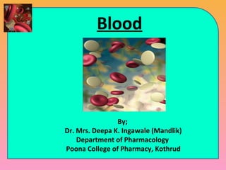
- 1. Blood By; Dr. Mrs. Deepa K. Ingawale (Mandlik) Department of Pharmacology Poona College of Pharmacy, Kothrud
- 2. Content in detail Definition of blood & hematology Functions & physical properties of blood Blood plasma & Formed elements Haemopoiesis Red blood cells & Erythropoiesis Anemia White blood cells & Platelets Mechanisms of homeostasis Pathways of blood clotting Blood groups Inflammation (Signs, types & mechanism) Types of Immunity Disorders of blood (Definitions only) 2
- 3. Learning Objectives To describe different functions of blood. To describe the physical characteristics of blood. To describe the principal components of blood. To explain the process of haemopoiesis. To describe the structure, functions, life cycle, & production of red blood cells. To describe the structure, functions & production of white blood cells. To describe the structure, function & origin of platelets. 3
- 4. Learning Objectives To describe the different mechanisms of hemostasis. To explain various stages of blood clotting. To describe the ABO and Rh system of blood grouping. To describe the components of innate immunity. To define adaptive immunity & origin of T cells and B cells. To explain the relationship between an antigen & an antibody. To compare the functions of cell-mediated immunity & antibody- mediated immunity. 4
- 5. Blood • Blood: A liquid connective tissue composed of extracellular matrix called as blood plasma that dissolves & suspends various cells & cell fragments. • Hematology: The branch of science that deals with the study of blood, blood-forming tissues & blood disorders. 5
- 6. Functions • Transport medium Oxygen, nutrients & waste material Hormones to their target glands Protective antibodies to the site of infection • Protection against infection • Regulation of pH • Maintenance of body temperature • Clot formation 6
- 7. Physical properties • Denser & viscous than water and sticky • Temperature is 38° • Slightly alkaline pH (7.35 to 7.45) • Color of blood varies with its oxygen content When it has a high oxygen content, it is bright red When it has a low oxygen content, it is dark red • Blood volume is 5 to 6 liters in an average adult male & 4 to 5 liters in an average adult female. 7
- 8. Components of Blood • Two components of blood: 1)Blood plasma (55%): Watery liquid extracellular matrix contains dissolved substances 2)Formed elements (45%): Cells & cell fragments 8
- 10. Blood Plasma • When the formed elements are removed from blood, a straw colored liquid is called as blood plasma. – Pale yellow color (91% water, 7% Proteins & 1.5% other solutes) – Albumin: Responsible for maintaining osmotic pressure of blood – Globulins: Responsible for Immune system – Fibrinogen: Responsible for formation of blood clots • Other regulatory substances are electrolytes, nutrients, enzymes, hormones, gases, & waste products (urea, uric acid, creatinine, ammonia, & bilirubin). 10
- 11. Components of Whole Blood Withdraw blood and place in tube 1 2 Centrifuge Plasma (55% of whole blood) Formed elements Buffy coat: leukocyctes & platelets (<1% of whole blood) Erythrocytes (45% of whole blood) 11
- 12. Formed Elements • Red blood cells (Erythrocytes) • White blood cells (Leukocytes) – Granulocytes Neutrophils Eosinophils Basophils – Agranulocytes Lymphocytes Monocytes • Platelets (Thrombocytes) 12
- 13. Haemopoiesis • The process of formation of blood cells called as hemopoiesis or hematopoiesis. • Red bone marrow is the primary site of haemopoiesis. • It is highly vascularized connective tissue located in the bone. • Present mainly in bones of Axial skeleton Pectoral & pelvic girdles Humerus & femur 13
- 15. Haemopoiesis 15
- 16. Red Blood Cells (RBCs) • Biconcave disc shaped • Male: 5.4 M/ mm3 of blood • Female: 4.8 M/ mm3 of blood • Have no nuclei • Functional for about 120 days • Production occurs in the red bone marrow Erythropoiesis Controlled by erythropoietin • Contains Hemoglobin (280 M/RBCs) • Function: Transport of oxygen from lungs to tissues & carbon dioxide from tissues to lungs 16
- 17. 19- 17 Hemoglobin • Consists of: – 4 globin molecules: Colorless protein (96%) – 4 heme molecules (4%): Transport of oxygen • Iron is required for oxygen transport Normal values: Female: 12 to 16 gm/100 ml of blood Male: 14 to 18 gm/100 ml of blood Infants: 14 to 20 gm/100 ml of blood
- 18. Hemoglobin • Lower hemoglobin values may be due to: • Anemia • Bleeding • Destruction of red blood cells • Leukemia • Malnutrition • Nutritional deficiencies of iron, folate, vitamin B12, vitamin B6 • Overhydration • Higher hemoglobin values may be due to: • Congenital heart disease • Dehydration • Erythrocytosis (Increase in RBCs) • Low blood oxygen levels (Hypoxia) • Pulmonary fibrosis • Polycythemia vera (Disorder of the bone marrow) 19- 18
- 19. Formation & Destruction of RBCs 19- 19
- 20. Formation & Destruction of RBCs • Red blood cells live only about 120 days. • Without a nucleus and other organelles, RBCs cannot synthesize new components to replace damaged ones. • Ruptured red blood cells are removed from circulation & destroyed by macrophages in the spleen and liver, and breakdown products are recycled. 1 Macrophages in spleen, liver, or red bone marrow phagocytize ruptured and worn-out red blood cells. 2 The globin and heme portions of hemoglobin are split apart. 3 Globin is broken down into amino acids, which can be reused to synthesize other proteins. 19- 20
- 21. Formation & Destruction of RBCs 4 Iron is removed in the form of Fe3, which associates with the plasma protein transferrin. 5 In muscle fibers, liver cells, and macrophages of the spleen and liver, Fe3 detaches from transferrin and attaches to an iron- storage protein called ferritin. 6 Upon release from a storage site or absorption from the gastrointestinal tract, Fe3 reattaches to transferrin. 7 The Fe3–transferrin complex is then carried to red bone marrow, where RBC precursor cells use it in hemoglobin synthesis. 8 Erythropoiesis in red bone marrow results in the production of red blood cells, which enter the circulation. 19- 21
- 22. Formation & Destruction of RBCs 9 When iron is removed from heme, the non-iron portion of heme is converted to biliverdin, a green pigment, and then into bilirubin, a yellow orange pigment. 10 Bilirubin enters the blood and is transported to the liver. 11 Within the liver, bilirubin is released by liver cells into bile which passes into the small intestine and then into the large intestine. 12 In the large intestine, bacteria convert bilirubin into urobilinogen 13 Some urobilinogen is absorbed back into the blood, converted to a yellow pigment called urobilin and excreted in urine. 14 Most urobilinogen is eliminated in feces in the form of a brown pigment called stercobilin, which gives feces its characteristic color. 19- 22
- 23. Definition of Anemia Deficiency in the oxygen-carrying capacity of blood due to a decrease in erythrocyte number. May be due to: Erythrocyte loss (bleeding) Decreased Erythrocyte production low erythropoietin Decreased bone marrow response to erythropoietin Increased Erythrocyte destruction (hemolysis) 23
- 24. Symptoms of Anemia • Decreased oxygenation – Exertional dyspnea – Dyspnea at rest – Fatigue – Lethargy, confusion • Decreased volume – Fatigue – Muscle cramps – Postural dizziness – Syncope 24
- 25. Types of Anemia Iron deficiency anemia Megaloblastic anemia Pernicious anemia Hemolytic anemia Aplastic anemia Sickle-cell anemia 25
- 26. Anemia Iron Deficiency Anemia: Inadequate absorption or excessive loss of iron Megaloblastic Anemia: Due to deficiency of folic acid & vitamin B12 Aplastic anemia: Destruction of red bone marrow Hemolytic anemia: Due to excessive breakdown of red blood cells Pernicious anemia: Due to impaired absorption of vitamin B12 because of a lack of intrinsic factor in gastric secretions. 26
- 27. Sickle-cell anemia • Body makes sickle-shaped red blood cells. • Sickle-shaped means crescent shape of RBC. • Normal RBCs are disc-shaped & move easily through blood vessels, contain an iron-rich protein called hemoglobin. • Sickle cells contain abnormal hemoglobin called sickle hemoglobin or hemoglobin S. • Sickle hemoglobin causes RBC to develop a sickle shape. • Sickle cells are sticky & tend to block blood flow. 27
- 29. White blood Cells (WBCs) • Range: 5000 – 10,000/mm3 of blood • Produced by leukopoiesis in red bone marrow, Contain nuclei • Functions – Defense against pathogens – Removal of toxins, wastes & damaged cells • Two types • Granulocytes: 75% of total WBC – Neutrophils – Eosinophils – Basophils • Agranulocytes: 25% of total WBC – Lymphocytes – Monocytes Eosinophilic granulocyte Neutrophilic granulocyte Basophilic granulocyte Lymphocyte Monocyte Monocyte 29
- 30. Leukocytosis & Leukopenia • Leukocytosis: An increase in the number of WBCs above 10,000, is called as leukocytosis. • Leukopenia: A decease in the number of white blood cells below 5000, is called as leukopenia. 30
- 31. Neutrophil • 60-70% of total WBC’s • Granules do not stain with dyes • Diameter: 10-12 μm • Nucleus: Usually 2-4 lobed Functions: • Neutrophils are phagocytic towards bacteria (1 neutrophil can phagocytize 5-20 bacteria) 31
- 32. Eosinophil • 2-4 % of total WBC’s • Granules stained by red acidic dyes • Diameter 10-12 μm • Nucleus: Usually 2 lobes • Functions: • Involved in allergic reactions & parasitic infections. • They destroy the antigen-antibody complexes & restrict the process of inflammation. 32
- 33. Basophil • 0.5- 1 % of total WBC’s • Granules stained with basic, purple blue color • Diameter 8-10 μm • Nucleus: Irregular and usually 2 lobes • Granules contain heparin & histamine • Functions: • At the site of infection basophils convert into mast cells • Basophils & mast cells release histamine, bradykinin & serotonin 33
- 34. Lymphocyte • 20-25 % of total WBC’s • Depending upon the site of production & their actions, divided into T, B cells & Natural killer cells • They are divided into • Small lymphocytes- Diameter 6-9 μm • Large Lymphocyte- Diameter 10-14 μm • Nucleus: Round • Functions: • Plays important role in immunity. 34
- 35. Monocyte • 3-8 % of total WBC’s • Diameter: 12-20 μm • Nucleus: Oval or kidney shaped • Monocytes are converted into macrophages of the tissues • Functions: • Phagocytosis 35
- 36. Thrombocytes • Range: 250,000-500,000 /mm3 of blood • Have no nuclei • Diameter: 2-4 μm • Life span: 10 -12 days • Function • Involved in blood clotting mechanism 36
- 37. 19- 37 Blood Group • It is determined by the presence of; • Antigens (Agglutinogen) present on surface of RBCs • Antibodies (Agglutinins) present in blood plasma • Antibodies can bind to RBC antigens, resulting in agglutination (clumping) or hemolysis of RBCs • 2 types of Blood groups – ABO & Rh
- 38. According to ABO blood grouping system: 4 types of blood groups: A B AB O ABO blood grouping system 38
- 39. Blood group A A antigens on the surface of RBCs B antibodies in the plasma. Blood group B B antigens on the surface of RBCs A antibodies in the plasma. ABO blood grouping system 39
- 40. Blood group AB A & B antigens on the surface of RBCs No A & B antibodies are present in plasma. Blood group O Neither A or B antigens on surface of RBCs Both A & B antibodies are present in plasma. ABO blood grouping system 40
- 41. Antigens & antibodies of ABO blood types 41
- 42. Determination of blood group 42
- 43. Blood group ‘O’ is called "universal donor“ because it has no antigens on RBC. Blood group ‘AB’ is called "universal receivers” because it has no anti- bodies in the plasma. 43
- 44. 19- 44 Rh Blood Group • First studied in Rhesus monkeys • Types – Rh positive: antigens present on surface of RBCs – Rh negative: antigens are absent on surface of RBCs • Hemolytic disease of the newborn (HDN) – Mother produces anti-Rh antibodies that cross placenta & cause agglutination & hemolysis of fetal RBCs
- 45. • A person with Rh - blood develops Rh antibodies in the blood if receives blood from Rh+ person. • There is devolvement of anti-Rh antibodies, that react with donor’s Rh antigens & aggutinate the blood. •A person with Rh+ blood can receive blood from a person with Rh- blood without any problems. 45
- 46. Hemostasis- Stoppage of bleeding • When the blood vessel get damaged, platelet plays a vital role in Hemostasis. • 3 mechanisms are involved in hemostasis; Vascular spasm Blood clotting Platelet plug formation 46
- 47. Hemostasis 4. Coagulation 1. Blood vessel injury 2. Vasoconstriction 3. Platelet plug formation 47
- 48. Vascular spasm • When arteries are damaged, the smooth muscle in the walls of arteries contracts immediately, a reaction is called as vascular spasm. • This reduces blood loss for several minutes to several hours. • The spasm is caused due to release of mediators from the activated platelets. 48
- 49. Blood clotting • Blood clot consists of network of insoluble protein fibers called as fibrin in which the formed elements (RBCs, WBCs & Platelets) of blood are trapped. • The process of clot formation is called as clotting. 49
- 50. Blood clotting Blood clotting factors: I.Fibrinogen II.Prothrombin III.Tissue factor IV.Calcium ions V.Labile factor- Proaccelerin VI.Absent VII.Stable factor- Proconvertin VIII.Antihaemophilic factor (A) IX.Christmas factor or AHF (B) X.Stuart factor XI.Plasma thromboplastin or AHF (C) XII.Hageman factor or AHF (D) XIII.Fibrin- stabilizing factor 50
- 51. 51
- 52. The Extrinsic Pathway • Fewer steps & occurs rapidly within a seconds • Tissue protein called tissue factor (TF) leaks into the blood from cells outside blood vessels & initiates the formation of prothrombinase. • TF is a complex mixture of lipoproteins & phospholipids released from the surfaces of damaged cells. • In the presence of Ca2 , TF begins a sequence of reactions that activates clotting factor X . • Once factor X is activated, it combines with factor V in the presence of Ca2 to form the enzyme prothrombinase, completing the extrinsic pathway. 52
- 53. The Intrinsic Pathway • More complex & occurs more slowly, requires several minutes. • Its activators are present either in direct contact with blood or contained within the blood. • Outside tissue damage is not needed. • If endothelial cells become damaged, blood can come in contact with collagen fibers of the blood vessel. • Trauma to endothelial cells causes damage to platelets, resulting in release of phospholipids by the platelets. 53
- 54. The Intrinsic Pathway • Contact with collagen fibers activates clotting factor XII, which begins a sequence of reactions that activates clotting factor X. • Platelet phospholipids & Ca2 can also participate in the activation of factor X. • Once factor X is activated, it combines with factor V to form the active enzyme prothrombinase, completing the intrinsic pathway. 54
- 55. The Common Pathway • The formation of prothrombinase starts the beginning of the common pathway. • In the second stage of blood clotting, prothrombinase & Ca2 catalyze the conversion of prothrombin to thrombin. • In the third stage, thrombin, in presence of Ca2 , converts fibrinogen (soluble), to loose fibrin threads (insoluble). • Thrombin also activates factor XIII (fibrin stabilizing factor), which strengthens and stabilizes the fibrin threads into a sturdy clot. 55
- 56. Platelet Plug formation 1. Platelet Adhesion 2. Platelet release reaction 3. Platelet aggregation 56
- 57. Platelet Plug formation • Inspite of having small size, platelets store lot many chemicals. • It contains ADP, ATP, Ca2 , serotonin, thromboxane A2, a prostaglandin, fibrin-stabilizing factor & platelet-derived growth factor (PDGF). 57
- 58. Platelet Plug formation • Initially, platelets stick to parts of a damaged blood vessel, such as collagen fibers of damaged endothelial cells. • This process is called as platelet adhesion. • Due to adhesion, the platelets become activated, & their characteristics change dramatically. • They extend many projections and they begin to liberate the contents of their vesicles. • This phase is as called platelet release reaction. 58
- 59. Platelet Plug formation • Liberated ADP & thromboxane A2 play a major role of activating nearby platelets. • Serotonin & thromboxane A2 function as vasoconstrictors, causing contraction of vascular smooth muscle, which decreases blood flow through the injured vessel. 59
- 60. Platelet Plug formation • The release of ADP makes other platelets sticky, & adhere to the originally activated platelets. • This gathering of platelets is called as platelet aggregation. • The accumulation & attachment of large numbers of platelets to the site of injury to form a solid mass called as platelet plug. 60
- 61. Inflammation • Inflammation is the reaction of vascularized living tissue to local injury. • This reaction results in accumulation of fluid & leucocytes in the extracellular tissues. • It is a defense mechanism to eliminate or limit the spread of injury or injurious agents. 61
- 62. Etiology 62
- 65. Types of Inflammation • Two types of inflammation; • Acute: Short duration (hours or day) • Chronic: Longer duration 65
- 66. Acute inflammation Short duration, lasting for minutes, hours, or days Its main characteristics: The exudation of fluid & plasma proteins (edema) The migration of leukocytes and neutrophils to the site of injury. Longer duration Its main characteristics: Associated with lymphocytes & macrophages migration, proliferation of blood vessels, fibrosis, & tissue necrosis. Chronic inflammation Acute & Chronic Inflammation 66
- 67. Tissues & cells in inflammation The circulating cells: Neutrophils, Monocytes, Eosinophils, Lymphocytes, Basophils, & Platelets The connective tissue cells: Mast cells, Fibroblasts, Macrophages & Lymphocytes The extracellular matrix: Fibrous proteins (Collagen, Elastin), glycoproteins (Fibronectin, Laminin, Collagen) & Proteoglycans 67
- 69. Inflammatory Mediators • Mediators have 2 origins; • Plasma Complimentary system Kinin System Clotting system • Cells Histamine Serotonin Lysosomal enzymes Prostaglandins Leukotrienes (IL1, IL 6, IL8) Cytokines Platelet activating factor 69
- 70. Mechanism of Inflammation 1. Vaso dilatation 2. Exudation - Edema 3. Emigration of cells 4. Chemotaxis 5. Phagocytosis 70
- 71. Vascular changes 1. Change in vascular blood flow • Transient vasoconstriction of the arterioles followed by vasodilatation. • Vasodilation involves arterioles first then microvascular bed (leading to redness & heat) • It leads to slowing of blood circulation (blood stasis) 71
- 72. Vascular changes 2. Increase in vascular permeability • Vascular permeability: capacity of a blood vessel wall to allow the entry of small molecules or lymphocytes in and out of the vessel. • Increase in hydrostatic pressure leading to leakage of fluid to the extravascualr space • Increase in osmotic pressure leading to leakage of protein-rich fluid • The end result is Edema. 72
- 73. Cellular events Divided into 5 steps; Margination Adhesion to endothelium Emigration Chemotaxis Phagocytosis 73
- 74. 74
- 75. Cellular Events 1. Margination: • Sticking of leucocytes to the endothelial lining of blood vessels (Pavementation) 2. Adhesion: • Mediators of inflammation activates the adhesion molecules on the surface of leucocytes & endothelial cells and facilitates their adhesion. • Adhesion molecules are bacterial toxins, complement fragments, chemotactic peptides and cytokines 75
- 76. Cellular Events 3. Emigration: • Emigration refers to the process by which leucocytes escape from blood vessels to the perivascular tissue. 4. Chemotaxis: • It is defined as unidirectional migration of leucocyte towards an attractant (chemotactic factors) 76
- 77. Cellular Events 5. Phagocytosis: • It is the process of engulfment of pathogens & damaged cells. • 3 distinct steps: Recognition & attachment Engulfment Killing or degradation 77
- 78. Phagocytosis 1. Recognition & attachment: • WBC’s have specific receptors on the surface. 2. Engulfment: • The particle is engulfed by pseudopodia & enclosed in membrane bound vesicle called as phagosome. 3. Killing & degradation: • Phagosome fuses to lysosome to form phagolysosome. Killing is facilitated by: • a. Oxygen free radicals (oxidative burst) • b. Lysosomal enzymes (myeloperoxidase) 78
- 79. Phagocytosis 79
- 80. 80 INFECTIONINFECTION IMMUNITYIMMUNITY Host BodyHost Body MICROBESMICROBES Immunity
- 81. Immunity • From Latin word immunis- free from burden • [Burden: disease caused by variety of MO.] • Immunity: It is the body's ability to fight off harmful micro-organisms –PATHOGENS- that invade the body. 81
- 82. 82 Immune system • Immune system: Body’s defense mechanism consists of specialized cells & proteins designed to destroy the foreign particles.
- 83. Terminology – Antibodies • Protein produced in response to foreign substances • Can destroy or neutralize antigens – Antigens • Substances that can elicit a specific immune response • Foreign substances. – Pathogens • Micro-organisms that can cause diseases 83
- 85. 85 Immunity Two types of immunity: 1) Innate immunity (Natural or Non specific) - First Line defense mechanism - Second Line defense mechanism 2) Acquired immunity (Adaptive or Specific) - Third Line defense mechanism
- 86. 86 Component of Innate Immunity Innate Immune system First line Second line 1) Mechanical barriers A- Cells 2) Chemical & biochemical inhibitors 1- Phagocytes 3) Normal flora 2- Natural killer B- Interferons C- Inflammatory barriers D- Complement System
- 87. 87 First line: Innate Immune system 1) Mechanical barriers - Intact skin - Mucous coat - Mucous secretion - Blinking reflex and tears - Coughing & sneezing reflex
- 88. 88 2) Chemical & biochemical inhibitors - Sweet and sebaceous secretion - Hydrolytic enzymes in saliva - HCl of stomach - Proteolytic enzyme in small intestine - Lysozyme in tears - Acidic pH in the vagina
- 89. 89 Normal microbial flora Location of normal microbial floraLocation of normal microbial flora Each of these areas of the body contain their own microenvironmentsEach of these areas of the body contain their own microenvironments
- 90. 90 Normal flora
- 91. Cells of the immune system 91
- 92. 92 Second line: A) cells 1- Phagocytes • Specialized cells for ingestion & destruction of invading m.o. • Phagocytes are produced from Bone marrow stem cells. • Phagocytes may be microphages or macrophages • Microphages: Neutrophils • Macrophages: Monocytes
- 93. 93 2- Natural killer (NK) Cells • NK Cells are agranular lymphocytes. • Play a major role in rejection of tumors & cells infected by viruses. • Also called Null cells. • Functions: • Cytotoxic for viral infected cells, bacterial, fungal, parasitic infection.
- 94. 94 B) Interferons • Proteins produced by virally infected cells which act as a antiviral * Types of interferons: 1- Alpha interferon: Secreted by Macrophages 2- Beta interferon: Secreted by Fibroblasts, Viruses 3- Gamma interferon: T- lymphocytes, Specific antigens Action of interferons: 1) Activate T-cells 2) Activate macrophages 3) Activate NK
- 95. 95 C) Inflammatory response * Tissue damage by injury or by invading pathogen * Inflammatory response: Tissue damage Release of chemical mediators from Leukocytes (Histamine, Fibrin, Kinin, Cytokines) Invading microbe Redness of tissue Tissue temperature Vasodilatation of capillaries Capillary permeability Influx of fluids Influx of phagocytes into tissues
- 96. Complement System • Complements (C) is a factors that present in serum & are activated by Ag-Ab interactions & shows biological effects. • It helps an antibodies & phagocytic cells to clear pathogens from an organism. 96
- 97. Adaptive Immunity • It is composed of highly specialized, systemic cells that eliminate the pathogenic microorganisms. • The adaptive immune consists of; • Antibody responses • Cell-mediated responses • Carried out by different lymphocyte cells, B cells and T cells, respectively. 97
- 98. Acquired Immunity Active Immunity Passive Immunity Natural Artificial Natural Artificial 98
- 99. ACTIVE VERSUS PASSIVE IMMUNITY Adaptive Immunity Artificially AcquiredNaturally Acquired 99
- 100. Adaptive immunity • The 3rd Line of Defense: Specific Defense Mechanisms 100 Adaptive immunity Humoral immunity Cellular immunity 100
- 101. Acquired Immune System 1. Antibody (Humoral) immunity – B-cells in body’s fluids produce antibodies in response to a particular antigen. 1. Cell-mediated immunity – Killer T-cells (Cytotoxic T-cells) destroy • Cancer cells • Virus & bacteria infected cells 101
Editor's Notes
- FG20_05A.JPG Title: White Blood Cells Notes: Comparison of leukocytes as seen in blood smears. (a)Neutrophil. (b)Eosinophil. (c)Basophil. (d)Monocyte. (e)Lymphocyte. Platelets are visible in part (e) as small cellular fragments between the RBC&apos;s. Keywords: white blood cells, neutrophil, eosinophil, basophil, monocyte, lymphocyte, red blood cell, platelet
- FG20_05B.JPG Title: White Blood Cells Notes: Comparison of leukocytes as seen in blood smears. (a)Neutrophil. (b)Eosinophil. (c)Basophil. (d)Monocyte. (e)Lymphocyte. Platelets are visible in part (e) as small cellular fragments between the RBC&apos;s. Keywords: white blood cells, neutrophil, eosinophil, basophil, monocyte, lymphocyte, red blood cell, platelet
- FG20_05C.JPG Title: White Blood Cells Notes: Comparison of leukocytes as seen in blood smears. (a)Neutrophil. (b)Eosinophil. (c)Basophil. (d)Monocyte. (e)Lymphocyte. Platelets are visible in part (e) as small cellular fragments between the RBC&apos;s. Keywords: white blood cells, neutrophil, eosinophil, basophil, monocyte, lymphocyte, red blood cell, platelet
- FG20_05E.JPG Title: White Blood Cells Notes: Comparison of leukocytes as seen in blood smears. (a)Neutrophil. (b)Eosinophil. (c)Basophil. (d)Monocyte. (e)Lymphocyte. Platelets are visible in part (e) as small cellular fragments between the RBC&apos;s. Keywords: white blood cells, neutrophil, eosinophil, basophil, monocyte, lymphocyte, red blood cell, platelet
- Title: White Blood Cells Notes: Comparison of leukocytes as seen in blood smears. (a)Neutrophil. (b)Eosinophil. (c)Basophil. (d)Monocyte. (e)Lymphocyte. Platelets are visible in part (e) as small cellular fragments between the RBC&apos;s. Keywords: white blood cells, neutrophil, eosinophil, basophil, monocyte, lymphocyte, red blood cell, platelet