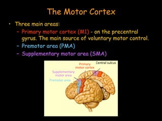
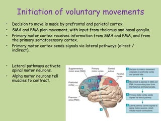
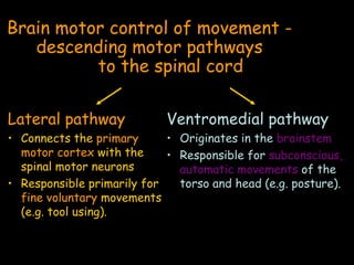
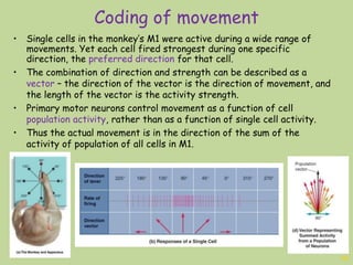
2. The Motor Cortex
• Three main areas:
– Primary motor cortex (M1) - on the precentral
gyrus. The main source of voluntary motor control.
– Premotor area (PMA)
– Supplementary motor area (SMA)
Central sulcusPrimary
motor cortex
Supplementary
motor area
Premotor area
3. Initiation of voluntary movements
18
• Decision to move is made by prefrontal and parietal cortex.
• SMA and PMA plan movement, with input from thalamus and basal ganglia.
• Primary motor cortex receives information from SMA and PMA, and from
the primary somatosensory cortex.
• Primary motor cortex sends signals via lateral pathways (direct /
indirect).
• Lateral pathways activate
spinal motor neurons.
• Alpha motor neurons tell
muscles to contract.
4. Brain motor control of movement -
descending motor pathways
to the spinal cord
Lateral pathway
• Connects the primary
motor cortex with the
spinal motor neurons
• Responsible primarily for
fine voluntary movements
(e.g. tool using).
Ventromedial pathway
• Originates in the brainstem
• Responsible for subconscious,
automatic movements of the
torso and head (e.g. posture).
5. Coding of movement
19
• What is coded in the primary motor cortex? And how?
• While our movements are generally very precise, each
neuron in M1 is active in a wide range of movements.
• This led Georgopoulos and his colleagues to propose the
idea that movement is encoded by populations of
neurons rather than individual cells.
6. Coding of movement
20
• Single cells in the monkey’s M1 were active during a wide range of
movements. Yet each cell fired strongest during one specific
direction, the preferred direction for that cell.
• The combination of direction and strength can be described as a
vector – the direction of the vector is the direction of movement, and
the length of the vector is the activity strength.
• Primary motor neurons control movement as a function of cell
population activity, rather than as a function of single cell activity.
• Thus the actual movement is in the direction of the sum of the
activity of population of all cells in M1.
7. Angiotensin II is produced in a reaction
involving an enzyme secreted by the
a. adrenal glands.
b. kidneys.
c. pituitary gland.
d. subfornical organs.
17. Brain structures associated with sleep:
• a. Dorsal raphe nuclei
• b. Thalamus
• c. Reticular formation
• d. Locus coeruleus
18. Falling asleep
Sleep debt – accumulation of serotonin in ventrolateral preoptic nucleus of
the hypothalamus, eventually deactivating the wakefulness circuits.
Adenosine – throughout the brain – is also related to sleep debt. Reminder:
caffeine is an adenosine antagonist!
19. Regarding osmotic pressure, the
intracellular fluid is hypertonic /
isotonic / hypotonic to the extracellular
fluid (circle the correct answer).
20. Progressive degeneration motor neurons
throughout the body characterize the
neurodegenerative disease
_______ _______ _______.ALS = Amyotrophic Lateral Sclerosis
21. Masculinization of the human brain
depend on:
• a. Aromatization
• b. Androgens
• c. Estradiol
• d. Placenta
22. Which of the following is correct about external
genitalia?
a. Ovarian hormones are responsible for development
of the labial folds, clitoris, and outer part of the
vagina.
b. The external genitalia are female-like in people
with androgen insensitivity syndrome (AIS).
c. The external genitals develop in males and females
from separate precursors.
d. AMH masculinizes the male external genitalia.
23. Testes produce small amounts of
estrogens including estradiol
• a. True
• b. False
24. Differentiation of external genitalia
occur at:
• a. 6 weeks
• b. 6-9 weeks
• c. 6-12 weeks
• d. None of the above
25. 3 Stages of Sexual Prenatal Development
1. Development of gonads (~6 weeks)
Until 6 weeks – identical primordial gonads
Genetic sexXY Chromosomes XX Chromosomes
Gene
expression
Sex-determining region of Y (SRY) gene
Testis-determining factor protein encoded
Testes development
No gene
expression
Ovaries development
No SRY expression
2. Differentiation of internal organs (9-12 weeks)
Until ~3 month both possess a male Wolffian system and a female Mullerian system
Testes secrete 2 hormones:
Testosterone (T; type of male androgen)
Anti-Mullerian hormone (AMH)
Promote development of Wolffian system
–> seminal vesicles, vas deferens, prostate
Absence of T ->
regression of Wolffian system
Degeneration of Mullerian system
Ovaries are not active in producing
female fetus hormones.
T and AMH are also not
produced.
Absence of AMH -> Mullerian system develops
-> uterus, vagina upper portion, fallopian tubes
3. Differentiation of external genitalia (6-12 weeks)
5-alpha-dihydrotestosterone (DHT)
hormone created from T
Development of male external genitalia:
penis, scrotum
Development of female external
genitalia: labia, clitoris, outer vagina
27. A variety of chemicals can also
activate nociceptors
Lactic acid
increase in hydrogen ions in the extra cellular fluid
activate nociceptors
send unpleasant messages of soreness to the brain.
Vanilloids, specifically Capsaicin (found in hot peppers).
Chemicals released when a cell is damaged (potassium
ions, enzymes, histamine, and ATP).
28. A tactile receptor, not encapsolated,
provide information about touch?
• a. Merkel’s discs
• b. Ruffini’s endings
• c. Meissner’s corpuscles
• d. None of the above
29. Four tactile receptors perceive touch
Type Encapsul-
ated?
Size of
receptive
field
Quality of
stimulus
Adaptation Level of
skin
Meissner’s
corpuscles
Yes Small touch fast-
adapting
Upper
levels
Pacinian
corpuscles
Yes Large vibration fast-
adapting
Lower
levels
Merkel’s
discs
No Small touch slow-
adapting
Upper
levels
Ruffini’s
endings
No Large stretch slow-
adapting
Lower
levels
+ free nerve endings
and endings wrapped around hair follicles
30. Thank you all for a great year,
I wish you all good luck!
Limor
Editor's Notes
ADH controls the retention of water Aldosterone controls the retention of sodium. Sodium is essential to maintain extracellular fluid, as without it the extracellular fluid would become hypotonic, and too much water would enter the cells. When blood volume is lost, treatments to increase extracellular sodium levels prevent further loss of water into cells.