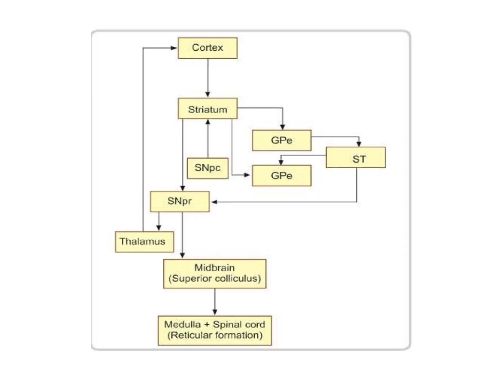
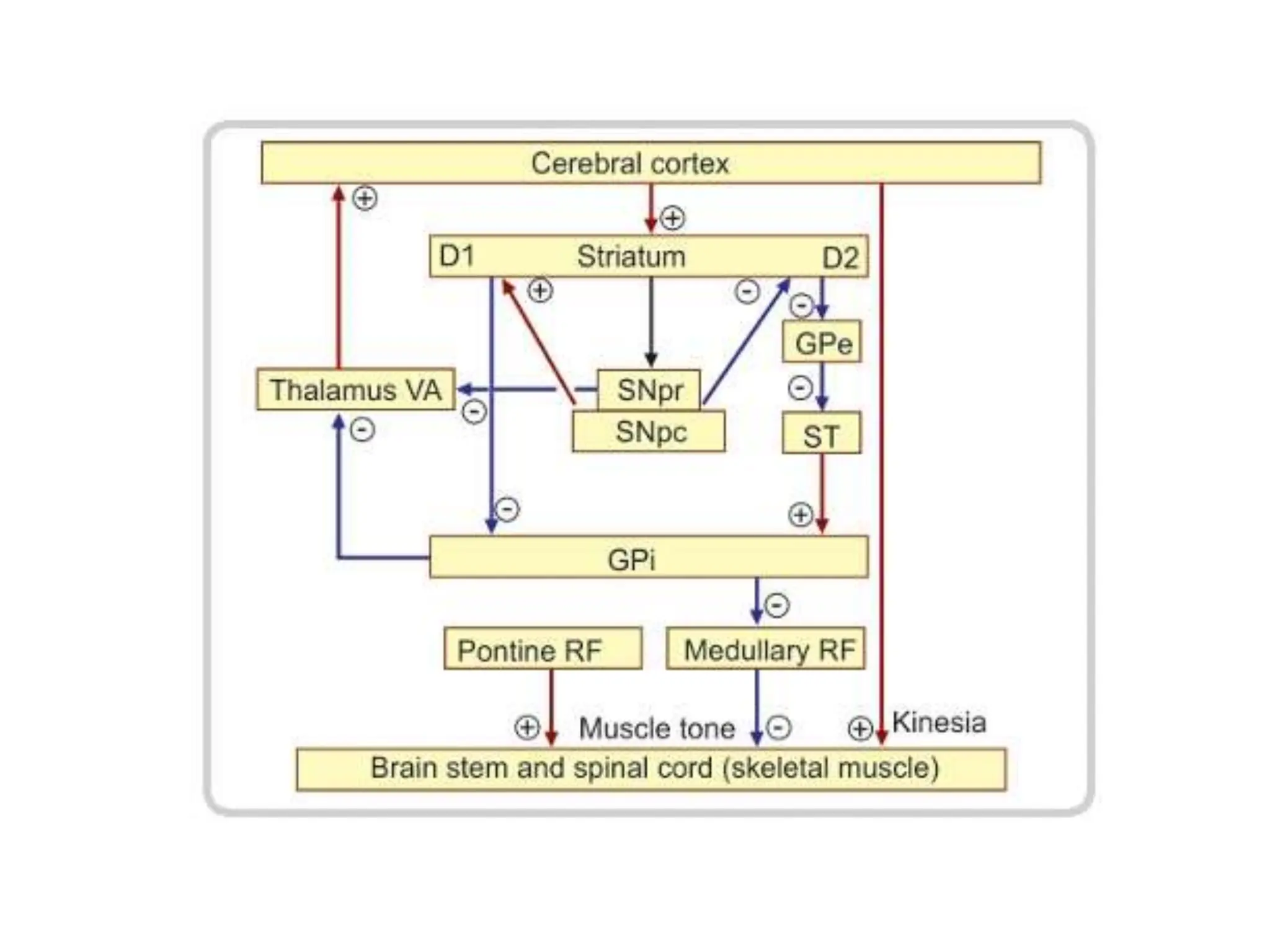
BASAL GANGLIA
The basal ganglia are
large masses of grey
matter situated in
cerebral hemisphere.
Structures
Anatomical Divisions
• Caudate nucleus
• Lentiform nucleus –
consists of putamen and
globus pallidus
• Amygdaloid nuclear
complex
• Claustrum`
CORPUS STRIATUM
CAUDATE NUCLEUS LENTIFORM NUCLEUS
PUTAMEN GLOBUS
PALLIDUS
(PALEOSTRIATUM)
STRIATUM
(NEOSTRIATUM)
Neostriatum (the striatum) – formed by caudate
nucleus and putamen
Paleostriatum – formed by pallidum (globus
pallidus)
Archistriatum – formed by Amygdaloid nuclear
complex and Claustrum
Functional divisions of basal ganglia
• Corpus striatum
• Subthalamic nucleus
• Substantia nigra
• Ventral striatum
• Ventral pallidum
It is a C shaped mass of grey matter
Derived from telencephalon.
Consists of large head, body and
thin tail.
Relations
It is intimately related to the lateral
ventricle.
•Head - bulges into anterior horn of
the ventricle and forms its floor. It is
continuous with the putamen.
•The body lies in the floor of the
central part of the ventricle
•Tail in the roof of the inferior horn
of the ventricle.
Caudate nucleus
The anterior part of the head is fused with the lentiform
nucleus inferiorly. This region is called fundus striati.
Fundus striati is continuous inferiorly with the anterior
perforated substance.
The tail lies in close relation to the amygdaloid complex.
Caudate nucleus
The body is related
medially to the thalamus
and laterally to the
internal capsule which
seperates it from the
lentiform nucleus.
Lentiform nucleus
Appears triangular in
the coronal section
with its tip directed
medially.
Divided by a thin
lamina of white matter
known as external
medullary lamina into
Lateral part- putamen
Medial part- globus
pallidus
Putamen
• Derived from
telencephalon.
• Lies medial to the insula
• Bounded laterally by the
fibres of external
capsule and medially by
the globus pallidus.
• Separated from the
caudate nucleus by the
anterior limb of the
internal capsule
Globus pallidus
• It is diencephalon in origin.
• It constitutes the inner
component of the lentiform
nucleus.
• The globus pallidus is
subdivided into external
(GPe) and internal (GPi)
segments by the internal
medullary lamina.
• Medial – posterior limb of
internal capsule seperates
from the thalamus
• Lateral – putamen.
Amygdaloid nuclear complex
• Lies in the temporal lobe close to
the temporal pole.
• Lies deep to the uncus
• Related to the anterior end of the
inferior horn of the lateral
ventricle.
Claustrum
• Thin lamina of grey matter lies
lateral to the lentiform nucleus. It
is separated from the latter by
the fibres of external capsule.
• Laterally it is separated by a thin
layer of white matter from the
cortex of insula.
• Its connections and functions are
not known.
Substantia Nigra
It consists of 2 parts
Pars reticulata – ventral cell
sparse portion, functionally
related to internal part of globus
pallidus. They use the inhibitory
neurotransmitter GABA.
Pars compacta – dorsal cell rich
portion. Neurons here are
pigmented because of the
presence of neuromelanin. These
cells contain dopamine.
Mesencephalic in origin.
It is a large motor nucleus present in the midbrain between the
tegmentum and the basis pedunculi.
Subthalamic nucleus
Derived from
diencephalon.
Lies dorsomedial to the
posterior limb of the
internal capsule and
dorsal to the substantia
nigra.
Lesion – hemiballismus
Ventral Striatum
• Masses of grey matter lying in the
region of anterior perforated
substance.
• The caudate nucleus and
lentiform nucleus fuse inferiorly
to form the fundus striati.
• Immediately below the fundus
striati is the olfactory tubercle in
the anterior perforated
substance.
• More medially is a mass of grey
matter called the nucleus
accumbens. It is closely related to
the caudate nucleus
superolaterally and to the septal
nuclei medially.
• The ventral striatum consists of
the olfactory tubercle and the
nucleus accumbens.
Ventral pallidum
In coronal section, the anterior commissure runs laterally
just below the head of the caudate nucleus. It cuts
through the globus pallidus.
The part of the globus
pallidus lying inferior to
the anterior commissure
is called the ventral
pallidum.
Thus,
Dorsal striatum – caudate nucleus and putamen
Ventral striatum – olfactory tubercle and nucleus
accumbens
Dorsal pallidum – globus pallidus
Ventral pallidum – part below the anterior perforated
substance
Blood supply
• Lenticulostriate
branches of middle and
anterior cerebral
arteries.
• Anterior choroidal
branch of internal
carotid artery.
Internal Organization
Others are medium aspiny neurons,
large spiny and aspiny neurons.
In striatum,
Zones that contain low density of
AChE enzymatic activity surrounded
by areas rich in AChE activity. AChE
rich areas are called matrix. AChE
poor areas are called striosomes.
Caudate nucleus and putamen are frequently referred together because
of their common charecteristics. They are composed of histologically
identical cells.
Most neurons are medium spiny neurons – medium sized cells that
possess spines on their dendrites.
Afferent connections
From cerebral cortex arising primarily from the
pyramidal cells of the layers V, VI via corticostriate
fibres. These are glutaminergic.
• sensorimotor cortex putamen
• association regions caudate nucleus
• Prefrontal regions head of the caudate nucleus
The intralaminar nucleus particularly central median
nucleus of thalamus via thalamostriate fibres
The pars compacta of the substantia nigra via
nigrostriate fibres. They are dopaminergic.
Afferent connections
• Noradrenergic fibres from the locus ceruleus
• Serotoninergic fibres from the raphe nuclei (in the
reticular formation of the midbrain)
• Afferents from limbic cortical areas, hippocampus
and the amygdala terminates in the ventral
striatum.
• The afferents from the cerebral cortex and the
thalamus provide the striatum with various
modalities of sensory information.
• Disruption of the input pathways is associated with
movement disorders like parkinsons disease.
Internal processing
• The direct pathway – increased motor activity
• The indirect pathway – decreased motor activity
CORTEX
STRIATUM
GPi
THALAMUS CORTEX
CORTEX
STRIATUM
GPi
GPe
STN
THALAMUS
Glu
GABA
Subs P
Glu
Glu
GABA
Enk
DIRECT
PATHWAY
INDIRECT
PATHWAY
Glu
GABA
Efferent connections
The main output is concentrated upon the pallidum and on
the substantia nigra.
Outflow from globus pallidus forms four bundles
1. Fasciculus lenticularis – arises from inner segment of
globus pallidus and enters the subthalamic region.
2. Ansa lenticularis – arises from both the inner and outer
segments of the globus pallidus and enters the
subthalamic region where it meets the dentato
rubrothalamic fibres and the fasciculus lenticularis. The
union of the three tracts is called fasciculus thalamicus
which terminates in the ventralis anterior, ventralis
lateral and centro median nuclei of the thalamus.
3. Subthalamic fasciculus- consists of reciprocal
connections between the globus pallidus and nucleus
subthalamicus.
4. Pallidonigral fibres – pass to the substantia nigra
Efferent connections
Substantia Nigra
Pars compacta
• Sends a dopaminergic projection to the striatum.
• A projection from striatum ends in pars reticularis.
• Pars reticulata receives fibres from pallidum directly or after
relay in the subthalamic nucleus or in the pedunculopontine
nucleus.
Pars reticularis
• Projects to the ventral lateral and ventral anterior nucleus of
the thalamus.These impulses are relayed to premotor and
prefrontal areas of the cortex.
• Other efferents reach the superior colliculus. They are
relayed from there to the reticular formation of the medulla
and to the spinal cord.
Circuitry of corpus striatum
• Starting from pars compacta, nigrostriate fibres liberate dopamine
at their terminal.
• Dopamine acts as a facilitatory neurotransmitter in the striatum
when the receptors are D1.
• It acts as a inhibitory neurotransmitter in the striatum when the
receptors are D2.
• Striatal D1 neurons inhibits GPi through a direct path.
• Striatal D2 neurons facilitates GPi through a indirect path. This
balances the impulses reaching the GPi.
• GPi normally inhibits the thalamus. Thalamus facilitates the the
cortex which causes the normal kinesia.
• GPi inhibits medullary reticular formation, which normally inhibits
the muscle tone.
• Impulses from pontine reticular formation increases muscle tone.
A fine balance between the pontine and medullary reticulospinal
tracts maintains normal muscular tone.
Circuitry of corpus striatum in
Parkinson’s disease.
• There is destruction of substantia nigra.
• The inhibitory impulses sent from GPi to medullary
reticular formation is stronger than normal causing
an imbalance between the pontine and medullary
reticulospinal tracts causing hypertonia.
• Inhibition of thalamo cortical pathway results in
hypokinesia.
• The pin rolling movements at rest is due to resting
discharge of dentate nucleus of cerebellum to the
VL nucleus of the thalamus.
Circuitry of corpus striatum in chorea
and ballismus
• The pathways for muscle tone and kinesia reverse
when compared to Parkinsons.
• Destruction of the D2 neurons in striatum causes
chorea.
Functions
• Pallidum eloborates the simple motor and postural
mechanisms of the brain stem into complex autonomic
motor behavior.
• Centre for expression gestures and reactive movements.
• The striatum eloborates and smoothens the coarser
motor integrations of the pallidum.
• The subthalamic nucleus, red nucleus, substantia nigra
have a synergistic action and integrates stereotyped
behavior and controls complex muscular movements.
• Also process information related to emotions,
motivations, and cognitive functions.
Clinical correlation
Various kinds of abnormal movements are seen in
neurological disorders involving the basal ganglia.
The movements could be
• Hyperkinetic, hypotonic (chorea, athetosis,
ballismus)
• Hypokinetic, hypertonic (parkinsons)
NEUROPSYCHIATRIC
DISORDERS
Parkinson’s disease
• A neurodegenerative disease associated with loss of
dopaminergic neurons in the Substantia Nigra pars
compacta.
• Parkinson's disease affects movement, producing motor
symptoms.
• Non-motor symptoms, which include
I. autonomic dysfunction,
II. neuropsychiatric problems (mood, cognition, behaviour
or thought alterations),
III. sensory and sleep difficulties, are also common. Some of
these non-motor symptoms are often present at the time
of diagnosis and can precede motor symptoms.
Characteristic triad
• Resting tremor
• Rigidity
• Bradykinesia
Others – gait and postural disturbances
Psychiatric manifestations
• Depression, apathy, anxiety, psychosis.
Huntington’s disease
• Huntington's disease (formerly Huntington's
chorea) is an autosomal dominant
neurodegenerative disorder characterized by
midlife onset, a progressive course, and a
combination of motor, psychiatric, and cognitive
symptoms.
• The disease is caused by a CAG (trinucleotide)
repeat expansion mutation in the huntingtin gene
on chromosome 4.
Primary involuntary movement abnormality
• Chorea or choreoathetosis
Decreased facial expression, difficulty in swallowing,
chewing, speaking, sleep disturbance, impairment in
cognition and memory, subcortical dementia
Psychiatric manifestations
•Higher rates of suicide
•Depression
•Mania
•Personality change
Wilson’s disease
Autosomal recessive disorder in which mutations in
the gene atp7b result in abnormal accumulation of
copper in the liver, basal ganglia, and other tissues.
Neuropsychiatric pathophysiology:
• When copper deposits in the basal ganglia specifically
in the lenticular nucleus, there is cell death.
Neurological findings
• Tremor, dystonia, rigidity, choreoathetosis,
bradykinesia, masked faces, micrographia, frontal
lobe disorder, subcortical dementia.
Psychiatric manifestations
• Personality changes, depression, suicidality, anxiety
disorders, psychotic disorders.
Fahr’s syndrome
• Idiopathic basal ganglia calcification
• Genetically dominant, inherited neurological disorder
most commonly affects lentiform nucleus and Gpi.
• Occurs in 3rd to 5th decade.
• Deteriorization of motor function and speech, seizure,
involuntary movement, dementia, headache, visual
impairment.
• Unsteady gait, slurring of speech, difficulty in
swallowing ,clumsiness, fatiguability.
Basal ganglia stroke
• Changes in the body movement, rigidity, stiffness, tremor,
ataxia, difficulty in swallowing, smiling, speech.
• Cognitive impairment – poor attention, memory
impairment
• Personality changes – frustration ,anxiety, loss of interest,
loss of motivation, depression, anger.
• Right basal ganglia stroke
• Anosognosia – unable to perceive the severity of deficit,
left side neglect, visuospatial hemineglect, constructional
apraxia.
• Left basal ganglia stroke
• Apathy (lack of interest), memory impairment, major
depression.
Chorea
• Rapid involuntary purposeless dancing movements
of the limbs.
• Chorea affects the proximal joints, while that of the
athetosis affects the distal joints.
• Sydenhams chorea- the pathology is seen in the
striatum
• Huntington’s chorea- autosomal dominant
degenerative disease of the striatum and cerebral
cortex
• Athetosis
• Continuous slow writhing movements with a
propensity to affect the arms and hands.
• Ballism
• Hemorrhagic ivolvement of the subthalamic nucleus.
• Involuntary and violent, flinging movements of large
amplitude
• Hemiballism – involves one half of the body , caused
by lesions in the subthalamic nucleus of the opposite
side. When restricted to one limb it is called as
monoballism.
OCD
• There is evidence of basal ganglia dysfunction in
imaging studies. Both increase and decreased
volumes of caudate nucleus is reported.
• Most patients showed an increased blood flow to
the caudate.
AUTISM
• Enlargement of caudate nucleus upto 8% . The
greater caudate volume is proportional to the
increased total brain volume.
• Motor, communicative, and social impairments are
associated with shape abnormalities in the basal
ganglia.
• Glutamate dysfunction in basal ganglia is associated
with autism.
ADHD
• Neuroimaging studies show evidence of striatal
dysfunction in ADHD.
• concluded that ADHD may be related to functional
abnormalities in the putamen.
• Boys with ADHD showed a significantly smaller
volume of basal ganglia compared with typically
developing boys.
ADDICTION
• Amygdala is a critical structure in addiction.
• Nucleus accumbens is also important in drug re
inforcement and addiction.
Thank You

Basal ganglia anatomy and physiology ppt

  • 1.
  • 2.
    The basal gangliaare large masses of grey matter situated in cerebral hemisphere. Structures Anatomical Divisions • Caudate nucleus • Lentiform nucleus – consists of putamen and globus pallidus • Amygdaloid nuclear complex • Claustrum`
  • 4.
    CORPUS STRIATUM CAUDATE NUCLEUSLENTIFORM NUCLEUS PUTAMEN GLOBUS PALLIDUS (PALEOSTRIATUM) STRIATUM (NEOSTRIATUM)
  • 5.
    Neostriatum (the striatum)– formed by caudate nucleus and putamen Paleostriatum – formed by pallidum (globus pallidus) Archistriatum – formed by Amygdaloid nuclear complex and Claustrum Functional divisions of basal ganglia • Corpus striatum • Subthalamic nucleus • Substantia nigra • Ventral striatum • Ventral pallidum
  • 6.
    It is aC shaped mass of grey matter Derived from telencephalon. Consists of large head, body and thin tail. Relations It is intimately related to the lateral ventricle. •Head - bulges into anterior horn of the ventricle and forms its floor. It is continuous with the putamen. •The body lies in the floor of the central part of the ventricle •Tail in the roof of the inferior horn of the ventricle. Caudate nucleus
  • 7.
    The anterior partof the head is fused with the lentiform nucleus inferiorly. This region is called fundus striati. Fundus striati is continuous inferiorly with the anterior perforated substance. The tail lies in close relation to the amygdaloid complex. Caudate nucleus The body is related medially to the thalamus and laterally to the internal capsule which seperates it from the lentiform nucleus.
  • 8.
    Lentiform nucleus Appears triangularin the coronal section with its tip directed medially. Divided by a thin lamina of white matter known as external medullary lamina into Lateral part- putamen Medial part- globus pallidus
  • 9.
    Putamen • Derived from telencephalon. •Lies medial to the insula • Bounded laterally by the fibres of external capsule and medially by the globus pallidus. • Separated from the caudate nucleus by the anterior limb of the internal capsule
  • 10.
    Globus pallidus • Itis diencephalon in origin. • It constitutes the inner component of the lentiform nucleus. • The globus pallidus is subdivided into external (GPe) and internal (GPi) segments by the internal medullary lamina. • Medial – posterior limb of internal capsule seperates from the thalamus • Lateral – putamen.
  • 11.
    Amygdaloid nuclear complex •Lies in the temporal lobe close to the temporal pole. • Lies deep to the uncus • Related to the anterior end of the inferior horn of the lateral ventricle. Claustrum • Thin lamina of grey matter lies lateral to the lentiform nucleus. It is separated from the latter by the fibres of external capsule. • Laterally it is separated by a thin layer of white matter from the cortex of insula. • Its connections and functions are not known.
  • 12.
    Substantia Nigra It consistsof 2 parts Pars reticulata – ventral cell sparse portion, functionally related to internal part of globus pallidus. They use the inhibitory neurotransmitter GABA. Pars compacta – dorsal cell rich portion. Neurons here are pigmented because of the presence of neuromelanin. These cells contain dopamine. Mesencephalic in origin. It is a large motor nucleus present in the midbrain between the tegmentum and the basis pedunculi.
  • 13.
    Subthalamic nucleus Derived from diencephalon. Liesdorsomedial to the posterior limb of the internal capsule and dorsal to the substantia nigra. Lesion – hemiballismus
  • 14.
    Ventral Striatum • Massesof grey matter lying in the region of anterior perforated substance. • The caudate nucleus and lentiform nucleus fuse inferiorly to form the fundus striati. • Immediately below the fundus striati is the olfactory tubercle in the anterior perforated substance. • More medially is a mass of grey matter called the nucleus accumbens. It is closely related to the caudate nucleus superolaterally and to the septal nuclei medially. • The ventral striatum consists of the olfactory tubercle and the nucleus accumbens.
  • 15.
    Ventral pallidum In coronalsection, the anterior commissure runs laterally just below the head of the caudate nucleus. It cuts through the globus pallidus. The part of the globus pallidus lying inferior to the anterior commissure is called the ventral pallidum.
  • 16.
    Thus, Dorsal striatum –caudate nucleus and putamen Ventral striatum – olfactory tubercle and nucleus accumbens Dorsal pallidum – globus pallidus Ventral pallidum – part below the anterior perforated substance
  • 18.
    Blood supply • Lenticulostriate branchesof middle and anterior cerebral arteries. • Anterior choroidal branch of internal carotid artery.
  • 19.
    Internal Organization Others aremedium aspiny neurons, large spiny and aspiny neurons. In striatum, Zones that contain low density of AChE enzymatic activity surrounded by areas rich in AChE activity. AChE rich areas are called matrix. AChE poor areas are called striosomes. Caudate nucleus and putamen are frequently referred together because of their common charecteristics. They are composed of histologically identical cells. Most neurons are medium spiny neurons – medium sized cells that possess spines on their dendrites.
  • 20.
    Afferent connections From cerebralcortex arising primarily from the pyramidal cells of the layers V, VI via corticostriate fibres. These are glutaminergic. • sensorimotor cortex putamen • association regions caudate nucleus • Prefrontal regions head of the caudate nucleus The intralaminar nucleus particularly central median nucleus of thalamus via thalamostriate fibres The pars compacta of the substantia nigra via nigrostriate fibres. They are dopaminergic.
  • 22.
    Afferent connections • Noradrenergicfibres from the locus ceruleus • Serotoninergic fibres from the raphe nuclei (in the reticular formation of the midbrain) • Afferents from limbic cortical areas, hippocampus and the amygdala terminates in the ventral striatum. • The afferents from the cerebral cortex and the thalamus provide the striatum with various modalities of sensory information. • Disruption of the input pathways is associated with movement disorders like parkinsons disease.
  • 23.
    Internal processing • Thedirect pathway – increased motor activity • The indirect pathway – decreased motor activity
  • 24.
  • 25.
    Efferent connections The mainoutput is concentrated upon the pallidum and on the substantia nigra. Outflow from globus pallidus forms four bundles 1. Fasciculus lenticularis – arises from inner segment of globus pallidus and enters the subthalamic region. 2. Ansa lenticularis – arises from both the inner and outer segments of the globus pallidus and enters the subthalamic region where it meets the dentato rubrothalamic fibres and the fasciculus lenticularis. The union of the three tracts is called fasciculus thalamicus which terminates in the ventralis anterior, ventralis lateral and centro median nuclei of the thalamus. 3. Subthalamic fasciculus- consists of reciprocal connections between the globus pallidus and nucleus subthalamicus. 4. Pallidonigral fibres – pass to the substantia nigra
  • 27.
    Efferent connections Substantia Nigra Parscompacta • Sends a dopaminergic projection to the striatum. • A projection from striatum ends in pars reticularis. • Pars reticulata receives fibres from pallidum directly or after relay in the subthalamic nucleus or in the pedunculopontine nucleus. Pars reticularis • Projects to the ventral lateral and ventral anterior nucleus of the thalamus.These impulses are relayed to premotor and prefrontal areas of the cortex. • Other efferents reach the superior colliculus. They are relayed from there to the reticular formation of the medulla and to the spinal cord.
  • 29.
    Circuitry of corpusstriatum • Starting from pars compacta, nigrostriate fibres liberate dopamine at their terminal. • Dopamine acts as a facilitatory neurotransmitter in the striatum when the receptors are D1. • It acts as a inhibitory neurotransmitter in the striatum when the receptors are D2. • Striatal D1 neurons inhibits GPi through a direct path. • Striatal D2 neurons facilitates GPi through a indirect path. This balances the impulses reaching the GPi. • GPi normally inhibits the thalamus. Thalamus facilitates the the cortex which causes the normal kinesia. • GPi inhibits medullary reticular formation, which normally inhibits the muscle tone. • Impulses from pontine reticular formation increases muscle tone. A fine balance between the pontine and medullary reticulospinal tracts maintains normal muscular tone.
  • 31.
    Circuitry of corpusstriatum in Parkinson’s disease. • There is destruction of substantia nigra. • The inhibitory impulses sent from GPi to medullary reticular formation is stronger than normal causing an imbalance between the pontine and medullary reticulospinal tracts causing hypertonia. • Inhibition of thalamo cortical pathway results in hypokinesia. • The pin rolling movements at rest is due to resting discharge of dentate nucleus of cerebellum to the VL nucleus of the thalamus.
  • 32.
    Circuitry of corpusstriatum in chorea and ballismus • The pathways for muscle tone and kinesia reverse when compared to Parkinsons. • Destruction of the D2 neurons in striatum causes chorea.
  • 33.
    Functions • Pallidum eloboratesthe simple motor and postural mechanisms of the brain stem into complex autonomic motor behavior. • Centre for expression gestures and reactive movements. • The striatum eloborates and smoothens the coarser motor integrations of the pallidum. • The subthalamic nucleus, red nucleus, substantia nigra have a synergistic action and integrates stereotyped behavior and controls complex muscular movements. • Also process information related to emotions, motivations, and cognitive functions.
  • 34.
    Clinical correlation Various kindsof abnormal movements are seen in neurological disorders involving the basal ganglia. The movements could be • Hyperkinetic, hypotonic (chorea, athetosis, ballismus) • Hypokinetic, hypertonic (parkinsons)
  • 35.
  • 36.
    Parkinson’s disease • Aneurodegenerative disease associated with loss of dopaminergic neurons in the Substantia Nigra pars compacta. • Parkinson's disease affects movement, producing motor symptoms. • Non-motor symptoms, which include I. autonomic dysfunction, II. neuropsychiatric problems (mood, cognition, behaviour or thought alterations), III. sensory and sleep difficulties, are also common. Some of these non-motor symptoms are often present at the time of diagnosis and can precede motor symptoms.
  • 37.
    Characteristic triad • Restingtremor • Rigidity • Bradykinesia Others – gait and postural disturbances Psychiatric manifestations • Depression, apathy, anxiety, psychosis.
  • 39.
    Huntington’s disease • Huntington'sdisease (formerly Huntington's chorea) is an autosomal dominant neurodegenerative disorder characterized by midlife onset, a progressive course, and a combination of motor, psychiatric, and cognitive symptoms. • The disease is caused by a CAG (trinucleotide) repeat expansion mutation in the huntingtin gene on chromosome 4.
  • 40.
    Primary involuntary movementabnormality • Chorea or choreoathetosis Decreased facial expression, difficulty in swallowing, chewing, speaking, sleep disturbance, impairment in cognition and memory, subcortical dementia Psychiatric manifestations •Higher rates of suicide •Depression •Mania •Personality change
  • 41.
    Wilson’s disease Autosomal recessivedisorder in which mutations in the gene atp7b result in abnormal accumulation of copper in the liver, basal ganglia, and other tissues. Neuropsychiatric pathophysiology: • When copper deposits in the basal ganglia specifically in the lenticular nucleus, there is cell death. Neurological findings • Tremor, dystonia, rigidity, choreoathetosis, bradykinesia, masked faces, micrographia, frontal lobe disorder, subcortical dementia. Psychiatric manifestations • Personality changes, depression, suicidality, anxiety disorders, psychotic disorders.
  • 42.
    Fahr’s syndrome • Idiopathicbasal ganglia calcification • Genetically dominant, inherited neurological disorder most commonly affects lentiform nucleus and Gpi. • Occurs in 3rd to 5th decade. • Deteriorization of motor function and speech, seizure, involuntary movement, dementia, headache, visual impairment. • Unsteady gait, slurring of speech, difficulty in swallowing ,clumsiness, fatiguability.
  • 44.
    Basal ganglia stroke •Changes in the body movement, rigidity, stiffness, tremor, ataxia, difficulty in swallowing, smiling, speech. • Cognitive impairment – poor attention, memory impairment • Personality changes – frustration ,anxiety, loss of interest, loss of motivation, depression, anger. • Right basal ganglia stroke • Anosognosia – unable to perceive the severity of deficit, left side neglect, visuospatial hemineglect, constructional apraxia. • Left basal ganglia stroke • Apathy (lack of interest), memory impairment, major depression.
  • 45.
    Chorea • Rapid involuntarypurposeless dancing movements of the limbs. • Chorea affects the proximal joints, while that of the athetosis affects the distal joints. • Sydenhams chorea- the pathology is seen in the striatum • Huntington’s chorea- autosomal dominant degenerative disease of the striatum and cerebral cortex
  • 46.
    • Athetosis • Continuousslow writhing movements with a propensity to affect the arms and hands. • Ballism • Hemorrhagic ivolvement of the subthalamic nucleus. • Involuntary and violent, flinging movements of large amplitude • Hemiballism – involves one half of the body , caused by lesions in the subthalamic nucleus of the opposite side. When restricted to one limb it is called as monoballism.
  • 47.
    OCD • There isevidence of basal ganglia dysfunction in imaging studies. Both increase and decreased volumes of caudate nucleus is reported. • Most patients showed an increased blood flow to the caudate.
  • 48.
    AUTISM • Enlargement ofcaudate nucleus upto 8% . The greater caudate volume is proportional to the increased total brain volume. • Motor, communicative, and social impairments are associated with shape abnormalities in the basal ganglia. • Glutamate dysfunction in basal ganglia is associated with autism.
  • 49.
    ADHD • Neuroimaging studiesshow evidence of striatal dysfunction in ADHD. • concluded that ADHD may be related to functional abnormalities in the putamen. • Boys with ADHD showed a significantly smaller volume of basal ganglia compared with typically developing boys.
  • 50.
    ADDICTION • Amygdala isa critical structure in addiction. • Nucleus accumbens is also important in drug re inforcement and addiction.
  • 51.