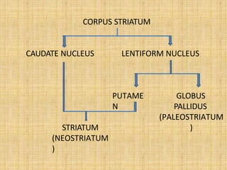
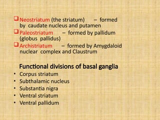
Neostriatum (the striatum)– formed
by caudate nucleus and putamen
Paleostriatum – formed by pallidum
(globus pallidus)
Archistriatum – formed by Amygdaloid
nuclear complex and Claustrum
Functional divisions of basal ganglia
• Corpus striatum
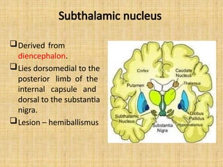
• Subthalamic nucleus
• Substantia nigra
• Ventral striatum
• Ventral pallidum
7.
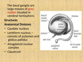
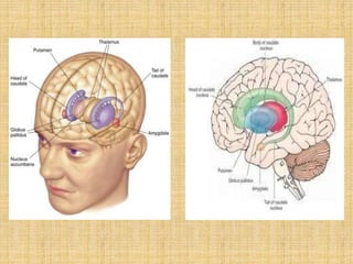
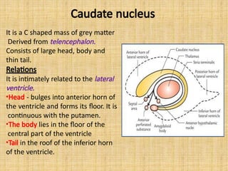
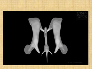
It is aC shaped mass of grey matter
Derived from telencephalon.
Consists of large head, body and
thin tail.
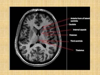
Relations
It is intimately related to the lateral
ventricle.
•Head - bulges into anterior horn of
the ventricle and forms its floor. It is
continuous with the putamen.
•The body lies in the floor of the
central part of the ventricle
•Tail in the roof of the inferior horn
of the ventricle.
Caudate nucleus
8.
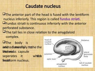
Caudate nucleus
The anteriorpart of the head is fused with the lentiform
nucleus inferiorly. This region is called fundus striati.
Fundus striati is continuous inferiorly with the anterior
perforated substance.
The tail lies in close relation to the amygdaloid
complex.
The body is
related mediallytothe
thalamus
and laterally to the
which
internal capsule
seperates it
from
the
lentiform nucleus.
9.
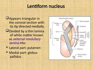
Lentiform nucleus
Appears triangularin
the coronal section with
its tip directed medially.
Divided by a thin lamina
of white matter known
as external medullary
lamina into
Lateral part- putamen
Medial part- globus
pallidus
10.
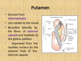
Putamen
• Derived from
telencephalon.
•Lies medial to the insula
• Bounded laterally by
the fibres of external
capsule and medially by
the globus pallidus.
• Separated from the
caudate nucleus by the
anterior limb of the
internal capsule
11.
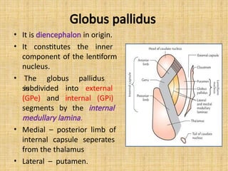
Globus pallidus
• Itis diencephalon in origin.
• It constitutes the inner
component of the lentiform
nucleus.
• The globus pallidus
is
subdivided into external
(GPe) and internal (GPi)
segments by the internal
medullary lamina.
• Medial – posterior limb of
internal capsule seperates
from the thalamus
• Lateral – putamen.
12.
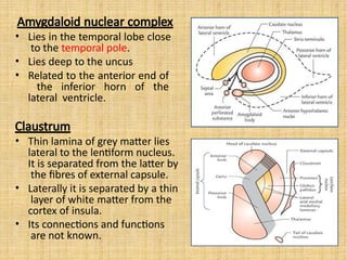
Amygdaloid nuclear complex
•Lies in the temporal lobe close
to the temporal pole.
• Lies deep to the uncus
• Related to the anterior end of
the inferior horn of the
lateral ventricle.
Claustrum
• Thin lamina of grey matter lies
lateral to the lentiform nucleus.
It is separated from the latter by
the fibres of external capsule.
• Laterally it is separated by a thin
layer of white matter from the
cortex of insula.
• Its connections and functions
are not known.
13.
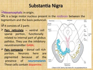
Substantia Nigra
Neurons
because
here are
ofthe
portion.
pigmented
presence of neuromelanin.
These cells contain dopamine.
Mesencephalic in origin.
It is a large motor nucleus present in the midbrain between the
tegmentum and the basis pedunculi.
It consists of 2 parts
Pars reticulata – ventral cell
sparse portion, functionally
related to internal part of globus
pallidus. They use the inhibitory
neurotransmitter GABA.
Pars compacta – dorsal cell rich
Ventral Striatum
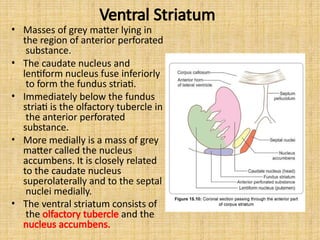
• Massesof grey matter lying in
the region of anterior perforated
substance.
• The caudate nucleus and
lentiform nucleus fuse inferiorly
to form the fundus striati.
• Immediately below the fundus
striati is the olfactory tubercle in
the anterior perforated
substance.
• More medially is a mass of grey
matter called the nucleus
accumbens. It is closely related
to the caudate nucleus
superolaterally and to the septal
nuclei medially.
• The ventral striatum consists of
the olfactory tubercle and the
nucleus accumbens.
16.
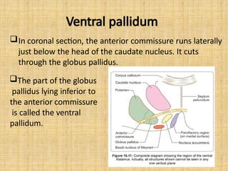
Ventral pallidum
In coronalsection, the anterior commissure runs laterally
just below the head of the caudate nucleus. It cuts
through the globus pallidus.
The part of the globus
pallidus lying inferior to
the anterior commissure
is called the ventral
pallidum.
17.
Thus,
Dorsal striatum –caudate nucleus and putamen
Ventral striatum – olfactory tubercle and nucleus
accumbens
Dorsal pallidum – globus pallidus
Ventral pallidum – part below the anterior perforated
substance
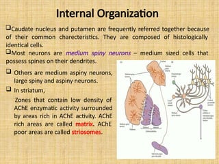
Internal Organization
Caudate nucleusand putamen are frequently referred together because
of their common charecteristics. They are composed of histologically
identical cells.
Most neurons are medium spiny neurons – medium sized cells that
possess spines on their dendrites.
Others are medium aspiny neurons,
large spiny and aspiny neurons.
In striatum,
Zones that contain low density of
AChE enzymatic activity surrounded
by areas rich in AChE activity. AChE
rich areas are called matrix. AChE
poor areas are called striosomes.
22.
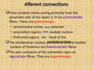
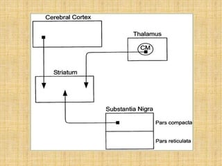
Afferent connections
From cerebralcortex arising primarily from the
pyramidal cells of the layers V, VI via corticostriate
fibres. These are glutaminergic.
• sensorimotor cortex
• association regions
• Prefrontal regions
putamen
caudate nucleus
head of the
caudate nucleus
The intralaminar nucleus particularly central median
nucleus of thalamus via thalamostriate fibres
The pars compacta of the substantia nigra via
nigrostriate fibres. They are dopaminergic.
24.
Afferent connections
• Noradrenergicfibres from the locus ceruleus
• Serotoninergic fibres from the raphe nuclei (in the
reticular formation of the midbrain)
• Afferents from limbic cortical areas, hippocampus
and the amygdala terminates in the ventral striatum.
• The afferents from the cerebral cortex and the
thalamus provide the striatum with various
modalities of sensory information.
• Disruption of the input pathways is associated with
movement disorders like parkinsons disease.
25.
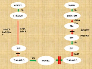
Internal processing
• Thedirect pathway – increased motor activity
• The indirect pathway – decreased motor activity
• The intrinsic circuitry of the basal ganglia is
disrupted by a severe loss of neurons in the
striatum in Huntington’s disease.
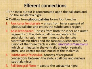
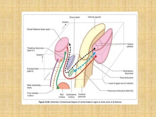
Efferent connections
The mainoutput is concentrated upon the pallidum and
on the substantia nigra.
Outflow from globus pallidus forms four bundles
1. Fasciculus lenticularis – arises from inner segment of
globus pallidus and enters the subthalamic region.
2. Ansa lenticularis – arises from both the inner and outer
segments of the globus pallidus and enters the
subthalamic region where it meets the dentato
rubrothalamic fibres and the fasciculus lenticularis. The
union of the three tracts is called fasciculus thalamicus
which terminates in the ventralis anterior, ventralis
lateral and centro median nuclei of the thalamus.
3. Subthalamic fasciculus- consists of reciprocal
connections between the globus pallidus and nucleus
subthalamicus.
4. Pallidonigral fibres – pass to the substantia nigra
29.
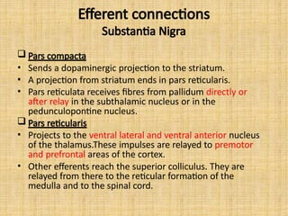
Efferent connections
Substantia Nigra
Pars compacta
• Sends a dopaminergic projection to the striatum.
• A projection from striatum ends in pars reticularis.
• Pars reticulata receives fibres from pallidum directly or
after relay in the subthalamic nucleus or in the
pedunculopontine nucleus.
Pars reticularis
• Projects to the ventral lateral and ventral anterior nucleus
of the thalamus.These impulses are relayed to premotor
and prefrontal areas of the cortex.
• Other efferents reach the superior colliculus. They are
relayed from there to the reticular formation of the
medulla and to the spinal cord.
31.
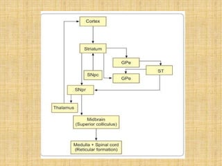
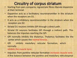
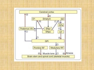
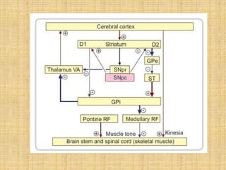
Circuitry of corpusstriatum
• Starting from pars compacta, nigrostriate fibres liberate dopamine
at their terminal.
• Dopamine acts as a facilitatory neurotransmitter in the striatum
when the receptors are D1.
• It acts as a inhibitory neurotransmitter in the striatum when the
receptors are D2.
• Striatal D1 neurons inhibits GPi through a direct path.
• Striatal D2 neurons facilitates GPi through a indirect path. This
balances the impulses reaching the GPi.
• GPi normally inhibits the thalamus. Thalamus facilitates the the
cortex which causes the normal kinesia.
• GPi inhibits medullary reticular formation, which
normally
inhibits the muscle tone.
• Impulses from pontine reticular formation increases muscle tone.
A fine balance between the pontine and medullary reticulospinal
33.
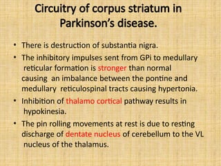
Circuitry of corpusstriatum in
Parkinson’s disease.
• There is destruction of substantia nigra.
• The inhibitory impulses sent from GPi to medullary
reticular formation is stronger than normal
causing an imbalance between the pontine and
medullary reticulospinal tracts causing hypertonia.
• Inhibition of thalamo cortical pathway results in
hypokinesia.
• The pin rolling movements at rest is due to resting
discharge of dentate nucleus of cerebellum to the VL
nucleus of the thalamus.
35.
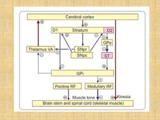
Circuitry of corpusstriatum in chorea
and ballismus
• The pathways for muscle tone and kinesia
reverse when compared to Parkinsons.
• Destruction of the D2 neurons in striatum
causes chorea.
37.
Functions
• Pallidum eloboratesthe simple motor and postural
mechanisms of the brain stem into complex autonomic
motor behavior.
• Centre for expression gestures and reactive
movements.
• The striatum eloborates and smoothens the coarser
motor integrations of the pallidum.
• The subthalamic nucleus, red nucleus, substantia nigra
have a synergistic action and integrates stereotyped
behavior and controls complex muscular movements.
• Also process information related to emotions,
motivations, and cognitive functions.
38.
Clinical correlation
Various kindsof abnormal movements are
seen in neurological disorders involving the
basal ganglia.
The movements could be
• Hyperkinetic, hypotonic (chorea, athetosis,
ballismus)
• Hypokinetic, hypertonic (parkinsons)
39.
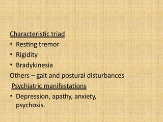
Tremors
• Regular oscillatingmovement about a joint due to
synchronous contraction of the agonist and antagonist
muscles.
• Types of tremors
1. Rest tremors
• Movements occurs in a relaxed supported extremity
• Reduced by ambulation
• Distinctive in parkinsonism
2. Postural tremors
• Sustained posture elicit tremors e.g outstretched
hands
• A coarse irregular rapid postural tremor is seen in
metabolic encephalopathy.
3. Intentional tremors
• Active limb oscillates more prominently on
reaching target
40.
Chorea
• Rapid involuntarypurposeless dancing
movements of the limbs.
• Chorea affects the proximal joints, while that of
the athetosis affects the distal joints.
• Sydenhams chorea- the pathology is seen in the
striatum
• Huntington’s chorea- autosomal dominant
degenerative disease of the striatum and cerebral
cortex
41.
Athetosis
Continuous slow writhingmovements with a propensity to
affect the arms and hands.
Ballism
Hemorrhagic ivolvement of the subthalamic nucleus.
Involuntary and violent, flinging movements of large
amplitude
Hemiballism – involves one half of the body , caused by
lesions in the subthalamic nucleus of the opposite
side. When restricted to one limb it is called as
monoballism. Dystonia
Sustained or repetitive involuntary muscle contractions
frequently causing twisting movements with abnormal
postures.
43.
Basal ganglia inpsychiatric disorders
OCD
• There is evidence of basal ganglia dysfunction
in imaging studies. Both increase and
decreased volumes of caudate nucleus is
reported.
• Most patients showed an increased blood
flow to the caudate.
• Increased caudate metabolism will reduce the
effect of the treatment of OCD.
44.
AUTISM
• Enlargement ofcaudate nucleus upto 8% . The
greater caudate volume is proportional to the
increased total brain volume.
• Motor, communicative, and social
impairments are associated with shape
abnormalities in the basal ganglia.
• Glutamate dysfunction in basal ganglia is
associated with autism.
45.
ADHD
• Neuroimaging studiesshow evidence of
striatal dysfunction in ADHD.
• Teicher and colleagues concluded that ADHD
may be related to functional abnormalities in
the putamen.
• Boys with ADHD showed a significantly smaller
volume of basal ganglia compared with
typically developing boys.
46.
SCHIZOPHRENIA
• In striatum,there are anomalies of dopamine,
synthesis, storage and release.
• Striatal dopaminergic system is overactive. There
is an increase in the presynaptic dopamine
function indicating an increase in the dopamine
synthesis capacity.
• There is an increase in the striatal D2 receptors.
• In substantia nigra, there is a higher variability of
tyrosine hydroxylase level, increase in
homovanillic acid, increase in glutamate receptor
subunits.
47.
DEPRESSION
• Functional imagingstudies have shown
pathological interactions in amygdala, ventral
striatum and prefrontal cortex in depression.
• Regional cerebral blood flow, glucose
metabolism in amygdala are increased.
• Nucleus accumbens is also involved in
affective disorders.
• Caudate hyperintensities are found in elderly
patients of depression
48.
ADDICTION
• Amygdala isa critical structure in addiction.
• Nucleus accumbens is also important in drug
re inforcement and addiction.
Parkinson’s disease
III.
• Aneurodegenerative disease associated with loss of
dopaminergic neurons in the Substantia Nigra pars
compacta.
• IN 1817, JAMES PARKINSON described paralysis agitans in
his “ESSAY ON THE SHAKING PALSY”.
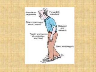
• Parkinson's disease affects movement, producing motor
symptoms.
• Non-motor symptoms, which include
I. autonomic dysfunction,
II. neuropsychiatric problems (mood, cognition, behaviour
or thought alterations),
sensory and sleep difficulties, are also common. Some of
these non-motor symptoms are often present at the time
of diagnosis and can precede motor symptoms.
Huntington’s disease
• Huntington'sdisease (formerly Huntington's chorea)
is an autosomal dominant neurodegenerative
disorder characterized by midlife onset, a progressive
course, and a combination of motor, psychiatric, and
cognitive symptoms.
• First described by George Huntington in 1872.
• The disease is caused by a CAG (trinucleotide) repeat
expansion mutation in the huntingtin gene on
chromosome 4.
54.
Primary involuntary movementabnormality
• Chorea or choreoathetosis
Associated voluntary movement abnormalities
• Visual tracking,
• fine motor movements,
• gait disturbances
Decreased facial expression, difficulty in swallowing,
chewing, speaking, sleep disturbance, impairment in
cognition and memory, subcortical dementia
Psychiatric manifestations
•Higher rates of suicide
•Depression
•Mania
•Personality change
55.
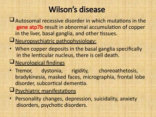
Wilson’s disease
Autosomal recessivedisorder in which mutations in the
gene atp7b result in abnormal accumulation of copper
in the liver, basal ganglia, and other tissues.
Neuropsychiatric pathophysiology:
• When copper deposits in the basal ganglia specifically
in the lenticular nucleus, there is cell death.
Neurological findings
• Tremor, dystonia, rigidity, choreoathetosis,
bradykinesia, masked faces, micrographia, frontal lobe
disorder, subcortical dementia.
Psychiatric manifestations
• Personality changes, depression, suicidality, anxiety
disorders, psychotic disorders.
56.
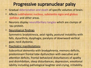
Progressive supranuclear palsy
•Gradual deterioration and death of specific volumes of brain.
• Affects subthalamic nucleus, substantia nigra and globus
pallidus and other areas.
• Neurons display neurofibrillary tangles which are clumps of
tau protein.
• Neurological findings
Symmetric bradykinesia, axial rigidiy, postural instability with
falls, dysarthria, dysphagia, paralysis of downward vertical
gaze, neck dystonia.
• Psychiatric manifestations
Subcortical dementia with bradyphrenia, memory deficits,
predominant frontal lobe dysfunction with executive and
attention deficits, frontal behavioral disturbances of apathy
and disinhibition, sleep disturbances, depression, emotional
lability including pathological laughter and crying, irritability.
57.
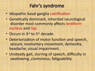
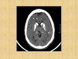
Fahr’s syndrome
• Idiopathicbasal ganglia calcification
• Genetically dominant, inherited neurological
disorder most commonly affects lentiform
nucleus and Gpi.
• Occurs in 3rd to 5th decade.
• Deteriorization of motor function and speech,
seizure, involuntary movement, dementia,
headache, visual impairment.
• Unsteady gait, slurring of speech, difficulty in
swallowing ,clumsiness, fatiguability.
59.
Basal ganglia stroke
•Changes in the body movement, rigidity, stiffness,
tremor, ataxia, difficulty in swallowing, smiling, speech.
• Cognitive impairment – poor attention, memory
impairment
• Personality changes – frustration ,anxiety, loss of
interest, loss of motivation, depression, anger.
• Right basal ganglia stroke
• Anosognosia – unable to perceive the severity of
deficit, left side neglect, visuospatial hemineglect,
constructional apraxia.
• Left basal ganglia stroke
• Apathy (lack of interest), memory impairment, major
depression.
60.
References
1. Comprehensive textbookof
Psychiatry - Kaplan and Sadocks.
2. Textbook of Neuroanatomy - I.B. Singh
3. Postgraduate Textbook of Psychiatry -
Ahuja