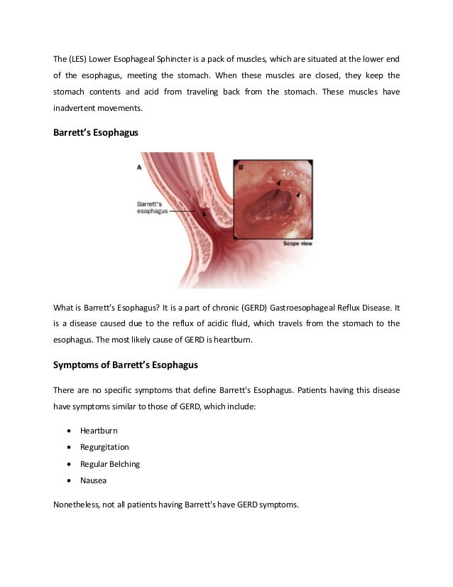
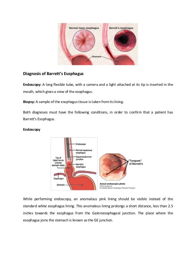
Barrett's esophagus is a condition where the lining of the esophagus is replaced by abnormal intestinal-type cells due to chronic acid reflux from the stomach. This occurs when stomach acid damages the normal esophageal lining over many years. The abnormal cells are pre-cancerous and increase the risk of developing esophageal adenocarcinoma. The main symptoms of Barrett's esophagus are heartburn and acid reflux. It is diagnosed through an endoscopy with biopsy to examine the abnormal cells under a microscope. The primary cause is gastroesophageal reflux disease which causes excessive stomach acid to back up into the esophagus.
This case study describes a 69-year old Filipino woman diagnosed with gastroesophageal reflux disease (GERD) and ischemic heart disease. She experienced symptoms like heartburn, acid indigestion, hiccups, and difficulty walking. Her medical history and examinations led doctors to diagnose her conditions. She was prescribed various medications to manage her diseases. Dietary interventions included a low salt, low fat, high fiber diet to help control her conditions and prevent further complications. Her nutritional status was assessed as mild underweight.
Presentation main surgery 123456nhnhnhnahkoHardikSiwach1
1. The document describes various abnormalities or anomalies of the oesophagus known as "bizarre oesophagus". These include conditions like esophageal webs, rings, diverticula, achalasia, and Barrett's esophagus.
2. Esophageal webs are thin membranes in the esophagus that cause narrowing and difficulty swallowing. Achalasia is a condition where the lower esophageal sphincter fails to relax properly, causing difficulty swallowing and food regurgitation.
3. The document provides details on the symptoms, diagnosis, and treatment of these different conditions of the bizarre oesophagus.
The document summarizes esophageal cancer, including its causes, risk factors, types, signs and symptoms, methods of diagnosis and treatment. It discusses that esophageal cancer occurs when cells in the esophagus grow abnormally and form tumors. Risk factors include GERD, smoking, Barrett's esophagus, alcohol consumption and not eating enough fruits and vegetables. Treatment options include surgery to remove the esophagus and nearby lymph nodes, radiation therapy, chemotherapy and immunotherapy. Prevention methods include avoiding smoking and excessive alcohol, and maintaining a healthy diet and weight.
Gastro esophageal Reflux Disease (GERD) and its managementDr. Ankit Gaur
In this presentation I have tried to explain in brief about gastro esophageal Reflux Disease (GERD), its etiology, risk factors, diagnosis, and its management via pharmacotherapy.
1. GERD is a common cause of regurgitation where the lower esophageal sphincter does not close properly, allowing stomach acid to back up into the esophagus.
2. Ingestion of caustic substances like strong acids or alkalis can burn the tissues of the upper GI tract and sometimes cause perforations, leading to symptoms like regurgitation.
3. Cancers of the esophagus such as adenocarcinoma or squamous cell carcinoma can cause regurgitation by blocking the esophagus. Barrett's esophagus is a precancerous condition caused by acid reflux.
Barrett's esophagus is a condition where the lining of the esophagus is replaced by intestinal-type cells due to chronic acid reflux. It increases the risk of esophageal adenocarcinoma. Diagnosis is typically made during endoscopy by identifying changes in esophageal cell type beyond the gastroesophageal junction. Treatment focuses on minimizing acid reflux through lifestyle changes and medications, with additional procedures used in cases of dysplasia to remove abnormal cells and further reduce cancer risk.
Peptic ulcer disease is a break in the inner lining of the stomach or first part of the small intestine. Common symptoms include abdominal pain that improves with eating, belching, vomiting, and weight loss. The most common causes are infection with Helicobacter pylori bacteria and use of non-steroidal anti-inflammatory drugs. Complications can include bleeding, perforation of the stomach wall, and blockage of the stomach. Diagnosis is usually confirmed through an endoscopy to visually identify ulcers. Treatment involves eliminating causes, medication to reduce stomach acid, and antibiotics to treat H. pylori infections.
Lecture 16 esophagus and stomach disorders - PathologyAreej Abu Hanieh
The document provides information on the esophagus and stomach disorders. It discusses the anatomy and physiology of the esophagus and stomach. Key points include that the esophagus transports food to the stomach through peristaltic movements. The lower esophageal sphincter separates the esophagus and stomach. Gastroesophageal reflux disease is caused by disruption of protective mechanisms in the esophagus. Common esophagus and stomach disorders mentioned include achalasia, hiatal hernia, Barrett's esophagus, dyspepsia, gastritis, and esophagitis. Treatment options focused on lifestyle changes and pharmacologic interventions like proton pump inhibitors.
Peptic ulcers form when the lining of the stomach or duodenum is corroded by acidic digestive juices. Common symptoms include abdominal pain relieved by food or antacids. While acid contributes to ulcer formation, infection with H. pylori bacteria is now believed to be the leading cause. Other risk factors include NSAID use, smoking, alcohol, and stress. Complications can include bleeding, perforation, and narrowing or obstruction of the stomach outlet. Endoscopy allows visualization and biopsy of ulcers, while treatment aims to eliminate H. pylori infection and reduce acid secretion.
GERD | Gastro esophageal reflux disease-a brief medical study martinshaji
Gastro esophageal reflux disease (GERD) is a chronic digestive disease. GERD occurs when stomach acid or, occasionally, stomach content, flows back into your food pipe (esophagus). The backwash (reflux) irritates the lining of your esophagus and causes GERD. This is a very common condition observed
please comment
thank you
GER occurs when stomach contents backflow into the esophagus. GERD is a chronic form of GER that occurs more than twice a week for several weeks and can lead to serious health issues over time if left untreated. GERD is caused by a weak lower esophageal sphincter or hiatal hernia. Common symptoms include heartburn, cough, asthma, and chest pain. GERD is typically diagnosed through upper endoscopy, pH monitoring, or upper GI series. Treatment involves lifestyle changes, medications like PPIs or H2 blockers, and sometimes surgery. Untreated GERD can cause complications such as esophagitis, strictures, respiratory issues, and Barrett's esophagus
Barrett's esophagus is a condition where the lining of the lower esophagus is replaced by abnormal columnar cells due to chronic acid exposure from gastroesophageal reflux disease (GERD). This puts patients at risk for developing esophageal adenocarcinoma. The condition is diagnosed via endoscopy with biopsy showing columnar epithelium in place of the normal squamous lining. Treatment depends on the presence and grade of any dysplasia, ranging from medication and endoscopic procedures to surgery.
Barrett's esophagus is a condition where the lining of the esophagus is replaced by abnormal cells similar to those found in the intestine, as an adaptation to chronic acid exposure from gastroesophageal reflux disease. It is associated with an increased risk of developing esophageal adenocarcinoma. Diagnosis requires endoscopy and biopsy to detect the presence of intestinal-type cells in the esophagus. Treatment depends on the grade of dysplasia found and ranges from medication and endoscopic procedures to surgery.
This document provides an overview of gastroesophageal reflux disease (GERD). It defines GERD as reflux of stomach and duodenal contents into the esophagus, characterized by symptoms, radiologic or endoscopic changes. The pathogenesis involves transient relaxation of the lower esophageal sphincter, allowing acid reflux. Complications can include esophageal stricture, ulcer, or Barrett's esophagus, a precursor to esophageal cancer. Diagnosis is based on symptoms and testing may include pH monitoring or endoscopy to assess esophageal damage.
Our Orlando Gastroenterologists are the leading experts when it comes to evaluating, diagnosing, and treating GI conditions and diseases.
http://gastro-specialists.com/
This document discusses intestinal obstruction, including its causes, classifications, symptoms, diagnosis and treatment. Some key points:
- Intestinal obstruction can be caused by adhesions, hernias, tumors, strictures and more. It is classified by the obstructed site and presence of blood flow issues.
- Symptoms include pain, vomiting, constipation and distension. Signs depend on obstruction location and duration. Strangulated obstructions require urgent surgery to prevent tissue death.
- Diagnosis involves medical history, physical exam, imaging like x-rays and CT scans. Treatment involves resuscitation, nasogastric drainage, and surgery to relieve the obstruction and address the underlying cause. S
The document discusses different types of intestinal volvulus including gastric, small bowel, large bowel, and combinations. Gastric volvulus is classified as organoaxial or mesenteroaxial depending on the axis of rotation. Small bowel volvulus often occurs due to midgut malrotation. Large bowel volvulus commonly affects the cecum or sigmoid colon. Sigmoid volvulus is the most common type of large bowel volvulus and presents as an inverted U-shape on imaging. Cecal volvulus results from torsion of the cecum around its own mesentery.
Achalasia is a rare disorder of the esophagus that results from damaged nerves that control food movement. It causes difficulty swallowing and food getting stuck. The document discusses the causes, symptoms, tests used to diagnose (endoscopy, manometry), and treatments of achalasia. Treatments include medications to relax muscles, botox injections, balloon dilation procedures, and surgeries like Heller myotomy to cut the lower esophageal sphincter muscle.
achalasia-oesophagus stomach body lining.pdfiwlucy9
Achalasia is a rare disorder of the esophagus that results from damaged nerves that control food movement. It causes difficulty swallowing food and liquid. The main symptoms are trouble swallowing, food getting stuck in the chest, and weight loss. While the cause is unknown, the immune system may destroy nerves in the esophagus wall. This prevents normal swallowing and relaxation of the lower esophageal sphincter. Treatment options include medications, botox injections, balloon dilation, and surgery to cut the lower sphincter muscle. The goal is to improve swallowing function, but full normal function is never regained.
This document provides information on esophageal cancer including statistics, risk factors, types, and histology. It notes that in 2013 there were about 18,000 new esophageal cancer cases diagnosed in the US resulting in about 15,000 deaths. The two main types are squamous cell carcinoma and adenocarcinoma. Risk factors include age, gender, gastroesophageal reflux disease, Barrett's esophagus, tobacco/alcohol use, obesity, and certain genetic conditions or injuries to the esophagus. The lining of the esophagus and the two main types of esophageal cancer are also described.
The document discusses the anatomy and pathologies of the esophagus. It describes the layers of the esophagus and contains sphincters. Lesions can cause dysphagia due to narrowing or obstruction. Gastroesophageal reflux disease is a common cause of heartburn. Varices are dilated veins caused by portal hypertension. Achalasia is a motility disorder causing difficulty swallowing. Barrett's esophagus is a complication of long-term reflux and increases cancer risk. Squamous cell carcinoma and adenocarcinoma are the two main types of esophageal cancer.
The document discusses several conditions related to the digestive system:
1) Diverticulosis is a condition where pouches called diverticula bulge out from weak spots in the colon wall, usually occurring where the colon joins the rectum. It becomes diverticulitis if the pouches become infected or rupture.
2) A hiatal hernia occurs when part of the stomach protrudes through the diaphragm into the chest cavity. This can cause acid reflux symptoms.
3) Peptic ulcers develop as sores or holes in the lining of the stomach, esophagus, or duodenum due to acidic digestive juices. Major causes are H. pylori bacteria infection and long
This document summarizes the anatomy, pathophysiology, diagnosis, and treatment of spontaneous esophageal rupture (SER), also known as Boerhaave's syndrome. It describes the anatomy of the esophagus and explains that SER usually occurs due to vomiting against a closed upper esophageal sphincter, which increases intraesophageal pressure and can cause a tear. Diagnosis involves considering the patient's history, symptoms of chest and abdominal pain, and findings on imaging tests. Treatment involves antibiotics, nothing by mouth, gastric decompression, and possibly surgery if symptoms do not improve with initial non-operative management. Early diagnosis and treatment are important for prognosis.
Toxic megacolon is an acute form of colonic distension characterized by a very dilated colon accompanied by abdominal distension and sometimes fever, abdominal pain, or shock. It occurs in 1-2.5% of patients with colon diseases like ulcerative colitis and has a high mortality risk if not treated promptly. Diagnostic criteria include radiographic evidence of large colon diameter and symptoms of fever, rapid heart rate, high white blood cell count, or low blood pressure.
The esophagus is a 10 inch muscular tube that extends from the pharynx to the stomach. It begins at the level of the cricoid cartilage and passes through the diaphragm at the level of the 10th thoracic vertebra. The muscles of the esophagus include an inner circular layer and outer longitudinal layer. Blood supply comes from branches of the inferior thyroid, descending thoracic aorta, and left gastric artery. Lymph drains to cervical, mediastinal and celiac nodes. Clinical notes discuss esophageal constrictions, achalasia, GERD, esophageal atresia, and various causes of esophagitis.
Dysphagia, or difficulty swallowing, can be caused by issues with the esophagus. The document discusses several diseases and conditions that cause dysphagia by obstructing the esophagus, including achalasia, esophageal cancer, and reflux esophagitis. It provides details on symptoms, diagnostics, and treatment for each condition. Esophageal spasms, tumors, diverticula, strictures, and foreign bodies can also obstruct the esophagus and cause dysphagia. The document examines each of these conditions and how they impact swallowing.
The esophagus connects the hypopharynx to the stomach and functions to transport food and fluid. Gastroesophageal reflux occurs when stomach contents reflux into the esophagus. This can be caused by increased stomach pressure or decreased lower esophageal sphincter tone. Chronic reflux can lead to esophagitis and complications like strictures or Barrett's esophagus. Diagnostic tests include endoscopy, pH monitoring, and manometry. Treatment involves lifestyle modifications and medications to reduce acid production like PPIs.
These lecture slides, by Dr Sidra Arshad, offer a quick overview of the physiological basis of a normal electrocardiogram.
Learning objectives:
1. Define an electrocardiogram (ECG) and electrocardiography
2. Describe how dipoles generated by the heart produce the waveforms of the ECG
3. Describe the components of a normal electrocardiogram of a typical bipolar lead (limb II)
4. Differentiate between intervals and segments
5. Enlist some common indications for obtaining an ECG
6. Describe the flow of current around the heart during the cardiac cycle
7. Discuss the placement and polarity of the leads of electrocardiograph
8. Describe the normal electrocardiograms recorded from the limb leads and explain the physiological basis of the different records that are obtained
9. Define mean electrical vector (axis) of the heart and give the normal range
10. Define the mean QRS vector
11. Describe the axes of leads (hexagonal reference system)
12. Comprehend the vectorial analysis of the normal ECG
13. Determine the mean electrical axis of the ventricular QRS and appreciate the mean axis deviation
14. Explain the concepts of current of injury, J point, and their significance
Study Resources:
1. Chapter 11, Guyton and Hall Textbook of Medical Physiology, 14th edition
2. Chapter 9, Human Physiology - From Cells to Systems, Lauralee Sherwood, 9th edition
3. Chapter 29, Ganong’s Review of Medical Physiology, 26th edition
4. Electrocardiogram, StatPearls - https://www.ncbi.nlm.nih.gov/books/NBK549803/
5. ECG in Medical Practice by ABM Abdullah, 4th edition
6. Chapter 3, Cardiology Explained, https://www.ncbi.nlm.nih.gov/books/NBK2214/
7. ECG Basics, http://www.nataliescasebook.com/tag/e-c-g-basics
Peptic ulcers form when the lining of the stomach or duodenum is corroded by acidic digestive juices. Common symptoms include abdominal pain relieved by food or antacids. While acid contributes to ulcer formation, infection with H. pylori bacteria is now believed to be the leading cause. Other risk factors include NSAID use, smoking, alcohol, and stress. Complications can include bleeding, perforation, and narrowing or obstruction of the stomach outlet. Endoscopy allows visualization and biopsy of ulcers, while treatment aims to eliminate H. pylori infection and reduce acid secretion.
GERD | Gastro esophageal reflux disease-a brief medical study martinshaji
Gastro esophageal reflux disease (GERD) is a chronic digestive disease. GERD occurs when stomach acid or, occasionally, stomach content, flows back into your food pipe (esophagus). The backwash (reflux) irritates the lining of your esophagus and causes GERD. This is a very common condition observed
please comment
thank you
GER occurs when stomach contents backflow into the esophagus. GERD is a chronic form of GER that occurs more than twice a week for several weeks and can lead to serious health issues over time if left untreated. GERD is caused by a weak lower esophageal sphincter or hiatal hernia. Common symptoms include heartburn, cough, asthma, and chest pain. GERD is typically diagnosed through upper endoscopy, pH monitoring, or upper GI series. Treatment involves lifestyle changes, medications like PPIs or H2 blockers, and sometimes surgery. Untreated GERD can cause complications such as esophagitis, strictures, respiratory issues, and Barrett's esophagus
Barrett's esophagus is a condition where the lining of the lower esophagus is replaced by abnormal columnar cells due to chronic acid exposure from gastroesophageal reflux disease (GERD). This puts patients at risk for developing esophageal adenocarcinoma. The condition is diagnosed via endoscopy with biopsy showing columnar epithelium in place of the normal squamous lining. Treatment depends on the presence and grade of any dysplasia, ranging from medication and endoscopic procedures to surgery.
Barrett's esophagus is a condition where the lining of the esophagus is replaced by abnormal cells similar to those found in the intestine, as an adaptation to chronic acid exposure from gastroesophageal reflux disease. It is associated with an increased risk of developing esophageal adenocarcinoma. Diagnosis requires endoscopy and biopsy to detect the presence of intestinal-type cells in the esophagus. Treatment depends on the grade of dysplasia found and ranges from medication and endoscopic procedures to surgery.
This document provides an overview of gastroesophageal reflux disease (GERD). It defines GERD as reflux of stomach and duodenal contents into the esophagus, characterized by symptoms, radiologic or endoscopic changes. The pathogenesis involves transient relaxation of the lower esophageal sphincter, allowing acid reflux. Complications can include esophageal stricture, ulcer, or Barrett's esophagus, a precursor to esophageal cancer. Diagnosis is based on symptoms and testing may include pH monitoring or endoscopy to assess esophageal damage.
Our Orlando Gastroenterologists are the leading experts when it comes to evaluating, diagnosing, and treating GI conditions and diseases.
http://gastro-specialists.com/
This document discusses intestinal obstruction, including its causes, classifications, symptoms, diagnosis and treatment. Some key points:
- Intestinal obstruction can be caused by adhesions, hernias, tumors, strictures and more. It is classified by the obstructed site and presence of blood flow issues.
- Symptoms include pain, vomiting, constipation and distension. Signs depend on obstruction location and duration. Strangulated obstructions require urgent surgery to prevent tissue death.
- Diagnosis involves medical history, physical exam, imaging like x-rays and CT scans. Treatment involves resuscitation, nasogastric drainage, and surgery to relieve the obstruction and address the underlying cause. S
The document discusses different types of intestinal volvulus including gastric, small bowel, large bowel, and combinations. Gastric volvulus is classified as organoaxial or mesenteroaxial depending on the axis of rotation. Small bowel volvulus often occurs due to midgut malrotation. Large bowel volvulus commonly affects the cecum or sigmoid colon. Sigmoid volvulus is the most common type of large bowel volvulus and presents as an inverted U-shape on imaging. Cecal volvulus results from torsion of the cecum around its own mesentery.
Achalasia is a rare disorder of the esophagus that results from damaged nerves that control food movement. It causes difficulty swallowing and food getting stuck. The document discusses the causes, symptoms, tests used to diagnose (endoscopy, manometry), and treatments of achalasia. Treatments include medications to relax muscles, botox injections, balloon dilation procedures, and surgeries like Heller myotomy to cut the lower esophageal sphincter muscle.
achalasia-oesophagus stomach body lining.pdfiwlucy9
Achalasia is a rare disorder of the esophagus that results from damaged nerves that control food movement. It causes difficulty swallowing food and liquid. The main symptoms are trouble swallowing, food getting stuck in the chest, and weight loss. While the cause is unknown, the immune system may destroy nerves in the esophagus wall. This prevents normal swallowing and relaxation of the lower esophageal sphincter. Treatment options include medications, botox injections, balloon dilation, and surgery to cut the lower sphincter muscle. The goal is to improve swallowing function, but full normal function is never regained.
This document provides information on esophageal cancer including statistics, risk factors, types, and histology. It notes that in 2013 there were about 18,000 new esophageal cancer cases diagnosed in the US resulting in about 15,000 deaths. The two main types are squamous cell carcinoma and adenocarcinoma. Risk factors include age, gender, gastroesophageal reflux disease, Barrett's esophagus, tobacco/alcohol use, obesity, and certain genetic conditions or injuries to the esophagus. The lining of the esophagus and the two main types of esophageal cancer are also described.
The document discusses the anatomy and pathologies of the esophagus. It describes the layers of the esophagus and contains sphincters. Lesions can cause dysphagia due to narrowing or obstruction. Gastroesophageal reflux disease is a common cause of heartburn. Varices are dilated veins caused by portal hypertension. Achalasia is a motility disorder causing difficulty swallowing. Barrett's esophagus is a complication of long-term reflux and increases cancer risk. Squamous cell carcinoma and adenocarcinoma are the two main types of esophageal cancer.
The document discusses several conditions related to the digestive system:
1) Diverticulosis is a condition where pouches called diverticula bulge out from weak spots in the colon wall, usually occurring where the colon joins the rectum. It becomes diverticulitis if the pouches become infected or rupture.
2) A hiatal hernia occurs when part of the stomach protrudes through the diaphragm into the chest cavity. This can cause acid reflux symptoms.
3) Peptic ulcers develop as sores or holes in the lining of the stomach, esophagus, or duodenum due to acidic digestive juices. Major causes are H. pylori bacteria infection and long
This document summarizes the anatomy, pathophysiology, diagnosis, and treatment of spontaneous esophageal rupture (SER), also known as Boerhaave's syndrome. It describes the anatomy of the esophagus and explains that SER usually occurs due to vomiting against a closed upper esophageal sphincter, which increases intraesophageal pressure and can cause a tear. Diagnosis involves considering the patient's history, symptoms of chest and abdominal pain, and findings on imaging tests. Treatment involves antibiotics, nothing by mouth, gastric decompression, and possibly surgery if symptoms do not improve with initial non-operative management. Early diagnosis and treatment are important for prognosis.
Toxic megacolon is an acute form of colonic distension characterized by a very dilated colon accompanied by abdominal distension and sometimes fever, abdominal pain, or shock. It occurs in 1-2.5% of patients with colon diseases like ulcerative colitis and has a high mortality risk if not treated promptly. Diagnostic criteria include radiographic evidence of large colon diameter and symptoms of fever, rapid heart rate, high white blood cell count, or low blood pressure.
The esophagus is a 10 inch muscular tube that extends from the pharynx to the stomach. It begins at the level of the cricoid cartilage and passes through the diaphragm at the level of the 10th thoracic vertebra. The muscles of the esophagus include an inner circular layer and outer longitudinal layer. Blood supply comes from branches of the inferior thyroid, descending thoracic aorta, and left gastric artery. Lymph drains to cervical, mediastinal and celiac nodes. Clinical notes discuss esophageal constrictions, achalasia, GERD, esophageal atresia, and various causes of esophagitis.
Dysphagia, or difficulty swallowing, can be caused by issues with the esophagus. The document discusses several diseases and conditions that cause dysphagia by obstructing the esophagus, including achalasia, esophageal cancer, and reflux esophagitis. It provides details on symptoms, diagnostics, and treatment for each condition. Esophageal spasms, tumors, diverticula, strictures, and foreign bodies can also obstruct the esophagus and cause dysphagia. The document examines each of these conditions and how they impact swallowing.
The esophagus connects the hypopharynx to the stomach and functions to transport food and fluid. Gastroesophageal reflux occurs when stomach contents reflux into the esophagus. This can be caused by increased stomach pressure or decreased lower esophageal sphincter tone. Chronic reflux can lead to esophagitis and complications like strictures or Barrett's esophagus. Diagnostic tests include endoscopy, pH monitoring, and manometry. Treatment involves lifestyle modifications and medications to reduce acid production like PPIs.
Similar to Barretts_Esophagus_Secrets_from_a_Guy_Who_Cured_Himself (1).pdf (20)
These lecture slides, by Dr Sidra Arshad, offer a quick overview of the physiological basis of a normal electrocardiogram.
Learning objectives:
1. Define an electrocardiogram (ECG) and electrocardiography
2. Describe how dipoles generated by the heart produce the waveforms of the ECG
3. Describe the components of a normal electrocardiogram of a typical bipolar lead (limb II)
4. Differentiate between intervals and segments
5. Enlist some common indications for obtaining an ECG
6. Describe the flow of current around the heart during the cardiac cycle
7. Discuss the placement and polarity of the leads of electrocardiograph
8. Describe the normal electrocardiograms recorded from the limb leads and explain the physiological basis of the different records that are obtained
9. Define mean electrical vector (axis) of the heart and give the normal range
10. Define the mean QRS vector
11. Describe the axes of leads (hexagonal reference system)
12. Comprehend the vectorial analysis of the normal ECG
13. Determine the mean electrical axis of the ventricular QRS and appreciate the mean axis deviation
14. Explain the concepts of current of injury, J point, and their significance
Study Resources:
1. Chapter 11, Guyton and Hall Textbook of Medical Physiology, 14th edition
2. Chapter 9, Human Physiology - From Cells to Systems, Lauralee Sherwood, 9th edition
3. Chapter 29, Ganong’s Review of Medical Physiology, 26th edition
4. Electrocardiogram, StatPearls - https://www.ncbi.nlm.nih.gov/books/NBK549803/
5. ECG in Medical Practice by ABM Abdullah, 4th edition
6. Chapter 3, Cardiology Explained, https://www.ncbi.nlm.nih.gov/books/NBK2214/
7. ECG Basics, http://www.nataliescasebook.com/tag/e-c-g-basics
These lecture slides, by Dr Sidra Arshad, offer a simplified look into the mechanisms involved in the regulation of respiration:
Learning objectives:
1. Describe the organisation of respiratory center
2. Describe the nervous control of inspiration and respiratory rhythm
3. Describe the functions of the dorsal and respiratory groups of neurons
4. Describe the influences of the Pneumotaxic and Apneustic centers
5. Explain the role of Hering-Breur inflation reflex in regulation of inspiration
6. Explain the role of central chemoreceptors in regulation of respiration
7. Explain the role of peripheral chemoreceptors in regulation of respiration
8. Explain the regulation of respiration during exercise
9. Integrate the respiratory regulatory mechanisms
10. Describe the Cheyne-Stokes breathing
Study Resources:
1. Chapter 42, Guyton and Hall Textbook of Medical Physiology, 14th edition
2. Chapter 36, Ganong’s Review of Medical Physiology, 26th edition
3. Chapter 13, Human Physiology by Lauralee Sherwood, 9th edition
share - Lions, tigers, AI and health misinformation, oh my!.pptxTina Purnat
• Pitfalls and pivots needed to use AI effectively in public health
• Evidence-based strategies to address health misinformation effectively
• Building trust with communities online and offline
• Equipping health professionals to address questions, concerns and health misinformation
• Assessing risk and mitigating harm from adverse health narratives in communities, health workforce and health system
Integrating Ayurveda into Parkinson’s Management: A Holistic ApproachAyurveda ForAll
Explore the benefits of combining Ayurveda with conventional Parkinson's treatments. Learn how a holistic approach can manage symptoms, enhance well-being, and balance body energies. Discover the steps to safely integrate Ayurvedic practices into your Parkinson’s care plan, including expert guidance on diet, herbal remedies, and lifestyle modifications.
One health condition that is becoming more common day by day is diabetes.
According to research conducted by the National Family Health Survey of India, diabetic cases show a projection which might increase to 10.4% by 2030.
Our backs are like superheroes, holding us up and helping us move around. But sometimes, even superheroes can get hurt. That’s where slip discs come in.
- Video recording of this lecture in English language: https://youtu.be/kqbnxVAZs-0
- Video recording of this lecture in Arabic language: https://youtu.be/SINlygW1Mpc
- Link to download the book free: https://nephrotube.blogspot.com/p/nephrotube-nephrology-books.html
- Link to NephroTube website: www.NephroTube.com
- Link to NephroTube social media accounts: https://nephrotube.blogspot.com/p/join-nephrotube-on-social-media.html
Cell Therapy Expansion and Challenges in Autoimmune DiseaseHealth Advances
There is increasing confidence that cell therapies will soon play a role in the treatment of autoimmune disorders, but the extent of this impact remains to be seen. Early readouts on autologous CAR-Ts in lupus are encouraging, but manufacturing and cost limitations are likely to restrict access to highly refractory patients. Allogeneic CAR-Ts have the potential to broaden access to earlier lines of treatment due to their inherent cost benefits, however they will need to demonstrate comparable or improved efficacy to established modalities.
In addition to infrastructure and capacity constraints, CAR-Ts face a very different risk-benefit dynamic in autoimmune compared to oncology, highlighting the need for tolerable therapies with low adverse event risk. CAR-NK and Treg-based therapies are also being developed in certain autoimmune disorders and may demonstrate favorable safety profiles. Several novel non-cell therapies such as bispecific antibodies, nanobodies, and RNAi drugs, may also offer future alternative competitive solutions with variable value propositions.
Widespread adoption of cell therapies will not only require strong efficacy and safety data, but also adapted pricing and access strategies. At oncology-based price points, CAR-Ts are unlikely to achieve broad market access in autoimmune disorders, with eligible patient populations that are potentially orders of magnitude greater than the number of currently addressable cancer patients. Developers have made strides towards reducing cell therapy COGS while improving manufacturing efficiency, but payors will inevitably restrict access until more sustainable pricing is achieved.
Despite these headwinds, industry leaders and investors remain confident that cell therapies are poised to address significant unmet need in patients suffering from autoimmune disorders. However, the extent of this impact on the treatment landscape remains to be seen, as the industry rapidly approaches an inflection point.
Local Advanced Lung Cancer: Artificial Intelligence, Synergetics, Complex Sys...Oleg Kshivets
Overall life span (LS) was 1671.7±1721.6 days and cumulative 5YS reached 62.4%, 10 years – 50.4%, 20 years – 44.6%. 94 LCP lived more than 5 years without cancer (LS=2958.6±1723.6 days), 22 – more than 10 years (LS=5571±1841.8 days). 67 LCP died because of LC (LS=471.9±344 days). AT significantly improved 5YS (68% vs. 53.7%) (P=0.028 by log-rank test). Cox modeling displayed that 5YS of LCP significantly depended on: N0-N12, T3-4, blood cell circuit, cell ratio factors (ratio between cancer cells-CC and blood cells subpopulations), LC cell dynamics, recalcification time, heparin tolerance, prothrombin index, protein, AT, procedure type (P=0.000-0.031). Neural networks, genetic algorithm selection and bootstrap simulation revealed relationships between 5YS and N0-12 (rank=1), thrombocytes/CC (rank=2), segmented neutrophils/CC (3), eosinophils/CC (4), erythrocytes/CC (5), healthy cells/CC (6), lymphocytes/CC (7), stick neutrophils/CC (8), leucocytes/CC (9), monocytes/CC (10). Correct prediction of 5YS was 100% by neural networks computing (error=0.000; area under ROC curve=1.0).
1. Barrett's Esophagus Secrets from a Guy Who Cured Himself
The human body contains many parts and each part works in a different way. If even one part is
slightly defected due to some disease, the human body slowly starts to shut down. It as simple
as when one part is affected, the other parts start to get affected too. Before we move on to
the actual disease, let us study a little about the “Esophagus”.
Esophagus
One of the most delicate parts of the human body is the “Esophagus”. It is a muscular tube,
which connects the pharynx (throat) with the stomach. It is about eight inches long, and is
covered in a moist pink tissue lining called mucosa. The esophagus tube runs behind the
trachea (windpipe), the heart, and in front of the spine. Before it enters the stomach, it passes
through the diaphragm.
The (UES) Upper Esophageal Sphincter is a pack of muscles at the topmost of the esophagus.
These muscles are under conscious control, and they help in belching, breathing, vomiting and
eating. They also prevent secretions and food from going down the windpipe.
2. The (LES) Lower Esophageal Sphincter is a pack of muscles, which are situated at the lower end
of the esophagus, meeting the stomach. When these muscles are closed, they keep the
stomach contents and acid from traveling back from the stomach. These muscles have
inadvertent movements.
Barrett’s Esophagus
What is Barrett’s Esophagus? It is a part of chronic (GERD) Gastroesophageal Reflux Disease. It
is a disease caused due to the reflux of acidic fluid, which travels from the stomach to the
esophagus. The most likely cause of GERD is heartburn.
Symptoms of Barrett’s Esophagus
There are no specific symptoms that define Barrett's Esophagus. Patients having this disease
have symptoms similar to those of GERD, which include:
• Heartburn
• Regurgitation
• Regular Belching
• Nausea
Nonetheless, not all patients having Barrett's have GERD symptoms.
3. The burning sensation felt behind a person’s breastbone is known as Heartburn, which usually
occurs in the lower part of the throat, but sometimes moves up towards the throat. This
burning sensation is accompanied by a particular pain or burning in the stomach or just
beneath the end of the breastbone.
The second common symptom of Barrett’s is regurgitation of acrimonious tasting fluid, which
comes back in to the mouth. GERD symptoms gradually get worse when a person is lying flat or
when a person has finished his or her meal.
The regurgitated, refluxed fluid infrequently enters the Larynx (voice box) or the lungs, which
results in extraesophageal manifestations of GERD, which are found at the outer side of the
esophagus. Some of these symptoms are:
• Sore throats
• Hoarseness
• Frequent Bronchitis
• New onset of adult Asthma
• Chronic cough
GERD might result in ulceration or strictures in the esophagus. The narrowing or stricture is
because of the fibrosis (scarring) the esophagus, which may cause “Dysphagia” (difficulty while
swallowing). Dysphagia is detected as a stopping or sticking of liquids or hard food, when it
passes from the esophagus into the chest, after the narrowing has become severe.
These strictures can be removed or treated by widening them during Endoscopy with dilators.
Untreated strictures might promote spillage of gastric fluids or food in to the lungs. It is rare,
but sometimes-huge Gastrointestinal (GI) bleeding is caused due to inflammation in the
esophagus. The consequences of this bleeding are maroon and black stools, or vomiting of
blood. However, inflamed esophagus may be the origin of slow bleeding, which is detected in
Anemic patients who have low red blood cell count. In this case, for detection of this disease,
stool tests are conducted.
4. Diagnosis of Barrett’s Esophagus
Endoscopy: A long flexible tube, with a camera and a light attached at its tip is inserted in the
mouth, which gives a view of the esophagus.
Biopsy: A sample of the esophagus tissue is taken from its lining.
Both diagnoses must have the following conditions, in order to confirm that a patient has
Barrett’s Esophagus.
Endoscopy
While performing endoscopy, an anomalous pink lining should be visible instead of the
standard white esophagus lining. This anomalous lining prolongs a short distance, less than 2.5
inches towards the esophagus from the Gastroesophageal junction. The place where the
esophagus joins the stomach is known as the GE junction.
5. Biopsy
From the biopsy, the microscopic evaluation must show an anomalous lining that covers the
usual lining cells of the esophagus. This anomalous lining will look like intestinal type lining cells,
which will also include “Goblet Cells” known as mucus-producing cells. Some other cells will
resemble the cells of the lining of the stomach. However, if no goblet cells are found, the
presence of Barrett's esophagus will be a negative.
The Cause of Barrett’s Esophagus
GERD - Gastroesophageal Reflux Disease
The main cause of Barrett’s Esophagus is GERD, also known as Gastroesophageal Reflux
Disease. In this disease, the esophagus which has lower esophageal sphincter, the tube that
passes food from the mouth to the stomach, is affected. The function of LES is to prevent
stomach contents from reverting to the esophagus from the stomach. Due to GERD, the
excessive acidic reflux makes the lower esophageal sphincter weak. This weakness may also be
caused due to the presence of Hiatal Hernia, which patients suffering from GERD also have.
6. Hiatal Hernia
Hiatal Hernia is a disease, in which the top few centimeters of the stomach keeps moving back
and forth, from the abdomen to the chest through the diaphragm. This sliding interferes with
the working of the sphincter, which acts as a barrier to the reflux condition that keeps the acid
from coming up, from the stomach to the esophagus.
Therefore, Barrett's Esophagus is caused due to this chronic motion and severe acid reflux. In
some cases of GERD, the repeated injury due to the reflux of the acidic fluid changes the type of
cell lining in the esophagus from “Squamous” (normal cells) to “Columnar” (intestinal type
cells). This transformation is called “Metaplasia”. Metaplasia is believed to be a defensive
response due to the specialized Columnar epithelium, which is more resilient to the injury from
acid reflux than the Squamous epithelium.
The fluid containing acid that is produced in the stomach, may also contain bile acids produced
by the liver, and enzymes that are produced by the pancreas. These acids may also have
refluxed back from the duodenum in to the stomach. Duodenum is the chief part of the small
intestine, which is just beyond the stomach. The acid, which refluxes back from the stomach to
the esophagus, is harmful to the esophagus. However, some evidence leads to the conclusion
that the pancreatic and bile enzymes when combining with the acid, are more harmful than
acid alone.
7. The crucial area where Barrett's is developed begins at the intersection of the esophageal
linings and the stomach. The esophagus generally is lined by squamous epithelium. The
Squamous epithelium has a pearly whitish appearance, while the lining in the intestine and the
stomach known as Columnar epithelium has a more salmon-pink color. The squamous
epithelium is made of Squamous cells that are identical to skin cells. The gastric or stomach
lining consists of tall columnar cells, when seen under the microscope.
The connection of the Squamous epithelium and the columnar epithelium occurs at the
intersection of the stomach and the esophagus where the lower esophageal sphincter is
situated. The common interface of both linings is frequently referred as the Z-Line, because it
has a zigzag appearance when it is examined during an endoscopy.
Barrett's Esophagus is often categorized into short or long term disease segment, which
normally is based on the esophagus length that is affected. Short term Barrett segment
generally involves three centimeters or less of the esophagus. While long term Barrett segment
means more than three centimeters of the esophagus is damaged. Once Barrett's Esophagus is
diagnosed, the metaplastic lining in the patient’s esophagus does not progress further if the
patient is currently under the treatment of GERD. Thus, over time, in Barrett's disease the
length of the affected area remains the same.
It is believed that the process of metaplasia is an adaptive or protective response to the injury
of the lining. Nevertheless, the downside of metaplasia in Barrett's esophagus is that it carries a
small risk of increase rate of turning into cancerous. However, not all metaplasia cases carry the
risk of cancer. For example, intestinal metaplasia of the stomach can lead to cancer, but
intestinal metaplasia of the duodenum does not.
Barrett’s Esophagus Cancer
The disease Barrett's Esophagus often leads to a cancer known as “Adenocarcinoma”. This
cancer arises in the esophagus from the Squamous lining and is called Squamous Cancer or
“Carcinoma”.
8. Excessive smoking and alcohol ingestion are two of the most definite risk factors that can cause
Squamous Cancer and Adenocarcinoma. However, it is not clear yet, whether alcohol and
smoking increases the risk of having Adenocarcinoma, which makes GERD more severe or if
alcohol and smoking has a more direct effect in causing Squamous Cancer. Since the
Adenocarcinoma diagnoses have risen, the cancer in the Squamous cells have become less
common.
Adenocarcinoma Connected With Gastroesophageal Junction (Cardia)
Cancer caused in the Gastroesophageal Junction is also known as Cardia Cancer, since the area
where the esophagus and the stomach connect, is called Cardia. The cancer is speculated to be
connected with chronic GERD. Therefore, it is said that Cardia Cancer begins with short-term
Barrette segment. If the diagnosis of the cancer is made late, the tumor spreads beyond the
short term Barrett segment and might appear in the Cardia. However, this speculation remains
yet to be proven.
Dysplasia
When a change is observed in the cells’ lining of the esophagus, in which the cells show an
anomalous change in appearance and structure, this change is known as “Dysplasia”. When
these changes go from low grade to high grade Dysplasia, these cells begin to appear as
malignant like cancer cells. Unlike cancer, these cells stay in their place and do not invade any
tissues outside the lining. During endoscopy, Dysplasia can be identified when a series of
changes in the esophagus are observed. This occurs when Barrett’s Esophagus advances to the
stage of Barrett’s associated cancer.
Biomarkers
Due to the development of molecular techniques, changes referred as “Biomarkers”, have been
detected in biopsy samples before Dysplasia develops. The changes in these samples are similar
to cancer. Biomarkers include vicissitudes in the chromosomes and genes, DNA content of cells,
and in growth factors. Sometimes, these Biomarkers appear during or before the occurrence of
9. Dysplasia. The eventual goal is to find out, which patients among those with no Dysplasia or low
grade Dysplasia are likely to develop cancer or high grade Dysplasia.
The possibility of Biomarkers would stratify Barrett's in patients based on the risk of cancer. The
stratification would permit the doctors to do Biopsy more often in patients who are at minority
and have a greater risk of developing cancer.
Treatment of Barrett’s Esophagus
Medical Therapy (Non-Surgical)
The treatment of Barrett’s Esophagus is similar to GERD. The backbone of the treatment of
GERD is the suppression of the acid. For slight reflux symptoms, medicines that are sold over
the count are normally used, which range from H2 blockers to low dose drugs known as H-2
receptor antagonists or antacids.
Examples of H2 blockers include:
• Famotidine (Pepcid)
• Cimetidine (Tagamet)
For tenacious symptoms, higher drug doses of H-2 receptor antagonist are used, which include:
• Famotidine (Pepcid)
• Cimetidine (Tagamet)
• Ranitidine (Zantac)
• Nizatidine (Axid)
For symptoms that are strong, and include bleeding strictures such as GERD, these require
ongoing therapy. In such cases, PPIs (Proton Pump Inhibitors) are used. These include:
• Lansoprazole (Prevacid)
• Esomeprazole (Nexium)
• Rabeprazole (Aciphex)
10. • Pantoprazole (Protonix)
• Omeprazole (Prilosec, Zegerid)
Proton Pump Inhibitors are strong inhibitors to stomach acid secretion. These are very effective
in healing esophageal ulcers and esophageal inflammation (Esophagitis), which are prompted
by acid reflux. They also help in relieving heartburn. The PPIs have a couple of side effects too.
When these medications are stopped, acid reflux symptoms frequently recur, occasionally with
bigger intensity. The intensity of these symptoms occurs, because of the rebound of excessive
secretion of acid. This is a reaction brought on by secretion of extra acid.
Long-term exposure of PPIs is reported to develop Carcinoids (stomach tumors) in elder female
rats, but the same symptoms were not found in people who had used PPI for a period of over
15 years. In some cases, people developed fundic gland polyps (small benign polyps) in the top
half of their stomach. People whose age exceeded from 50 years, they had increased rate of hip
fracture and Vitamin B12 absorption became less. The PPI therapy must be decreased bit by bit
after anti-reflux surgery (Fundoplication), which is an operation for treating GERD.
Along with the drug therapy, some lifestyle changes are important, which include:
• Change in diet
• Losing weight
• Eating foods that contain less fat and acidic fluids
• No chocolates or caffeine
• Avoid alcohol
• No smoking
• Avoid fluids or food for a period of 2 hours before sleeping
• Upper body must be in an elevated position when you lie in bed
Adjunctive drug therapy that had been used in the past was for those patients whose
symptoms were not controlled, even with a double dose of PPI. These supplementary drugs
normally used are known as “Prokinetics”. This medicine works by speeding up the gastric
11. functions and emptying it, so that the stomach is left with fewer fluids or food. This means no
acid reflux.
Surgical Treatment
GERD connected with Barrett's Esophagus is sometimes cured by anti-reflux surgery. The
operation is called Fundoplication, and is performed to put a stop to the acid reflux. It is not yet
confirmed that this surgery has a decreased risk of esophagus cancer.
Patients with GERD who are possible candidates for Fundoplication must have:
• Severe complications, like recurring strictures
• A high dose of acid destroying medicines
Newer Treatments
Numerous ways have been developed for removing the anomalous dysplastic cells from the
esophagus lining or even primary cancers that affect the esophagus lining. These treatments
include:
1. Laser Therapy
A laser instrument is inserted in the esophagus that kills the anomalous cells. Due to
recent refinement of this technology, now it is called Photodynamic Therapy.
In this laser therapy, the patient is given some medicines that make their esophagus
lining cells sensitive to light for a certain period of hours, which causes gastroscopy.
During the procedure, the anomalous section of the esophagus is exposed to a laser
light. The cells, which have been sensitized due to the use of the medicine, are
destroyed during this procedure, which causes the nearby cells to multiply, and they
take place of the recently destroyed cells. The possible side effects of PDT include
narrowing of the esophagus, which causes a stricture that may affect swallowing. Some
people develop skin reactions due to the medicines.
12. 2. Epithelial Radiofrequency Ablation (EFA)
In this treatment, a radiofrequency energy coil is used, which leads to gastroscopy. In
this procedure, towards the anomalous section of the esophagus, a small coil is guided
in which heat energy is emitted and the anomalous cells are destroyed. Again, the
nearby normal cells multiply and take the place of the destroyed anomalous cells.
3. Argon Plasma Coagulation
In this treatment, a jet of argon gas is used to burn away the dysplastic cells. The gas is
combined with electric current charges.
4. Endoscopic Mucosal Resection (EMR)
In this procedure, the affected inner lining of esophagus is stripped off, which is done
via instruments that are passed down horizontal of a gastroscope.
Research is still being done out to find, which treatment is best, if a person is diagnosed
with Barrett's Esophagus. Regular checkup and endoscopy is required to track the
progress of this disease to stop it from turning into a cancer.
Jay Holt, in his book, has revealed his herbal secrets along with two formulas and one
special cure, which will help patients to get rid of Barrett’s Esophagus without any
surgical treatment or medicines. In his book, he explains how his life ultimately changed
when he did not receive a positive response from the doctors. He explains the process in
13. various steps, from which, with the help of the herbs, he got rid of his Barrett’s
Esophagus. The medicine helps in the following steps:
• Relief from high blood pressure and anxiety
• Gut pressure relieving
• Gut valve tightening
• Fat melting
• Quick relief from pain
• Cancer busting duo
Treatment of Barrett’s Esophagus from Several Herbs
Chinese Herbs for Tea
Both Western and Chinese herbs offer relief from Barrett’s Esophagus’s acid reflux. For
thousands of years, the use of Chinese herbs points to anecdotal evidence of their safety and
efficacy. All these Chinese and Western herbs are found in health food stores and various shops
but, doctors advice to obtain these herbs from apothecaries who specialize in Asian products or
from Oriental medicine doctors. Try not to take any of these Chinese herbs without consulting a
herbalist or natural health practitioner.
For a long period, doctors of oriental medicine have recommended to use of herbs in teas for
various diseases. The main point to remember is that, these herbs must be used fresh or dried.
Herbs sitting on the counter for a longer period may have lost their potency due to the
extended period of time they had been kept untouched.
14. • Licorice Root
Licorice root is an old herb, which has been used in China for centuries. It coats the esophagus
and the throat with mucilage that soothes any kind of burn. Mucilage is a thin mucus film,
which is created when the tea made with this herb encounters the digestive tract lining. Mucus
is produced when the compounds derived from licorice raise the prostaglandin levels and
create additional mucus that contributes in cellular healing.
• Green Tea
Green Tea contains natural antioxidants, which are called polyphenols. These antioxidants are
reported to protect the esophagus against cancer. Green tea also helps to stimulate the lower
esophageal sphincter, which prevents acid reflux from the stomach, back into the esophagus.
15. • Oldenlandia Diffusa
Oldenlandia Diffusa is another herb, which has been used by Chinese to treat the cancer of the
esophagus. This herb gets rid of the toxins that are present in the body and removes heat,
which emanates from the body. Chinese Rhizome and Rhubarb are also used for the treatment
of Barrett’s Esophagus.
• Herba Selaginella Doederleinii
Herba Selaginella Doederleinii stops the growth of tumors that lead to cancer in the esophagus.
This herb can also be incorporated in an herbal regimen during radiation and chemotherapy to
accelerate reduction of cancerous tumors.