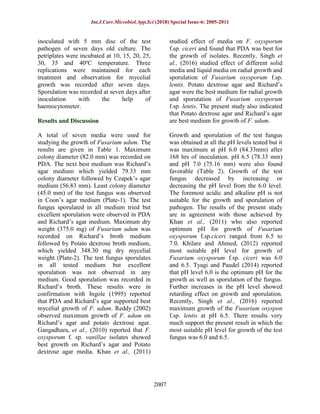
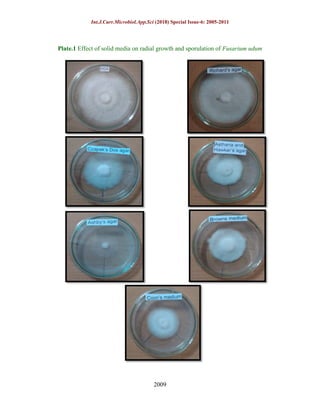
Int.J.Curr.Microbiol.App.Sci (2018) Special Issue-6: 2005-2011
2005
Original Research Article
Effect of Different Media, pH and Temperature on Growth and Sporulation
of Fusarium udum Causing Wilt of Pigeonpea
Balkishan Chaudhary1
*, Sanjeev Kumar1
, Ratan Lal Sharma1
and Shish Ram Jakhar2
1
Department of Plant Pathology, College of Agriculture,
2
Department of Soil Science and Agricultural Chemistry, College of Agriculture,
Jawaharlal Nehru Krishi Vishwa Vidyalaya, Jabalpur, MP-482004, India
*Corresponding author
A B S T R A C T
Introduction
Pigeon pea is an important pulse crop of
India. It is a demanding and always in need
food crop in India. India holds a major
contribution of 90% of total world
production. India engages an area of 3.63
million ha with a production of 2.76 million
tonnes and the productivity of 751 kg/ha
(Singh and Singh, 2014). The crop is prone
to many serious diseases caused by fugal,
bacterial viral and nematode. High
sensitivity of the crop to the attack of
diseases appears to be the main reason for
low yields. Pigeonpea is known to be
affected by more than hundred pathogens
(Nene et al., 1989). However, only a few of
them cause economic losses. The diseases of
considerable economic importance at
present are sterility mosaic, Fusarium wilt,
Phytophthora blight, Macrophomina root
rot, stem canker and Alternaria blight.
Wilt caused by Fusarium udum Butler is the
most important disease caused to this crop
(Kannaiyan et al., 1984). Fusarium udum is
a soil born pathogen and also the pigeonpea
specific wilt pathogen (Kannaiyan et al.,
1985). It causes significant loss to the crop
as well as economic loss to the farmers. The
pathogen has an ability to survive in the
infected crop debris for 3–4 years and cause
serious economic loss, sometimes upto
100% in the susceptible cultivars (Kiprop et
al., 2002). The economic loss caused by
Fusarium wilt in Pigeonpea is 470,000
tonnes in India (Joshi et al., 2001).
Environmental factors such as temperature,
water activity and pH have a great influence
on fungal development (Yadav et al., 2014).
Variation in the type of carbon and nitrogen
sources besides changes in pH, temperature,
incubation period, shaking and inoculum
International Journal of Current Microbiology and Applied Sciences
ISSN: 2319-7692 Special Issue-6 pp. 2005-2011
Journal homepage: http://www.ijcmas.com
Laboratory studies were conducted to study the effect of different culture media, pH and
temperature on mycelia growth, sporulation and dry mycelia weight of Fusarium udum.
The fungus grew the best on PDA and Richard’s agar media among seven culture media
were tested. The most suitable pH level for growth of fungus was 6.0 and 6.5 with excellent
sporulation. Growth of F. udum was maximum at 300
C after seven days of inoculation,
which was reduced drastically below 100
C and above 350
C.
Keywords
Media, pH,
temperature, F.
udum, Wilt,
Pigeonpea
Int.J.Curr.Microbiol.App.Sci (2018) Special Issue-6: 2005-2011
2006
size have great influence on the growth of
pathogen (Tyagi and Paudel, 2014; Dubey,
2016). Present work depicts the role of
different pH and media to understand
ecological survival of pathogen which will
be helpful in management strategy in the
field.
Materials and Methods
Isolate of Fusarium udm was recovered
from diseased pigeonpea plants from
research farm of Jwaharlal Nehru Krishi
Vishwa Vidyalaya- Jabalpur. Small pieces
of discolored vascular tissue from roots of
diseased plants were placed on potato
dextrose agar (PDA) and incubated at 28 ±
1°C in the dark for four days. Isolate was
identified as Fusarium udum by
morphological criteria (Leslie and
Summerell, 2006). A single macroconidial
culture was prepared from isolate. Studies of
the following physiological aspects of
Fusarium udum isolates were conducted in
laboratory.
Effect of culture media
Following seven culture media were used to
find out the most suitable one for the
mycelial growth and sporulation. Each
culture medium was prepared in 1 liter of
water and autoclaved at 121.6ºC at 15 psi for
20 min. These were cooled to 45ºC and then
poured in 90 mm Petri dishes for
solidification.
1. Potato Dextrose agar (PDA) medium
(Peeled and sliced potato 200g, Dextrose
20g, Agar-agar 20g).
2. Richards’s agar (RA) medium (Potassium
nitrate 10g, Potassium monobasic phosphate
5g, Magnesium sulphate 2.5g, Ferric
chloride 0.02g, Sucrose 50g, Agar-agar
20g).
3. Czapeks Dox agar (CDA) medium
(Sodium nitrate 2g, Di potassium hydrogen
phosphate 1g, Magnesium sulphate 0.5g,
Potassium chloride 0.5g, Ferrous sulphate
0.01g, Sucrose 30g, Agar-agar 20g).
4. Asthana and Hawker’s medium (D-
Glucose 5g, Potassium nitrate 3.50g,
Potassium dihydrogen Phosphate 1.75g,
Magnesium sulphate 0.75g, Agar- agar 20g).
5. Ashby’s agar medium (Mannitol 20g, Di
potassium phosphate 0.2g, Magnesium
sulphate 0.2g, Sodium chloride 0.2g,
Potassium sulphate 0.1g, Calcium carbonate
5g, Agar-agar 15g, final pH (at 25°C)
7.4±0.2).
6. Browns agar (BA) medium (Dextrose 2g,
Tri basic potassium phosphate 1.25g,
Magnesium sulphate 0.75g, Agar-agar 20g).
7. Coon’s agar (CA) medium (Sucrose 7.2 g,
Dextrose 3.60g, Magnesium sulphate 1.23g,
Potassium nitrate 2.02g, Potassium di-
phosphate2.72g, Agar- agar 15g).
Effect of pH
There were eight different pH level ranging
from 5.0 to 8.5 with a difference of 0.5 were
prepared by using pH meter and by using
either N/10 HCl or NaOH before
autoclaving the PDA medium. For each pH
value, three replications were maintained.
The Petriplates containing sterilized medium
was inoculated with 5mm mycelium disc
and incubated at 28 + 1ºC. At the interval of
24hrs, the linear growth was measured till 7
days. The range of sporulation test ranges on
various pH was recorded after 7 days.
Sporulation was calculated with the help of
haemocytometer.
Effect of temperature
The experiments were conducted to find out,
the most suitable temperature for mycelial
growth and sporulation of F. udum. The
sterilized poured petriplates with PDA were
Int.J.Curr.Microbiol.App.Sci (2018) Special Issue-6: 2005-2011
2007
inoculated with 5 mm disc of the test
pathogen of seven days old culture. The
petriplates were incubated at 10, 15, 20, 25,
30, 35 and 40ºC temperature. Three
replications were maintained for each
treatment and observation for mycelial
growth was recorded after seven days.
Sporulation was recorded at seven days after
inoculation with the help of
haemocytometer.
Results and Discussion
A total of seven media were used for
studying the growth of Fusarium udum. The
results are given in Table 1. Maximum
colony diameter (82.0 mm) was recorded on
PDA. The next best medium was Richard’s
agar medium which yielded 79.33 mm
colony diameter followed by Czapek’s agar
medium (56.83 mm). Least colony diameter
(45.0 mm) of the test fungus was observed
in Coon’s agar medium (Plate-1). The test
fungus sporulated in all medium tried but
excellent sporulation were observed in PDA
and Richard’s agar medium. Maximum dry
weight (375.0 mg) of Fusarium udum was
recorded on Richard’s broth medium
followed by Potato dextrose broth medium,
which yielded 348.30 mg dry mycelial
weight (Plate-2). The test fungus sporulates
in all tested medium but excellent
sporulation was not observed in any
medium. Good sporulation was recorded in
Richard’s broth. These results were in
confirmation with Ingole (1995) reported
that PDA and Richard’s agar supported best
mycelial growth of F. udum. Reddy (2002)
observed maximum growth of F. udum on
Richard’s agar and potato dextrose agar.
Gangadhara, et al., (2010) reported that F.
oxysporum f. sp. vanillae isolates showed
best growth on Richard’s agar and Potato
dextrose agar media. Khan et al., (2011)
studied effect of media on F. oxysporum
f.sp. ciceri and found that PDA was best for
the growth of isolates. Recently, Singh et
al., (2016) studied effect of different solid
media and liquid media on radial growth and
sporulation of Fusarium oxysporum f.sp.
lentis. Potato dextrose agar and Richard’s
agar were the best medium for radial growth
and sporutation of Fusarium oxysporum
f.sp. lentis. The present study also indicated
that Potato dextrose agar and Richard’s agar
are best medium for growth of F. udum.
Growth and sporulation of the test fungus
was obtained at all the pH levels tested but it
was maximum at pH 6.0 (84.33mm) after
168 hrs of inoculation. pH 6.5 (78.33 mm)
and pH 7.0 (75.16 mm) were also found
favorable (Table 2). Growth of the test
fungus decreased by increasing or
decreasing the pH level from the 6.0 level.
The foremost acidic and alkaline pH is not
suitable for the growth and sporulation of
pathogen. The results of the present study
are in agreement with those achieved by
Khan et al., (2011) who also reported
optimum pH for growth of Fusarium
oxysporum f.sp.ciceri ranged from 6.5 to
7.0. Khilare and Ahmed, (2012) reported
most suitable pH level for growth of
Fusarium oxysporum f.sp. ciceri was 6.0
and 6.5. Tyagi and Paudel (2014) reported
that pH level 6.0 is the optimum pH for the
growth as well as sporulation of the fungus.
Further increases in the pH level showed
retarding effect on growth and sporulation.
Recently, Singh et al., (2016) reported
maximum growth of the Fusarium oxyspon
f.sp. lentis at pH 6.5. There results very
much support the present result in which the
most suitable pH level for growth of the test
fungus was 6.0 and 6.5.
Int.J.Curr.Microbiol.App.Sci (2018) Special Issue-6: 2005-2011
2008
Table.1 Effect of solid and liquid media on radial growth and sporulation of Fusarium udum
*Average of 3 replications
Table.2 Effect of various pH on radial growth and sporulation of Fusarium udum
S. No. pH Colony diameter (mm) after 168 hrs* Sporulation
1 5.0 63.00 ++
2 5.5 70.33 +++
3 6.0 84.33 ++++
4 6.5 78.33 ++++
5 7.0 75.16 +++
6 7.5 51.00 ++
7 8.0 45.00 ++
8 8.5 20.00 --
CD (0.05) 1.773
*Average of 3 replications
Table.3 Effect of different temperature on radial growth and sporulation of F. udum
S. No. Temp. o
C Colony diameter (mm) after 168 hrs* Sporulation
1 10 11.66 --
2 15 27.33 +
3 20 49.67 ++
4 25 66.33 +++
5 30 89.23 ++++
6 35 21.83 +
7 40 0 --
CD (0.05) 2.441
*Average of 3 replications
S. No. Name of the medium Solid Media Liquid Media
Colony diameter (mm)
after 168 hrs*
Sporulation
Dry mycelial
weight (mg)
after 21 day*
Sporulation
1 Potato dextrose agar 82.0 ++++ 348.3 ++
2 Richard’s agar 79.3 ++++ 375.0 +++
3 Czapek’s Dox agar 56.8 +++ 302.1 ++
4 Asthana and Hawker’s agar 49.6 ++ 202.2 ++
5 Browns medium 47.0 ++ 166.1 ++
6 Ashby’s agar 46.3 + 125.6 +
7 Coon’s medium 45.0 + 75.0 +
CD (0.05) 2.331 1.951
Int.J.Curr.Microbiol.App.Sci (2018) Special Issue-6: 2005-2011
2009
Plate.1 Effect of solid media on radial growth and sporulation of Fusarium udum
Int.J.Curr.Microbiol.App.Sci (2018) Special Issue-6: 2005-2011
2010
Plate.2 Effect of liquid media on dry mycelia weight of Fusarium udum
Growth of F. udum was studied from 10 to
40ºC temperature and result is presented in
Table 3. It was seen that there was quite a
large variation in the growth of these isolate
at different temperature after 7 days. The
maximum mycelial growth was recorded at
30ºC (88.23mm) followed by 25ºC
(66.83mm), 20ºC (49.67mm) and 35ºC
(21.00mm), while no mycelial growth was
recorded at 10 and 40ºC. Temperatures from
25 to 35ºC were most favorable for the
growth of target pathogen. The highest
growth of pathogen was recorded at 30ºC
with higher sporulation. Reddy (2002)
reported that growth of 40 isolates of F.
udum differed in their temperature
requirement which varied from 20ºC to
35ºC. Scott, et al., (2010) studied effect of
temperature on Fusarium wilt of lettuce
(Lactuca sativa), caused by F. oxysporum f.
sp. lactucae, were observed to increase in
radial growth from 10ºC up to an apparent
maximum near 25ºC. Khilare and Ahmed
(2012) reported most suitable temperatures
level for growth of Fusarium oxysporum
f.sp. ciceri was 30ºC after seven days of
inoculation, which was reduced drastically
below 15ºC and above 35ºC. Recently,
Desai et al., (2016) reported maximum
growth of the Fusarium udum at 28ºC.
There results very much support the present
studies in which most suitable temperatures
level for growth of the test fungus was 25
and 30ºC.
References
Anjaneya, R. B. (2002). Variability of
Fusarium udum and evaluation of
pigeonpea (Cajanus cajan (L). Mills)
genotypes. M.Sc (Agri) Thesis, Univ.
Agril. Sci. Bangalore, pp.115.
Desai, U. A., Andoji Y. S. and Shivaji S. S.
(2016). Influence of temperature and
different culture media on growth of
Fusarium udum (Butler), causal
organism of wilt of pigeonpea.
International Journal of Biological
Research, 4 (1): 42-45.
Dubey, S.C. (2016). Race profiling, genetic
diversity, diagnostics and management
of Fusarium oxysporum f.sp. ciceris
Int.J.Curr.Microbiol.App.Sci (2018) Special Issue-6: 2005-2011
2011
causindg will of chickpea. Indian
Phytopathology, 69(3): 210-217.
Gangadhara, N. B., Nagaraja, R., Basavaraja
M. K. and Krishna N. R. (2010).
Variability studies of Fusarium
oxysporum f. sp. vanillae isolates.
International Journal of Science and
Nature, 1(1):12-16.
Ingole, M. N. (1995). Estimation of losses,
variability among isolates and
management of pigeon pea wilt caused
by Fusarium udum Butler. M.Sc. (Ag.)
Thesis, Dr. PDKV, Akola, pp.146.
Joshi, P. K., Parthasarathy, Rao. P., Gowda, C.
L. L., Jones, R. B., Silim, S. N., Saxena,
K. B. and Kumar, J. (2001). The world
Chickpea and Pigeonpea Economies:
Facts, Trends, And Outlook. In:
Shiferaw B, Silim S, Muricho G, Audi
P, Mligo J, Lyimo S, You L. and Chris-
tiansen JL. 2005. Assessment of the
Adoption and Impact of Improved
Pigeonpea Varieties in Tanzania.
Journal of SAT Agricultural Research
3(1).
Khan, I. H. S., Saifulla, M., Mahesh, S. B. and
Pallavi, M. S. (2011). Effect of different
media and enviromental conditions on
the growth of Fusarium oxysporum
f.sp.ciceri causing Fusarium wilt of
chickpea. Inter. J. Sci. and Nat. 2: 402-
404.
Khilare, V. C. and Rafi A. (2012). Effect of
different media, ph and temperature on
the growth of Fusarium oxysporum f.sp.
ciceri causing chickpea wilt. Inter. J.
Adv. Biol. Res., Vol. 2(1): 99-102.
Kiprop, E. K., Baudoin, J. P., Mwang'ombe,
A. W., Kimani, P. M, Mer-geai, G. and
Maquet, A. (2002). Characterization of
Kenyan isolates of Fusarium Udum
from Pigeonpea [Cajanus Cajan (L.)
Millsp.] cultural characteristics,
Aggressiveness and AFLP Analysis. J.
Phyto-pathol.150: 517-527.
Kannaiyan, J., Nene, Y. L. and Raju, T. N.
(1985). Host Specificity of Pigeonpea
Wilt Pathogen Fusarium udum. Indian
Phytopath. 38: 553.
Kannaiyan, J., Nene, Y. L., Reddy, M. V.,
Ryan, J. G. and Raju, T. N. (1984).
Prevalence of Pigeonpea Diseases and
Associated Crop Losses in Asia, Africa
and America. Tropical Pest
Management 30: 62–71.
Nene, Y. L., Sheila, V. K. and Sharma, S. B.
(1989). A world List of Chickpea and
Pigeonpea Pathogens. Legume
Pathology Progress Report, 7: 23.
Leslie, J. F. and Summerell, B. A. (2006). The
Fusarium, Laboratory Manual,
Blackwell Publishing, pp. 1 – 388.
Scott, J. C., Gordon, T. R., Shaw, D. V. and
Koike, S. T. (2010). Effect of
temperature on severity of Fusarium
wilt of lettuce caused by Fusarium
oxysporum f. sp. lactucae. Plant
Disease, 94 (1): 13-17.
Singh, J. P. and Singh, F. (2014). Scenario of
pigeonpea research in India. National
Conference of Pulses: Challenges and
Opportunities under changing climate
scenariao 29th
Sept. -1st
Oct. JNKVV
Jabalpur, MP.
Singh R, Laxmikant and Singh M. (2016).
Management of root rot of pea (Pisum
sativum L.) through commercially
available bioagent formulation and
fungicide. 6th
international Conference
“Plant, Pathogens and People” February
23-27, New Delhi, India.
Tyagi, S. and Paudel, R. (2014). Effect of
different pH on the growth and
sporulation of
Fusarium oxysporum: The causal organism of
wilt disease of Tomato. Int. J. Bas.
and Appl. Biol. (2) 103–106.
Yadav, R. S., Tyagi S., Javeria S. and
Gangwar R. K. (2014). Effect of
different cultural condition on the
growth of Fusarium moniliforme
causing Bakanae disease. European
Journal of Molecular Biotechnology,
4(2): 95-100.

Balkishan chaudhary, et al

  • 1.
    Int.J.Curr.Microbiol.App.Sci (2018) SpecialIssue-6: 2005-2011 2005 Original Research Article Effect of Different Media, pH and Temperature on Growth and Sporulation of Fusarium udum Causing Wilt of Pigeonpea Balkishan Chaudhary1 *, Sanjeev Kumar1 , Ratan Lal Sharma1 and Shish Ram Jakhar2 1 Department of Plant Pathology, College of Agriculture, 2 Department of Soil Science and Agricultural Chemistry, College of Agriculture, Jawaharlal Nehru Krishi Vishwa Vidyalaya, Jabalpur, MP-482004, India *Corresponding author A B S T R A C T Introduction Pigeon pea is an important pulse crop of India. It is a demanding and always in need food crop in India. India holds a major contribution of 90% of total world production. India engages an area of 3.63 million ha with a production of 2.76 million tonnes and the productivity of 751 kg/ha (Singh and Singh, 2014). The crop is prone to many serious diseases caused by fugal, bacterial viral and nematode. High sensitivity of the crop to the attack of diseases appears to be the main reason for low yields. Pigeonpea is known to be affected by more than hundred pathogens (Nene et al., 1989). However, only a few of them cause economic losses. The diseases of considerable economic importance at present are sterility mosaic, Fusarium wilt, Phytophthora blight, Macrophomina root rot, stem canker and Alternaria blight. Wilt caused by Fusarium udum Butler is the most important disease caused to this crop (Kannaiyan et al., 1984). Fusarium udum is a soil born pathogen and also the pigeonpea specific wilt pathogen (Kannaiyan et al., 1985). It causes significant loss to the crop as well as economic loss to the farmers. The pathogen has an ability to survive in the infected crop debris for 3–4 years and cause serious economic loss, sometimes upto 100% in the susceptible cultivars (Kiprop et al., 2002). The economic loss caused by Fusarium wilt in Pigeonpea is 470,000 tonnes in India (Joshi et al., 2001). Environmental factors such as temperature, water activity and pH have a great influence on fungal development (Yadav et al., 2014). Variation in the type of carbon and nitrogen sources besides changes in pH, temperature, incubation period, shaking and inoculum International Journal of Current Microbiology and Applied Sciences ISSN: 2319-7692 Special Issue-6 pp. 2005-2011 Journal homepage: http://www.ijcmas.com Laboratory studies were conducted to study the effect of different culture media, pH and temperature on mycelia growth, sporulation and dry mycelia weight of Fusarium udum. The fungus grew the best on PDA and Richard’s agar media among seven culture media were tested. The most suitable pH level for growth of fungus was 6.0 and 6.5 with excellent sporulation. Growth of F. udum was maximum at 300 C after seven days of inoculation, which was reduced drastically below 100 C and above 350 C. Keywords Media, pH, temperature, F. udum, Wilt, Pigeonpea
  • 2.
    Int.J.Curr.Microbiol.App.Sci (2018) SpecialIssue-6: 2005-2011 2006 size have great influence on the growth of pathogen (Tyagi and Paudel, 2014; Dubey, 2016). Present work depicts the role of different pH and media to understand ecological survival of pathogen which will be helpful in management strategy in the field. Materials and Methods Isolate of Fusarium udm was recovered from diseased pigeonpea plants from research farm of Jwaharlal Nehru Krishi Vishwa Vidyalaya- Jabalpur. Small pieces of discolored vascular tissue from roots of diseased plants were placed on potato dextrose agar (PDA) and incubated at 28 ± 1°C in the dark for four days. Isolate was identified as Fusarium udum by morphological criteria (Leslie and Summerell, 2006). A single macroconidial culture was prepared from isolate. Studies of the following physiological aspects of Fusarium udum isolates were conducted in laboratory. Effect of culture media Following seven culture media were used to find out the most suitable one for the mycelial growth and sporulation. Each culture medium was prepared in 1 liter of water and autoclaved at 121.6ºC at 15 psi for 20 min. These were cooled to 45ºC and then poured in 90 mm Petri dishes for solidification. 1. Potato Dextrose agar (PDA) medium (Peeled and sliced potato 200g, Dextrose 20g, Agar-agar 20g). 2. Richards’s agar (RA) medium (Potassium nitrate 10g, Potassium monobasic phosphate 5g, Magnesium sulphate 2.5g, Ferric chloride 0.02g, Sucrose 50g, Agar-agar 20g). 3. Czapeks Dox agar (CDA) medium (Sodium nitrate 2g, Di potassium hydrogen phosphate 1g, Magnesium sulphate 0.5g, Potassium chloride 0.5g, Ferrous sulphate 0.01g, Sucrose 30g, Agar-agar 20g). 4. Asthana and Hawker’s medium (D- Glucose 5g, Potassium nitrate 3.50g, Potassium dihydrogen Phosphate 1.75g, Magnesium sulphate 0.75g, Agar- agar 20g). 5. Ashby’s agar medium (Mannitol 20g, Di potassium phosphate 0.2g, Magnesium sulphate 0.2g, Sodium chloride 0.2g, Potassium sulphate 0.1g, Calcium carbonate 5g, Agar-agar 15g, final pH (at 25°C) 7.4±0.2). 6. Browns agar (BA) medium (Dextrose 2g, Tri basic potassium phosphate 1.25g, Magnesium sulphate 0.75g, Agar-agar 20g). 7. Coon’s agar (CA) medium (Sucrose 7.2 g, Dextrose 3.60g, Magnesium sulphate 1.23g, Potassium nitrate 2.02g, Potassium di- phosphate2.72g, Agar- agar 15g). Effect of pH There were eight different pH level ranging from 5.0 to 8.5 with a difference of 0.5 were prepared by using pH meter and by using either N/10 HCl or NaOH before autoclaving the PDA medium. For each pH value, three replications were maintained. The Petriplates containing sterilized medium was inoculated with 5mm mycelium disc and incubated at 28 + 1ºC. At the interval of 24hrs, the linear growth was measured till 7 days. The range of sporulation test ranges on various pH was recorded after 7 days. Sporulation was calculated with the help of haemocytometer. Effect of temperature The experiments were conducted to find out, the most suitable temperature for mycelial growth and sporulation of F. udum. The sterilized poured petriplates with PDA were
  • 3.
    Int.J.Curr.Microbiol.App.Sci (2018) SpecialIssue-6: 2005-2011 2007 inoculated with 5 mm disc of the test pathogen of seven days old culture. The petriplates were incubated at 10, 15, 20, 25, 30, 35 and 40ºC temperature. Three replications were maintained for each treatment and observation for mycelial growth was recorded after seven days. Sporulation was recorded at seven days after inoculation with the help of haemocytometer. Results and Discussion A total of seven media were used for studying the growth of Fusarium udum. The results are given in Table 1. Maximum colony diameter (82.0 mm) was recorded on PDA. The next best medium was Richard’s agar medium which yielded 79.33 mm colony diameter followed by Czapek’s agar medium (56.83 mm). Least colony diameter (45.0 mm) of the test fungus was observed in Coon’s agar medium (Plate-1). The test fungus sporulated in all medium tried but excellent sporulation were observed in PDA and Richard’s agar medium. Maximum dry weight (375.0 mg) of Fusarium udum was recorded on Richard’s broth medium followed by Potato dextrose broth medium, which yielded 348.30 mg dry mycelial weight (Plate-2). The test fungus sporulates in all tested medium but excellent sporulation was not observed in any medium. Good sporulation was recorded in Richard’s broth. These results were in confirmation with Ingole (1995) reported that PDA and Richard’s agar supported best mycelial growth of F. udum. Reddy (2002) observed maximum growth of F. udum on Richard’s agar and potato dextrose agar. Gangadhara, et al., (2010) reported that F. oxysporum f. sp. vanillae isolates showed best growth on Richard’s agar and Potato dextrose agar media. Khan et al., (2011) studied effect of media on F. oxysporum f.sp. ciceri and found that PDA was best for the growth of isolates. Recently, Singh et al., (2016) studied effect of different solid media and liquid media on radial growth and sporulation of Fusarium oxysporum f.sp. lentis. Potato dextrose agar and Richard’s agar were the best medium for radial growth and sporutation of Fusarium oxysporum f.sp. lentis. The present study also indicated that Potato dextrose agar and Richard’s agar are best medium for growth of F. udum. Growth and sporulation of the test fungus was obtained at all the pH levels tested but it was maximum at pH 6.0 (84.33mm) after 168 hrs of inoculation. pH 6.5 (78.33 mm) and pH 7.0 (75.16 mm) were also found favorable (Table 2). Growth of the test fungus decreased by increasing or decreasing the pH level from the 6.0 level. The foremost acidic and alkaline pH is not suitable for the growth and sporulation of pathogen. The results of the present study are in agreement with those achieved by Khan et al., (2011) who also reported optimum pH for growth of Fusarium oxysporum f.sp.ciceri ranged from 6.5 to 7.0. Khilare and Ahmed, (2012) reported most suitable pH level for growth of Fusarium oxysporum f.sp. ciceri was 6.0 and 6.5. Tyagi and Paudel (2014) reported that pH level 6.0 is the optimum pH for the growth as well as sporulation of the fungus. Further increases in the pH level showed retarding effect on growth and sporulation. Recently, Singh et al., (2016) reported maximum growth of the Fusarium oxyspon f.sp. lentis at pH 6.5. There results very much support the present result in which the most suitable pH level for growth of the test fungus was 6.0 and 6.5.
  • 4.
    Int.J.Curr.Microbiol.App.Sci (2018) SpecialIssue-6: 2005-2011 2008 Table.1 Effect of solid and liquid media on radial growth and sporulation of Fusarium udum *Average of 3 replications Table.2 Effect of various pH on radial growth and sporulation of Fusarium udum S. No. pH Colony diameter (mm) after 168 hrs* Sporulation 1 5.0 63.00 ++ 2 5.5 70.33 +++ 3 6.0 84.33 ++++ 4 6.5 78.33 ++++ 5 7.0 75.16 +++ 6 7.5 51.00 ++ 7 8.0 45.00 ++ 8 8.5 20.00 -- CD (0.05) 1.773 *Average of 3 replications Table.3 Effect of different temperature on radial growth and sporulation of F. udum S. No. Temp. o C Colony diameter (mm) after 168 hrs* Sporulation 1 10 11.66 -- 2 15 27.33 + 3 20 49.67 ++ 4 25 66.33 +++ 5 30 89.23 ++++ 6 35 21.83 + 7 40 0 -- CD (0.05) 2.441 *Average of 3 replications S. No. Name of the medium Solid Media Liquid Media Colony diameter (mm) after 168 hrs* Sporulation Dry mycelial weight (mg) after 21 day* Sporulation 1 Potato dextrose agar 82.0 ++++ 348.3 ++ 2 Richard’s agar 79.3 ++++ 375.0 +++ 3 Czapek’s Dox agar 56.8 +++ 302.1 ++ 4 Asthana and Hawker’s agar 49.6 ++ 202.2 ++ 5 Browns medium 47.0 ++ 166.1 ++ 6 Ashby’s agar 46.3 + 125.6 + 7 Coon’s medium 45.0 + 75.0 + CD (0.05) 2.331 1.951
  • 5.
    Int.J.Curr.Microbiol.App.Sci (2018) SpecialIssue-6: 2005-2011 2009 Plate.1 Effect of solid media on radial growth and sporulation of Fusarium udum
  • 6.
    Int.J.Curr.Microbiol.App.Sci (2018) SpecialIssue-6: 2005-2011 2010 Plate.2 Effect of liquid media on dry mycelia weight of Fusarium udum Growth of F. udum was studied from 10 to 40ºC temperature and result is presented in Table 3. It was seen that there was quite a large variation in the growth of these isolate at different temperature after 7 days. The maximum mycelial growth was recorded at 30ºC (88.23mm) followed by 25ºC (66.83mm), 20ºC (49.67mm) and 35ºC (21.00mm), while no mycelial growth was recorded at 10 and 40ºC. Temperatures from 25 to 35ºC were most favorable for the growth of target pathogen. The highest growth of pathogen was recorded at 30ºC with higher sporulation. Reddy (2002) reported that growth of 40 isolates of F. udum differed in their temperature requirement which varied from 20ºC to 35ºC. Scott, et al., (2010) studied effect of temperature on Fusarium wilt of lettuce (Lactuca sativa), caused by F. oxysporum f. sp. lactucae, were observed to increase in radial growth from 10ºC up to an apparent maximum near 25ºC. Khilare and Ahmed (2012) reported most suitable temperatures level for growth of Fusarium oxysporum f.sp. ciceri was 30ºC after seven days of inoculation, which was reduced drastically below 15ºC and above 35ºC. Recently, Desai et al., (2016) reported maximum growth of the Fusarium udum at 28ºC. There results very much support the present studies in which most suitable temperatures level for growth of the test fungus was 25 and 30ºC. References Anjaneya, R. B. (2002). Variability of Fusarium udum and evaluation of pigeonpea (Cajanus cajan (L). Mills) genotypes. M.Sc (Agri) Thesis, Univ. Agril. Sci. Bangalore, pp.115. Desai, U. A., Andoji Y. S. and Shivaji S. S. (2016). Influence of temperature and different culture media on growth of Fusarium udum (Butler), causal organism of wilt of pigeonpea. International Journal of Biological Research, 4 (1): 42-45. Dubey, S.C. (2016). Race profiling, genetic diversity, diagnostics and management of Fusarium oxysporum f.sp. ciceris
  • 7.
    Int.J.Curr.Microbiol.App.Sci (2018) SpecialIssue-6: 2005-2011 2011 causindg will of chickpea. Indian Phytopathology, 69(3): 210-217. Gangadhara, N. B., Nagaraja, R., Basavaraja M. K. and Krishna N. R. (2010). Variability studies of Fusarium oxysporum f. sp. vanillae isolates. International Journal of Science and Nature, 1(1):12-16. Ingole, M. N. (1995). Estimation of losses, variability among isolates and management of pigeon pea wilt caused by Fusarium udum Butler. M.Sc. (Ag.) Thesis, Dr. PDKV, Akola, pp.146. Joshi, P. K., Parthasarathy, Rao. P., Gowda, C. L. L., Jones, R. B., Silim, S. N., Saxena, K. B. and Kumar, J. (2001). The world Chickpea and Pigeonpea Economies: Facts, Trends, And Outlook. In: Shiferaw B, Silim S, Muricho G, Audi P, Mligo J, Lyimo S, You L. and Chris- tiansen JL. 2005. Assessment of the Adoption and Impact of Improved Pigeonpea Varieties in Tanzania. Journal of SAT Agricultural Research 3(1). Khan, I. H. S., Saifulla, M., Mahesh, S. B. and Pallavi, M. S. (2011). Effect of different media and enviromental conditions on the growth of Fusarium oxysporum f.sp.ciceri causing Fusarium wilt of chickpea. Inter. J. Sci. and Nat. 2: 402- 404. Khilare, V. C. and Rafi A. (2012). Effect of different media, ph and temperature on the growth of Fusarium oxysporum f.sp. ciceri causing chickpea wilt. Inter. J. Adv. Biol. Res., Vol. 2(1): 99-102. Kiprop, E. K., Baudoin, J. P., Mwang'ombe, A. W., Kimani, P. M, Mer-geai, G. and Maquet, A. (2002). Characterization of Kenyan isolates of Fusarium Udum from Pigeonpea [Cajanus Cajan (L.) Millsp.] cultural characteristics, Aggressiveness and AFLP Analysis. J. Phyto-pathol.150: 517-527. Kannaiyan, J., Nene, Y. L. and Raju, T. N. (1985). Host Specificity of Pigeonpea Wilt Pathogen Fusarium udum. Indian Phytopath. 38: 553. Kannaiyan, J., Nene, Y. L., Reddy, M. V., Ryan, J. G. and Raju, T. N. (1984). Prevalence of Pigeonpea Diseases and Associated Crop Losses in Asia, Africa and America. Tropical Pest Management 30: 62–71. Nene, Y. L., Sheila, V. K. and Sharma, S. B. (1989). A world List of Chickpea and Pigeonpea Pathogens. Legume Pathology Progress Report, 7: 23. Leslie, J. F. and Summerell, B. A. (2006). The Fusarium, Laboratory Manual, Blackwell Publishing, pp. 1 – 388. Scott, J. C., Gordon, T. R., Shaw, D. V. and Koike, S. T. (2010). Effect of temperature on severity of Fusarium wilt of lettuce caused by Fusarium oxysporum f. sp. lactucae. Plant Disease, 94 (1): 13-17. Singh, J. P. and Singh, F. (2014). Scenario of pigeonpea research in India. National Conference of Pulses: Challenges and Opportunities under changing climate scenariao 29th Sept. -1st Oct. JNKVV Jabalpur, MP. Singh R, Laxmikant and Singh M. (2016). Management of root rot of pea (Pisum sativum L.) through commercially available bioagent formulation and fungicide. 6th international Conference “Plant, Pathogens and People” February 23-27, New Delhi, India. Tyagi, S. and Paudel, R. (2014). Effect of different pH on the growth and sporulation of Fusarium oxysporum: The causal organism of wilt disease of Tomato. Int. J. Bas. and Appl. Biol. (2) 103–106. Yadav, R. S., Tyagi S., Javeria S. and Gangwar R. K. (2014). Effect of different cultural condition on the growth of Fusarium moniliforme causing Bakanae disease. European Journal of Molecular Biotechnology, 4(2): 95-100.