
Recommended
More Related Content
Similar to Integumentary system .ppsx
Similar to Integumentary system .ppsx (20)
More from lumaGhaziALzamel
More from lumaGhaziALzamel (15)
Recently uploaded
Recently uploaded (20)
Integumentary system .ppsx
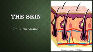
- 1. THE SKIN Dr. Luma Alzamel
- 2. Structure of the skin The skin has a surface area of about 1.5 to 2 m2 in adults and it contains glands, hair and nails. There are two main layers: • epidermis • dermis. Between the skin and underlying structures there is a layer of subcutaneous fat.
- 4. Epidermis The epidermis is the most superficial layer of the skin and is composed of stratified keratinized squamous epithelium which varies in thickness in different parts of the body. It is thickest on the palms of the hands and soles of the feet. There are no blood vessels or nerve endings in the epidermis, but its deeper layers are bathed in interstitial fluid from the dermis, which provides oxygen and nutrients, and is drained away as lymph. There are several layers (strata) of cells in the epidermis which extend from the deepest germinative layer to the surface stratum corneum (a thick horny layer). The cells on the surface are flat, thin, non-nucleated, dead cells in which the cytoplasm has been replaced by the fibrous protein keratin. These cells are constantly being rubbed off and replaced by cells which originated in the germinative layer and have undergone gradual change as they progressed towards the surface. Complete replacement of the epidermis takes about 40 days.
- 5. The maintenance of healthy epidermis depends upon three processes being synchronized: The skin showing the main layers of the epidermis. • desquamation (shedding) of the keratinized cells from the surface • effective keratinization of the cells approaching the Surface. • continual cell division in the deeper layers with newly formed cells being pushed to the surface. Hairs, secretions from sebaceous glands and ducts of sweat glands pass through the epidermis to reach the surface. The surface of the epidermis is ridged by projections of cells in the dermis called the papillae.
- 6. The color of the skin is affected by three main factors. • Melanin, a dark pigment derived from the amino acid tyrosine and secreted by melanocytes in the deep germinative layer, is absorbed by surrounding epithelial cells. The amount is genetically determined and varies between different parts of the body, between members of the same race and between races. The number of melanocytes is constant so the differences in color depend on the amount of melanin secreted. It protects the skin from the harmful effects of sunlight. Exposure to sunlight promotes synthesis of increased amounts of melanin. • The level of oxygenation of hemoglobin and the amount of blood circulating in the dermis give the skin its pink color. • Bile pigments in blood and carotenes in subcutaneous fat give the skin a yellowish color.
- 7. Dermis The dermis is tough and elastic. It is formed from connective tissue and the matrix contains collagen fibers interlaced with elastic fibers. Rupture of elastic fibers occurs when the skin is overstretched, resulting in permanent striae, or stretch marks, that may be found in pregnancy and obesity. Collagen fibers bind water and give the skin its tensile strength, but as this ability declines with age, wrinkles develop. Fibroblasts, macrophages and mast cells are the main cells found in the dermis. Underlying its deepest layer there is areolar tissue and varying amounts of adipose tissue (fat).
- 8. The structures in the dermis are: • blood vessels • lymph vessels • sensory (somatic) nerve endings which are sensitive to touch, change in temperature, pressure and pain are widely distributed in the dermis. • sweat glands and their ducts are found widely distributed throughout the skin and are most numerous in the palms of the hands, soles of the feet, axillae and groins. They are composed of epithelial cells. Glands opening into hair follicles do not become active until puberty. In the axilla they secrete an odorless milky fluid which, if decomposed by surface microbes, causes an unpleasant odor. The functions of this secretion are not known. Sweat glands are stimulated by sympathetic nerves in response to raised body temperature and fear.
- 9. The most important function of sweat secreted by glands opening on to the skin surface is in the regulation of body temperature. Evaporation of sweat from body surfaces takes heat from the body and the amount of sweat produced is governed by the temperature-regulating center in the hypothalamus. Excessive sweating may lead to dehydration and serious depletion of body sodium chloride unless intake of water and salt is appropriately increased. After 7 to 10 days' exposure to high environmental temperatures the amount of salt lost is substantially reduced but water loss remains high. • hairs, arrector pili muscles and sebaceous glands.
- 10. Hairs These are formed by a down-growth of epidermal cells into the dermis or subcutaneous tissue, called hair follicles. At the base of the follicle is a cluster of cells called the bulb. The hair is formed by multiplication of cells of the bulb and as they are pushed upwards, away from them source of nutrition, the cells die and become keratinized. The part of the hair above the skin is the shaft and the remainder, the root. The color of the hair is genetically determined and depends on the amount of melanin present. White hair is the result of the replacement of melanin by tiny air bubbles. Accessory organs of the skin
- 11. Nails The nails are derived from the same cells as epidermis and hair and consist of a hard, horny keratin plate. They protect the tips of the fingers and toes. The nail plate of the nail is the exposed part that has grown out from the germinative zone of the epidermis called the nail bed. Fingernails grow more quickly than toenails and growth is quicker when the environmental temperature is high. Subcutaneous gland Sweat gland