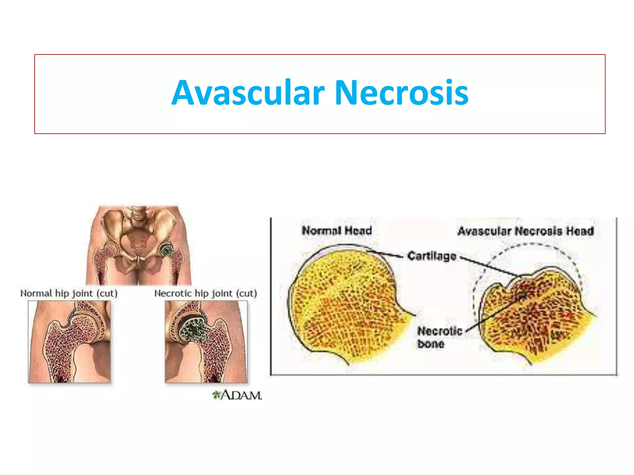
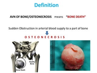
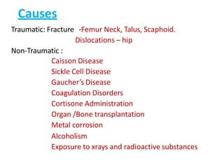
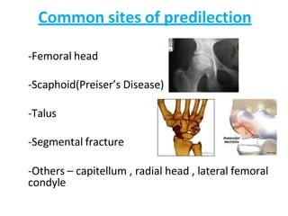
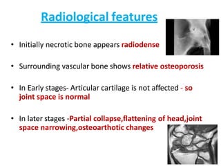
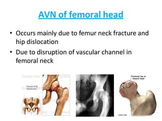
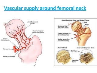
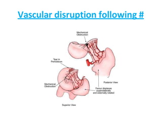
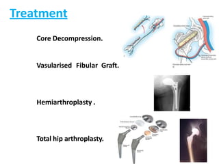
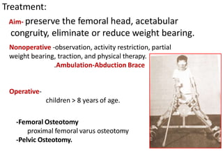
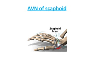
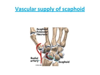
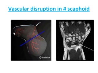
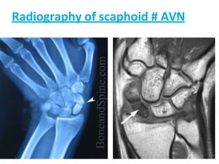
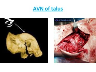
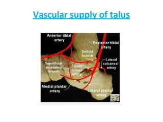
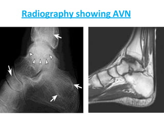
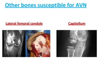
Avascular Necrosis
Definition
AVN OF BONE/OSTEONECROSIS means “BONE DEATH”
Sudden Obstruction in arterial blood supply to a part of bone
O S T E O N E C R O S I S
Causes
Traumatic: Fracture -Femur Neck, Talus, Scaphoid.
Dislocations – hip
Non-Traumatic :
Caisson Disease
Sickle Cell Disease
Gaucher’s Disease
Coagulation Disorders
Cortisone Administration
Organ /Bone transplantation
Metal corrosion
Alcoholism
Exposure to xrays and radioactive substances
Common sites of predilection
-Femoral head
-Scaphoid(Preiser’s Disease)
-Talus
-Segmental fracture
-Others – capitellum , radial head , lateral femoral
condyle
Pathogenesis
BONE ISCHAEMIA is due to
- Interruption of arterial inflow
- Occlusion of Venous Outflow
- Intravascular Blockage of arterioles & capillaries
- Increase in marrow pressure
Repair process in a cancellous bone
• Proliferating capillaries and fibrous stroma
penetrate marrow space of dead bone
• Phagocytes remove the marrow debris
• Osteoblasts lay down immature woven bone
• All these process further increases the
radiodensity of necrotic bone
• Later osteoclastic resorption removes the woven
bone as well as the old trabaeculae
• This is replaced by well organised lamellar bone
• This process of apposition of new bone on some
surface and osteoclastic resorption on other
surface is called CREEPING SUBSTITUTION
Repair process in cortical bone
• Excavation of haversian canal by osteoclastic
resorption proceeds
• This enlarges the canal which multiple in
number and uniform in size
• Later osteoblasts lay down concentric rings of
new bone
Pathology
• Microscopically 4 stages are recognised
-Stage of marrow necrosis and cell death.
-Reactive vascularisation and infiltration.
-Distortion of shape by collapse and compression of trabeculae
-Subchondral collapse
Deformation of articular cartilage
Dysbaric osteonecrosis
 Caisson disease /Decompressionsickness/
Aeroembolism.
 Seen in deep sea divers , tunnel workers,
working in unpressurized aircrafts.
 Nitrogen gas bubbles liberated in a
concentration that cannot be readily
absorbed by blood stream or excreted by
lungs.
 As a result gas bubbles accumulate in tissues
causing local ischemia or intravascular
occlusion
Corticosteroid induced AVN
1. Fat embolism theory :
• Fat accumulates in liver in patients treated with steroid , and serum lipid
concentration also increases
• It gives rise to fat embolism and AVN
2. Subchondral osteoporotic fracture :
Steroids induce protein catabolism
Resulting in generalised osteoporosis
Produces subchondral fractures and aseptic necrosis
Clinical Features
Early stages: Pain-near joint
Later stages: Stiffness, Limitation of movements
Advanced stage: Fixed Deformities
Radiological features
• Initially necrotic bone appears radiodense
• Surrounding vascular bone shows relative osteoporosis
• In Early stages- Articular cartilage is not affected - so
joint space is normal
• In later stages -Partial collapse,flattening of head,joint
space narrowing,osteoarthotic changes
AVN of femoral head
• Occurs mainly due to femur neck fracture and
hip dislocation
• Due to disruption of vascular channel in
femoral neck
Vascular supply around femoral neck
Vascular disruption following #
Radiography of femur head - AVN
Treatment
Core Decompression.
Vasularised Fibular Graft.
Hemiarthroplasty .
Total hip arthroplasty.
Legg-calve perthe’s disease
AVN of ossification centre of capital epiphysis of femoral
head.
3-12 age group.
c/f: Limp, antalgic gait, limited motion
HIP DEFORMITY
xray: Early findings include Medial joint space widening
irregularity of femoral head ossification
cresent sign (represents a subchondral fracture)
Treatment:
Aim- preserve the femoral head, acetabular
congruity, eliminate or reduce weight bearing.
Nonoperative -observation, activity restriction, partial
weight bearing, traction, and physical therapy.
.Ambulation-Abduction Brace
Operative-
children > 8 years of age.
-Femoral Osteotomy
proximal femoral varus osteotomy
-Pelvic Osteotomy.
AVN of scaphoid
Vascular supply of scaphoid
Vascular disruption in # scaphoid
Radiography of scaphoid # AVN
AVN of talus
Vascular supply of talus
Radiography showing AVN
Other bones susceptible for AVN
Lateral femoral condyle Capitellum
The goal in treating avascular
necrosis
Is to improve the patient's use of the
affected joint,
Stop further damage to the bone
and
Ensure bone and joint survival.
• Various methods for delaying disease progression
• Non surgical –
• Bisphosphonate
• Anticoagulants
• Vasodilators
• Biophysical modalities
• Surgical –
• Core decompression
• Vascularised bone graft/muscle pedicle graft
Usually arthroplasty is awaited without any big
surgical intervention
avascularnecrosisugclass-160330183140.pptx

avascularnecrosisugclass-160330183140.pptx

  • 1.
  • 2.
    Definition AVN OF BONE/OSTEONECROSISmeans “BONE DEATH” Sudden Obstruction in arterial blood supply to a part of bone O S T E O N E C R O S I S
  • 3.
    Causes Traumatic: Fracture -FemurNeck, Talus, Scaphoid. Dislocations – hip Non-Traumatic : Caisson Disease Sickle Cell Disease Gaucher’s Disease Coagulation Disorders Cortisone Administration Organ /Bone transplantation Metal corrosion Alcoholism Exposure to xrays and radioactive substances
  • 4.
    Common sites ofpredilection -Femoral head -Scaphoid(Preiser’s Disease) -Talus -Segmental fracture -Others – capitellum , radial head , lateral femoral condyle
  • 5.
    Pathogenesis BONE ISCHAEMIA isdue to - Interruption of arterial inflow - Occlusion of Venous Outflow - Intravascular Blockage of arterioles & capillaries - Increase in marrow pressure
  • 6.
    Repair process ina cancellous bone • Proliferating capillaries and fibrous stroma penetrate marrow space of dead bone • Phagocytes remove the marrow debris • Osteoblasts lay down immature woven bone • All these process further increases the radiodensity of necrotic bone
  • 7.
    • Later osteoclasticresorption removes the woven bone as well as the old trabaeculae • This is replaced by well organised lamellar bone • This process of apposition of new bone on some surface and osteoclastic resorption on other surface is called CREEPING SUBSTITUTION
  • 8.
    Repair process incortical bone • Excavation of haversian canal by osteoclastic resorption proceeds • This enlarges the canal which multiple in number and uniform in size • Later osteoblasts lay down concentric rings of new bone
  • 9.
    Pathology • Microscopically 4stages are recognised -Stage of marrow necrosis and cell death. -Reactive vascularisation and infiltration. -Distortion of shape by collapse and compression of trabeculae -Subchondral collapse Deformation of articular cartilage
  • 10.
    Dysbaric osteonecrosis  Caissondisease /Decompressionsickness/ Aeroembolism.  Seen in deep sea divers , tunnel workers, working in unpressurized aircrafts.  Nitrogen gas bubbles liberated in a concentration that cannot be readily absorbed by blood stream or excreted by lungs.  As a result gas bubbles accumulate in tissues causing local ischemia or intravascular occlusion
  • 11.
    Corticosteroid induced AVN 1.Fat embolism theory : • Fat accumulates in liver in patients treated with steroid , and serum lipid concentration also increases • It gives rise to fat embolism and AVN 2. Subchondral osteoporotic fracture : Steroids induce protein catabolism Resulting in generalised osteoporosis Produces subchondral fractures and aseptic necrosis
  • 12.
    Clinical Features Early stages:Pain-near joint Later stages: Stiffness, Limitation of movements Advanced stage: Fixed Deformities
  • 13.
    Radiological features • Initiallynecrotic bone appears radiodense • Surrounding vascular bone shows relative osteoporosis • In Early stages- Articular cartilage is not affected - so joint space is normal • In later stages -Partial collapse,flattening of head,joint space narrowing,osteoarthotic changes
  • 14.
    AVN of femoralhead • Occurs mainly due to femur neck fracture and hip dislocation • Due to disruption of vascular channel in femoral neck
  • 15.
  • 16.
  • 17.
  • 18.
    Treatment Core Decompression. Vasularised FibularGraft. Hemiarthroplasty . Total hip arthroplasty.
  • 19.
    Legg-calve perthe’s disease AVNof ossification centre of capital epiphysis of femoral head. 3-12 age group. c/f: Limp, antalgic gait, limited motion HIP DEFORMITY xray: Early findings include Medial joint space widening irregularity of femoral head ossification cresent sign (represents a subchondral fracture)
  • 20.
    Treatment: Aim- preserve thefemoral head, acetabular congruity, eliminate or reduce weight bearing. Nonoperative -observation, activity restriction, partial weight bearing, traction, and physical therapy. .Ambulation-Abduction Brace Operative- children > 8 years of age. -Femoral Osteotomy proximal femoral varus osteotomy -Pelvic Osteotomy.
  • 21.
  • 22.
  • 23.
  • 24.
  • 25.
  • 26.
  • 27.
  • 28.
    Other bones susceptiblefor AVN Lateral femoral condyle Capitellum
  • 29.
    The goal intreating avascular necrosis Is to improve the patient's use of the affected joint, Stop further damage to the bone and Ensure bone and joint survival.
  • 30.
    • Various methodsfor delaying disease progression • Non surgical – • Bisphosphonate • Anticoagulants • Vasodilators • Biophysical modalities • Surgical – • Core decompression • Vascularised bone graft/muscle pedicle graft Usually arthroplasty is awaited without any big surgical intervention