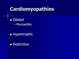
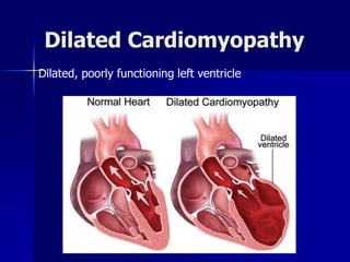
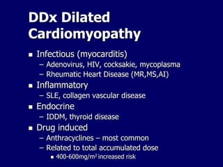
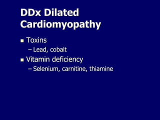
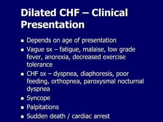
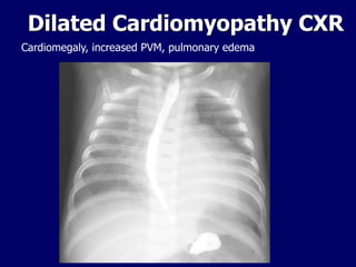
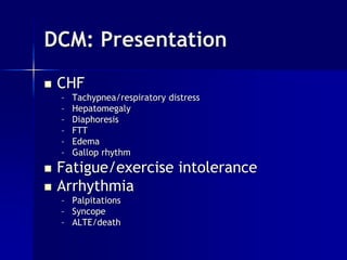
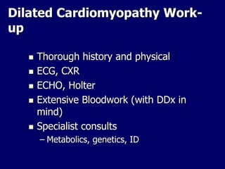
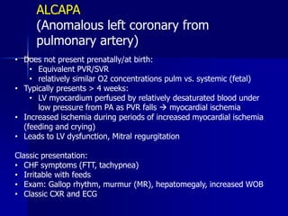
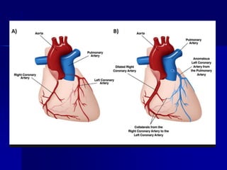
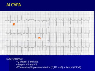
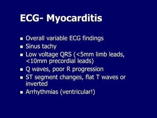
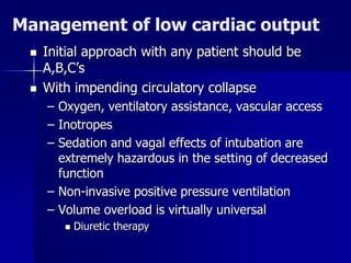
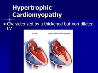
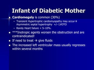
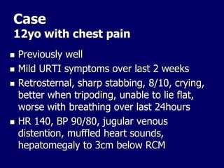
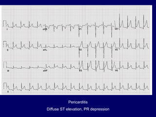
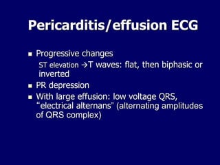
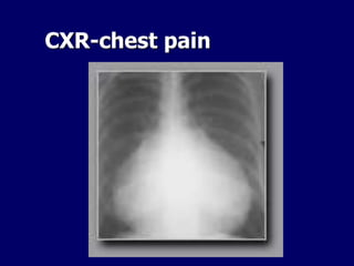
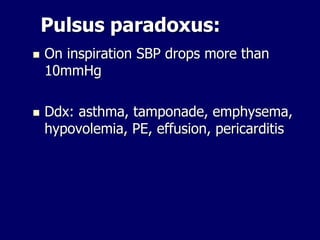
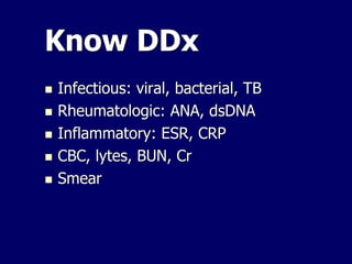
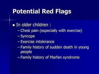
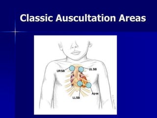
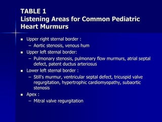
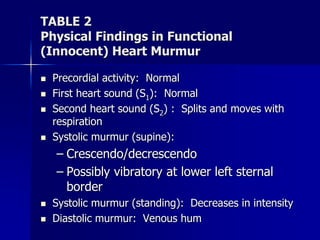
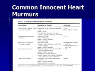
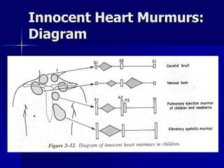
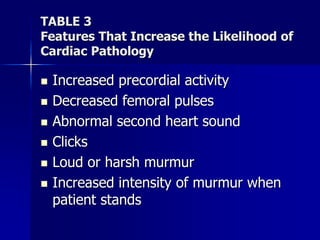
Cardiology Review
Part 2
Oct 2018
Cardiomyopathies
 Dilated
– Myocarditis
 Hypertrophic
 Restrictive
Dilated Cardiomyopathy
Dilated, poorly functioning left ventricle
DDx Dilated
Cardiomyopathy
 Cardiomyopathy
– Idiopathic – 60% of cases
– Familial-usually AD
 Congenital heart disease
 Coronary abnormalities
– Kawasaki, ALCAPA (anomalous left coronary
artery from the pulmonary artery)
 Arrhythmias-incessant SVT (babies)
 Metabolic disorders/rare syndromes
 Neuromuscular disorders
– Duchenne MD
DDx Dilated
Cardiomyopathy
 Infectious (myocarditis)
– Adenovirus, HIV, cocksakie, mycoplasma
– Rheumatic Heart Disease (MR,MS,AI)
 Inflammatory
– SLE, collagen vascular disease
 Endocrine
– IDDM, thyroid disease
 Drug induced
– Anthracyclines – most common
– Related to total accumulated dose
 400-600mg/m2 increased risk
DDx Dilated
Cardiomyopathy
 Toxins
– Lead, cobalt
 Vitamin deficiency
– Selenium, carnitine, thiamine
Dilated CHF – Clinical
Presentation
 Depends on age of presentation
 Vague sx – fatigue, malaise, low grade
fever, anorexia, decreased exercise
tolerance
 CHF sx – dyspnea, diaphoresis, poor
feeding, orthopnea, paroxysmal nocturnal
dyspnea
 Syncope
 Palpitations
 Sudden death / cardiac arrest
Dilated Cardiomyopathy CXR
Cardiomegaly, increased PVM, pulmonary edema
Figure 33: 13-day-old infant with intrauterine infection, cardiomegaly
DCM: Presentation
 CHF
– Tachypnea/respiratory distress
– Hepatomegaly
– Diaphoresis
– FTT
– Edema
– Gallop rhythm
 Fatigue/exercise intolerance
 Arrhythmia
– Palpitations
– Syncope
– ALTE/death
Dilated Cardiomyopathy Work-
up
 Thorough history and physical
 ECG, CXR
 ECHO, Holter
 Extensive Bloodwork (with DDx in
mind)
 Specialist consults
– Metabolics, genetics, ID
ALCAPA
(Anomalous left coronary from
pulmonary artery)
• Does not present prenatally/at birth:
• Equivalent PVR/SVR
• relatively similar O2 concentrations pulm vs. systemic (fetal)
• Typically presents > 4 weeks:
• LV myocardium perfused by relatively desaturated blood under
low pressure from PA as PVR falls  myocardial ischemia
• Increased ischemia during periods of increased myocardial ischemia
(feeding and crying)
• Leads to LV dysfunction, Mitral regurgitation
Classic presentation:
• CHF symptoms (FTT, tachypnea)
• Irritable with feeds
• Exam: Gallop rhythm, murmur (MR), hepatomegaly, increased WOB
• Classic CXR and ECG


ECG FINDINGS:
- Q waves- I and AVL
- deep in V5 and V6
-ST elevation/depression inferior (II,III, avF) + lateral (V5,V6)
ALCAPA
ECG- Myocarditis
 Overall variable ECG findings
 Sinus tachy
 Low voltage QRS (<5mm limb leads,
<10mm precordial leads)
 Q waves, poor R progression
 ST segment changes, flat T waves or
inverted
 Arrhythmias (ventricular!)
Management of low cardiac output
 Initial approach with any patient should be
A,B,C’s
 With impending circulatory collapse
– Oxygen, ventilatory assistance, vascular access
– Inotropes
– Sedation and vagal effects of intubation are
extremely hazardous in the setting of decreased
function
– Non-invasive positive pressure ventilation
– Volume overload is virtually universal
 Diuretic therapy
Congestive Heart Failure
General Management
–ACE Inhibitors
 potential for benefit with  afterload to increase
systemic blood flow
 may not be necessary where surgery available,
however some studies showed improved
symptoms and growth
 Renal insufficiency in shunt lesions
–B-Blockers
 Improved neurohormonal profile
  symptoms and improved growth
 May not be necessary where surgery is available
 Digoxin – mixed results in studies
If stable off
inotropes, can
introduce oral
therapy slowly
DCM: Treatment
 Anti-failure therapy
– Diuretics, after-load reduction, ACEi, spironolactone, -
blockers,
 Anticoagulation if EF < 25%
 Treat specific cause when possible
– SVT  anti-arrhythmics
– Myocarditis  supportive care, anti-inflammatory treatment
– AI  valve replacement
 Complication specific
– Anti-arrhythmics, defibrillators
 Heart transplant
DCM - Outcomes
 Significant variability depending on
etiology
 1 year survival – 60-90%
 5 year survival – 20-60%
 Best prognosis if preceding (<3mos) viral
infection – probably recovering viral
myocarditis
Hypertrophic
Cardiomyopathy
 Characterized by a thickened but non-dilated
LV
Hypertrophic Cardiomyopathy
Etiology
Genetic- Autosomal dominant mostly
Metabolic disease
– Storage disease
 Glycogen storage disease –Pompe’s disease
 Hunter-Hurler
– Mitochondrial myopathies
Other Syndromes
– Noonan syndrome (~25%)
Infants of diabetic mothers
– Generally regresses over time
– Outflow tract obstruction uncommon
Neuromuscular disorders
– Friedrich Ataxia
Presentation
 Asymptomatic
 Easy fatiguability/SOBOE
 Syncope
 Murmur
– Outflow tract obstruction – HOCM
 Arrhythmia/palpitations
– Vtach
Presentation
 Syncope
– Outflow tract obstruction
– Coronary insufficiency
– VT/Vfib
 Chest pain
– myocardial bridging of coronary arteries
– Subendocardial ischemia
 ALTE/sudden cardiac death
Treatment
Medical
 Medications
– B-blockers
– disopyramide
– Verapamil
 Surgery
– Resection of heart muscle
 Disease specific
– Enzyme replacement - Pompe disease
– Carnitine Coenzyme Q - Mitochondrial disease
– Idebenone - Freidrich’s ataxia
 Complication specific
– Anti-arrhythmic
– Implantable cardioverter-defibrillator (ICD)
Infant of Diabetic Mother
 Cardiomegaly is common (30%)
– Transient Hypertrophic cardiomyopathy may occur
Asymmetric septal hypertrophy +/- LVOTO
– Rarely Heart failure < 5–10%.
 ***Inotropic agents worsen the obstruction and are
contraindicated!
 If need to treat  give fluids
 The increased left ventricular mass usually regresses
within several months
PERICARDITIS
Case
12yo with chest pain
 Previously well
 Mild URTI symptoms over last 2 weeks
 Retrosternal, sharp stabbing, 8/10, crying,
better when tripoding, unable to lie flat,
worse with breathing over last 24hours
 HR 140, BP 90/80, jugular venous
distention, muffled heart sounds,
hepatomegaly to 3cm below RCM
Pericarditis
Diffuse ST elevation, PR depression
Pericarditis/effusion ECG
 Progressive changes
ST elevation T waves: flat, then biphasic or
inverted
 PR depression
 With large effusion: low voltage QRS,
“electrical alternans” (alternating amplitudes
of QRS complex)
CXR-chest pain
Pulsus paradoxus:
 On inspiration SBP drops more than
10mmHg
 Ddx: asthma, tamponade, emphysema,
hypovolemia, PE, effusion, pericarditis
Know DDx
 Infectious: viral, bacterial, TB
 Rheumatologic: ANA, dsDNA
 Inflammatory: ESR, CRP
 CBC, lytes, BUN, Cr
 Smear
Pericarditis: Treatment
 Depends on etiology
 Typically NSAIDS
– Ibuprofen 2-3 weeks
– Occasionally colchicine, especially if recurrent
– Occasionally steroids or other anti-inflammatory
 Recurrence up to 15-30%
 Postpericardiotomy syndrome
– Up to 30% of patients post heart surgery but
less frequent in <2yo
– Fever the first week after surgery, malaise,
chest pain, irritabilty,+rub
– ASA 50-75mg/kg for 4-6weeks (indomethacin
or NSAIDs)
– Steroids: prednisone 1-2mg/kg/day
– 20% recurrence rate
MURMURS
Innocent Murmurs
 More than 80% of children have an innocent heart
murmur of one type or another sometime during
childhood
 All innocent heart murmurs are accentuated in
high-output states (usually a febrile illness)
 A left ventricular false tendon may be associated
(normal variant)
 Associated with normal ECG and X-ray findings
Potential Red Flags
 Cardiac pathology should be suspected in
infants when:
– Poor feeding
– Failure to thrive
– Unexplained respiratory symptoms
– Cyanosis
– Associated syndrome (e.g., Down syndrome)
Potential Red Flags
 In older children :
– Chest pain (especially with exercise)
– Syncope
– Exercise intolerance
– Family history of sudden death in young
people
– Family history of Marfan syndrome
Classic Auscultation Areas
TABLE 1
Listening Areas for Common Pediatric
Heart Murmurs
 Upper right sternal border :
– Aortic stenosis, venous hum
 Upper left sternal border:
– Pulmonary stenosis, pulmonary flow murmurs, atrial septal
defect, patent ductus arteriosus
 Lower left sternal border :
– Still's murmur, ventricular septal defect, tricuspid valve
regurgitation, hypertrophic cardiomyopathy, subaortic
stenosis
 Apex :
– Mitral valve regurgitation
TABLE 2
Physical Findings in Functional
(Innocent) Heart Murmur
 Precordial activity: Normal
 First heart sound (S1): Normal
 Second heart sound (S2) : Splits and moves with
respiration
 Systolic murmur (supine):
– Crescendo/decrescendo
– Possibly vibratory at lower left sternal
border
 Systolic murmur (standing): Decreases in intensity
 Diastolic murmur: Venous hum
Common Innocent Heart
Murmurs
Innocent Heart Murmurs:
Diagram
TABLE 3
Features That Increase the Likelihood of
Cardiac Pathology
 Increased precordial activity
 Decreased femoral pulses
 Abnormal second heart sound
 Clicks
 Loud or harsh murmur
 Increased intensity of murmur when
patient stands

Myocarditis.ppt

  • 1.
  • 2.
  • 3.
    Dilated Cardiomyopathy Dilated, poorlyfunctioning left ventricle
  • 4.
    DDx Dilated Cardiomyopathy  Cardiomyopathy –Idiopathic – 60% of cases – Familial-usually AD  Congenital heart disease  Coronary abnormalities – Kawasaki, ALCAPA (anomalous left coronary artery from the pulmonary artery)  Arrhythmias-incessant SVT (babies)  Metabolic disorders/rare syndromes  Neuromuscular disorders – Duchenne MD
  • 5.
    DDx Dilated Cardiomyopathy  Infectious(myocarditis) – Adenovirus, HIV, cocksakie, mycoplasma – Rheumatic Heart Disease (MR,MS,AI)  Inflammatory – SLE, collagen vascular disease  Endocrine – IDDM, thyroid disease  Drug induced – Anthracyclines – most common – Related to total accumulated dose  400-600mg/m2 increased risk
  • 6.
    DDx Dilated Cardiomyopathy  Toxins –Lead, cobalt  Vitamin deficiency – Selenium, carnitine, thiamine
  • 7.
    Dilated CHF –Clinical Presentation  Depends on age of presentation  Vague sx – fatigue, malaise, low grade fever, anorexia, decreased exercise tolerance  CHF sx – dyspnea, diaphoresis, poor feeding, orthopnea, paroxysmal nocturnal dyspnea  Syncope  Palpitations  Sudden death / cardiac arrest
  • 8.
    Dilated Cardiomyopathy CXR Cardiomegaly,increased PVM, pulmonary edema
  • 9.
    Figure 33: 13-day-oldinfant with intrauterine infection, cardiomegaly
  • 10.
    DCM: Presentation  CHF –Tachypnea/respiratory distress – Hepatomegaly – Diaphoresis – FTT – Edema – Gallop rhythm  Fatigue/exercise intolerance  Arrhythmia – Palpitations – Syncope – ALTE/death
  • 11.
    Dilated Cardiomyopathy Work- up Thorough history and physical  ECG, CXR  ECHO, Holter  Extensive Bloodwork (with DDx in mind)  Specialist consults – Metabolics, genetics, ID
  • 12.
    ALCAPA (Anomalous left coronaryfrom pulmonary artery) • Does not present prenatally/at birth: • Equivalent PVR/SVR • relatively similar O2 concentrations pulm vs. systemic (fetal) • Typically presents > 4 weeks: • LV myocardium perfused by relatively desaturated blood under low pressure from PA as PVR falls  myocardial ischemia • Increased ischemia during periods of increased myocardial ischemia (feeding and crying) • Leads to LV dysfunction, Mitral regurgitation Classic presentation: • CHF symptoms (FTT, tachypnea) • Irritable with feeds • Exam: Gallop rhythm, murmur (MR), hepatomegaly, increased WOB • Classic CXR and ECG
  • 14.
      ECG FINDINGS: - Qwaves- I and AVL - deep in V5 and V6 -ST elevation/depression inferior (II,III, avF) + lateral (V5,V6) ALCAPA
  • 15.
    ECG- Myocarditis  Overallvariable ECG findings  Sinus tachy  Low voltage QRS (<5mm limb leads, <10mm precordial leads)  Q waves, poor R progression  ST segment changes, flat T waves or inverted  Arrhythmias (ventricular!)
  • 16.
    Management of lowcardiac output  Initial approach with any patient should be A,B,C’s  With impending circulatory collapse – Oxygen, ventilatory assistance, vascular access – Inotropes – Sedation and vagal effects of intubation are extremely hazardous in the setting of decreased function – Non-invasive positive pressure ventilation – Volume overload is virtually universal  Diuretic therapy
  • 17.
    Congestive Heart Failure GeneralManagement –ACE Inhibitors  potential for benefit with  afterload to increase systemic blood flow  may not be necessary where surgery available, however some studies showed improved symptoms and growth  Renal insufficiency in shunt lesions –B-Blockers  Improved neurohormonal profile   symptoms and improved growth  May not be necessary where surgery is available  Digoxin – mixed results in studies If stable off inotropes, can introduce oral therapy slowly
  • 18.
    DCM: Treatment  Anti-failuretherapy – Diuretics, after-load reduction, ACEi, spironolactone, - blockers,  Anticoagulation if EF < 25%  Treat specific cause when possible – SVT  anti-arrhythmics – Myocarditis  supportive care, anti-inflammatory treatment – AI  valve replacement  Complication specific – Anti-arrhythmics, defibrillators  Heart transplant
  • 19.
    DCM - Outcomes Significant variability depending on etiology  1 year survival – 60-90%  5 year survival – 20-60%  Best prognosis if preceding (<3mos) viral infection – probably recovering viral myocarditis
  • 20.
  • 21.
    Hypertrophic Cardiomyopathy Etiology Genetic- Autosomaldominant mostly Metabolic disease – Storage disease  Glycogen storage disease –Pompe’s disease  Hunter-Hurler – Mitochondrial myopathies Other Syndromes – Noonan syndrome (~25%) Infants of diabetic mothers – Generally regresses over time – Outflow tract obstruction uncommon Neuromuscular disorders – Friedrich Ataxia
  • 22.
    Presentation  Asymptomatic  Easyfatiguability/SOBOE  Syncope  Murmur – Outflow tract obstruction – HOCM  Arrhythmia/palpitations – Vtach
  • 23.
    Presentation  Syncope – Outflowtract obstruction – Coronary insufficiency – VT/Vfib  Chest pain – myocardial bridging of coronary arteries – Subendocardial ischemia  ALTE/sudden cardiac death
  • 24.
    Treatment Medical  Medications – B-blockers –disopyramide – Verapamil  Surgery – Resection of heart muscle  Disease specific – Enzyme replacement - Pompe disease – Carnitine Coenzyme Q - Mitochondrial disease – Idebenone - Freidrich’s ataxia  Complication specific – Anti-arrhythmic – Implantable cardioverter-defibrillator (ICD)
  • 25.
    Infant of DiabeticMother  Cardiomegaly is common (30%) – Transient Hypertrophic cardiomyopathy may occur Asymmetric septal hypertrophy +/- LVOTO – Rarely Heart failure < 5–10%.  ***Inotropic agents worsen the obstruction and are contraindicated!  If need to treat  give fluids  The increased left ventricular mass usually regresses within several months
  • 26.
  • 27.
    Case 12yo with chestpain  Previously well  Mild URTI symptoms over last 2 weeks  Retrosternal, sharp stabbing, 8/10, crying, better when tripoding, unable to lie flat, worse with breathing over last 24hours  HR 140, BP 90/80, jugular venous distention, muffled heart sounds, hepatomegaly to 3cm below RCM
  • 28.
  • 29.
    Pericarditis/effusion ECG  Progressivechanges ST elevation T waves: flat, then biphasic or inverted  PR depression  With large effusion: low voltage QRS, “electrical alternans” (alternating amplitudes of QRS complex)
  • 30.
  • 31.
    Pulsus paradoxus:  Oninspiration SBP drops more than 10mmHg  Ddx: asthma, tamponade, emphysema, hypovolemia, PE, effusion, pericarditis
  • 32.
    Know DDx  Infectious:viral, bacterial, TB  Rheumatologic: ANA, dsDNA  Inflammatory: ESR, CRP  CBC, lytes, BUN, Cr  Smear
  • 33.
    Pericarditis: Treatment  Dependson etiology  Typically NSAIDS – Ibuprofen 2-3 weeks – Occasionally colchicine, especially if recurrent – Occasionally steroids or other anti-inflammatory  Recurrence up to 15-30%
  • 34.
     Postpericardiotomy syndrome –Up to 30% of patients post heart surgery but less frequent in <2yo – Fever the first week after surgery, malaise, chest pain, irritabilty,+rub – ASA 50-75mg/kg for 4-6weeks (indomethacin or NSAIDs) – Steroids: prednisone 1-2mg/kg/day – 20% recurrence rate
  • 35.
  • 36.
    Innocent Murmurs  Morethan 80% of children have an innocent heart murmur of one type or another sometime during childhood  All innocent heart murmurs are accentuated in high-output states (usually a febrile illness)  A left ventricular false tendon may be associated (normal variant)  Associated with normal ECG and X-ray findings
  • 37.
    Potential Red Flags Cardiac pathology should be suspected in infants when: – Poor feeding – Failure to thrive – Unexplained respiratory symptoms – Cyanosis – Associated syndrome (e.g., Down syndrome)
  • 38.
    Potential Red Flags In older children : – Chest pain (especially with exercise) – Syncope – Exercise intolerance – Family history of sudden death in young people – Family history of Marfan syndrome
  • 39.
  • 40.
    TABLE 1 Listening Areasfor Common Pediatric Heart Murmurs  Upper right sternal border : – Aortic stenosis, venous hum  Upper left sternal border: – Pulmonary stenosis, pulmonary flow murmurs, atrial septal defect, patent ductus arteriosus  Lower left sternal border : – Still's murmur, ventricular septal defect, tricuspid valve regurgitation, hypertrophic cardiomyopathy, subaortic stenosis  Apex : – Mitral valve regurgitation
  • 41.
    TABLE 2 Physical Findingsin Functional (Innocent) Heart Murmur  Precordial activity: Normal  First heart sound (S1): Normal  Second heart sound (S2) : Splits and moves with respiration  Systolic murmur (supine): – Crescendo/decrescendo – Possibly vibratory at lower left sternal border  Systolic murmur (standing): Decreases in intensity  Diastolic murmur: Venous hum
  • 42.
  • 43.
  • 44.
    TABLE 3 Features ThatIncrease the Likelihood of Cardiac Pathology  Increased precordial activity  Decreased femoral pulses  Abnormal second heart sound  Clicks  Loud or harsh murmur  Increased intensity of murmur when patient stands