SlideShare a Scribd company logo
ARHTROSCOPY OF
SHOULDER
Shoulder is a very complex joint and therefore prone to develop
problems like pain, weakness, instability or restriction of
movements. Although your shoulder surgeon can get to
diagnosis based on your symptoms, history, examination , X-ray
and various scans, in some cases he has to use arthroscopy to
determine pin point the problem and if possible at the same time
perform corrective surgery to remedy the problem.
Arthroscopy also commonly called as keyhole surgery as this is
performed though 2-4 small key hole incisions of around 5 mm
length. The surgeon uses a small telescope with a camera to
look inside the joint and video is seen on the screen in theatre.
Through the other holes specialized instruments are passed to
do the required intervention. As compared to open surgery this
procedure has several advantages. There are no big skin
incisions and less scarring. There is also no muscle cutting
involved leading to minimal pain post-operative period with a
quicker recovery. All this also reduces the incidence of overall
complication rates. Most of the patients are able to go home
same day following arthroscopic surgery.
Biceps Problems
• The famous Biceps tendon in front of your arm takes its
origin in the shoulder. The pathologies affecting this
tendon are one of the common cause of pain in the front of
shoulder. The inflammation an degenerations of this tendon
are called as biceps tendinitis. Ina advanced cases the
tendon is torn from the shoulder with resultant bruising
and a bulge in the mid arm causing weakness of the muscle
and pain with cosmetic deformity.
The treatment consists of rest, avoiding activities bringing
on the pain and supervised physiotherapy. An injection in
the tendon sheath also is of immense help. If all these non-
operative measures fail to give relief then surgery provides
excellent pain relief where your surgeon reattach the
tendon on the top end of your arm bone, which can be
performed arthroscopically ( key hole) or open surgery.
PHOTO- anatomical, pappoyi and injection.
Occasionally, in younger age group specially, this tendon
gets pulled off from its far attachment in front of elbow
causing pain, bruising and marked weakness in this area.
This requires very early surgery to reattach this tendon to
prevent permanent loss of function of this important
muscle.
Calcific Tendonitis
• One of the common and most painful condition in 30-60 age
group affecting the shoulder. This is due to intense reaction
to the build up of calcium in the shoulder muscles/ tendons
(rotator cuff). The exact cause of this calcium deposit is
unknown but repetitive micro injury to the tendon or
degeneration can be one of reasons .This calcium deposits
triggers an extremely painful response resulting in swelling
and pain. This reduces the space available in the shoulder
joint to move. This can be diagnosed on clinical examination
after taking careful history and confirmed on X-rays. The small
amount of calcium not visible on xrays. can be seen on
ultrasound scans which are very sensitive to presence of
calcium and associated damage to shoulder tendon/muscle.
PHOTOS OF CALCIUM ON XRAY.
Fractured around the shoulder
CLAVICLE (COLLAR BONE)
FRACTURE & AC JOINT
DISRUPTION-
• A fractured collarbone and acromio-clavicular joint (ACJ) separation
are common injuries involving all age groups who fall on the side of
their shoulder or on the outstretched hand. Historically, these injuries
were treated non surgically with slings or splints, but the long follow
up in these cases have shown compromised functions at shoulder and
persistence of pain in cases of severe displacement of clavicle fracture,
resulting shortening of clavicle bone or in case of joint disruption.
Results specially in elderly females are poor with non-operative
measures. Therefore, severe displaced fractures of clavicle, bony spike
right under skin threatening to come through the skin, soft tissue
interposition, multi fragmented fracture ( comminuted) or acromio-
clavicular joint separation should be treated with surgical repair to get
the normal anatomy back and ensuring normal function of the shoulder
joint. Injuries in this region specially sustained in high velocity
accidents canalso involve associated injuries to chest, neurovascular
structures ( subclavian artery or brachial plexus) which can be fatal.

More Related Content

What's hot

Calcaneum fracture- pathoanatomy & various fracture pattern
Calcaneum fracture- pathoanatomy & various fracture patternCalcaneum fracture- pathoanatomy & various fracture pattern
Calcaneum fracture- pathoanatomy & various fracture pattern
Girish Motwani
 
Meniscus repair
Meniscus repairMeniscus repair
Meniscus repair
sfkneerobot
 
Pathologica l fractures and sprain
Pathologica l fractures and sprainPathologica l fractures and sprain
Pathologica l fractures and sprain
Dr. Anshu Sharma
 
Skeletal trauma imaging
Skeletal trauma imagingSkeletal trauma imaging
Skeletal trauma imaging
radiologyoffice
 
Fractures Presentation
Fractures PresentationFractures Presentation
Fractures Presentation
kingsley ubawike
 
Lecture ucmc pilon plafond fracture distal tibia
Lecture ucmc pilon plafond fracture distal tibiaLecture ucmc pilon plafond fracture distal tibia
Lecture ucmc pilon plafond fracture distal tibia
Spiro Antoniades
 
Pilon Fractures
Pilon FracturesPilon Fractures
Pilon Fracturesjfreshour
 
Fracture of Radial and/or Ulnar Bones
Fracture of Radial and/or Ulnar BonesFracture of Radial and/or Ulnar Bones
Fracture of Radial and/or Ulnar Bones
Redzwan Abdullah
 
Arthoplasty workshop dhulikhel 2018
Arthoplasty workshop dhulikhel 2018Arthoplasty workshop dhulikhel 2018
Arthoplasty workshop dhulikhel 2018
SureshPandey32
 
Calcaneal fracture
Calcaneal fractureCalcaneal fracture
Calcaneal fracture
MohammedMawash
 
Calcaneal fractures new
Calcaneal fractures newCalcaneal fractures new
Calcaneal fractures newThanh Nguyen
 
radiological signs of in orthopaedics
radiological signs of in orthopaedicsradiological signs of in orthopaedics
radiological signs of in orthopaedics
sguruprasad311286
 
Hip Injuries In The Athelete+Arthroscopy Of The Hip
Hip Injuries In The Athelete+Arthroscopy Of The HipHip Injuries In The Athelete+Arthroscopy Of The Hip
Hip Injuries In The Athelete+Arthroscopy Of The Hip
Advanced Orthopaedics, LLC
 
Banda de Tensión
Banda de TensiónBanda de Tensión
Banda de Tensión
Rolando Castillo Ovalle
 
Orthopedic surgery 11th bone tumors
Orthopedic surgery 11th bone  tumorsOrthopedic surgery 11th bone  tumors
Orthopedic surgery 11th bone tumors
RamiAboali
 
Common fractures svh jmo
Common fractures   svh jmoCommon fractures   svh jmo
Common fractures svh jmo
Nicola Walsh
 
Basics of orthopedic imaging
Basics of orthopedic imagingBasics of orthopedic imaging
Basics of orthopedic imaging
aviralchalise
 
Elbow injuries - upper limb update from Melbourne Arm Clinic
Elbow injuries - upper limb update from Melbourne Arm ClinicElbow injuries - upper limb update from Melbourne Arm Clinic
Elbow injuries - upper limb update from Melbourne Arm Clinic
Dr Devinder Garewal
 

What's hot (20)

Calcaneum fracture- pathoanatomy & various fracture pattern
Calcaneum fracture- pathoanatomy & various fracture patternCalcaneum fracture- pathoanatomy & various fracture pattern
Calcaneum fracture- pathoanatomy & various fracture pattern
 
Meniscus repair
Meniscus repairMeniscus repair
Meniscus repair
 
Pathologica l fractures and sprain
Pathologica l fractures and sprainPathologica l fractures and sprain
Pathologica l fractures and sprain
 
Lecture 14 shah fracture talus
Lecture 14 shah fracture talusLecture 14 shah fracture talus
Lecture 14 shah fracture talus
 
Skeletal trauma imaging
Skeletal trauma imagingSkeletal trauma imaging
Skeletal trauma imaging
 
Fractures Presentation
Fractures PresentationFractures Presentation
Fractures Presentation
 
Lecture ucmc pilon plafond fracture distal tibia
Lecture ucmc pilon plafond fracture distal tibiaLecture ucmc pilon plafond fracture distal tibia
Lecture ucmc pilon plafond fracture distal tibia
 
Pilon Fractures
Pilon FracturesPilon Fractures
Pilon Fractures
 
Fracture of Radial and/or Ulnar Bones
Fracture of Radial and/or Ulnar BonesFracture of Radial and/or Ulnar Bones
Fracture of Radial and/or Ulnar Bones
 
Arthoplasty workshop dhulikhel 2018
Arthoplasty workshop dhulikhel 2018Arthoplasty workshop dhulikhel 2018
Arthoplasty workshop dhulikhel 2018
 
Forearm Fracture
Forearm FractureForearm Fracture
Forearm Fracture
 
Calcaneal fracture
Calcaneal fractureCalcaneal fracture
Calcaneal fracture
 
Calcaneal fractures new
Calcaneal fractures newCalcaneal fractures new
Calcaneal fractures new
 
radiological signs of in orthopaedics
radiological signs of in orthopaedicsradiological signs of in orthopaedics
radiological signs of in orthopaedics
 
Hip Injuries In The Athelete+Arthroscopy Of The Hip
Hip Injuries In The Athelete+Arthroscopy Of The HipHip Injuries In The Athelete+Arthroscopy Of The Hip
Hip Injuries In The Athelete+Arthroscopy Of The Hip
 
Banda de Tensión
Banda de TensiónBanda de Tensión
Banda de Tensión
 
Orthopedic surgery 11th bone tumors
Orthopedic surgery 11th bone  tumorsOrthopedic surgery 11th bone  tumors
Orthopedic surgery 11th bone tumors
 
Common fractures svh jmo
Common fractures   svh jmoCommon fractures   svh jmo
Common fractures svh jmo
 
Basics of orthopedic imaging
Basics of orthopedic imagingBasics of orthopedic imaging
Basics of orthopedic imaging
 
Elbow injuries - upper limb update from Melbourne Arm Clinic
Elbow injuries - upper limb update from Melbourne Arm ClinicElbow injuries - upper limb update from Melbourne Arm Clinic
Elbow injuries - upper limb update from Melbourne Arm Clinic
 

Similar to Arhtroscopy of shoulder

Clavicle fracture
Clavicle fractureClavicle fracture
Clavicle fractureHarshita89
 
Medical Coding for Laminectomy – A Common Orthopedic Surgical Procedure
Medical Coding for Laminectomy – A Common Orthopedic Surgical ProcedureMedical Coding for Laminectomy – A Common Orthopedic Surgical Procedure
Medical Coding for Laminectomy – A Common Orthopedic Surgical Procedure
Outsource Strategies International
 
Pelvic ring for md1
Pelvic ring for md1Pelvic ring for md1
Pelvic ring for md1
mostafa hegazy
 
Cubitus varus deformity
Cubitus varus deformityCubitus varus deformity
Cubitus varus deformity
ramachandra reddy
 
Colles #
Colles #Colles #
Colles #henrian
 
Calcaneus Fractures
Calcaneus FracturesCalcaneus Fractures
Calcaneus Fracturesjfreshour
 
Fractures of radius and ulna .
Fractures of radius and ulna . Fractures of radius and ulna .
Fractures of radius and ulna .
Kimi Faasua
 
Fracture of the distal radius
Fracture of the distal radiusFracture of the distal radius
Fracture of the distal radius
Md Ashiqur Rahman
 
Osteoarthritis of Knee Joint
Osteoarthritis of Knee JointOsteoarthritis of Knee Joint
Osteoarthritis of Knee Joint
Dr.Anshu Sharma
 
Ankle and foot injuries
Ankle and foot injuriesAnkle and foot injuries
Ankle and foot injuries
Amardeep kaur
 
Present ortho korat
Present ortho koratPresent ortho korat
Present ortho korat
Eve Kritima
 
Apophyseal injuries of elbow , medial epicondyle avulsion fractures
Apophyseal injuries of elbow , medial epicondyle avulsion fractures Apophyseal injuries of elbow , medial epicondyle avulsion fractures
Apophyseal injuries of elbow , medial epicondyle avulsion fractures
ORTHO RIFLE
 
approach-to-fractures-managment-in-elderly-1-_1_.ppt
approach-to-fractures-managment-in-elderly-1-_1_.pptapproach-to-fractures-managment-in-elderly-1-_1_.ppt
approach-to-fractures-managment-in-elderly-1-_1_.ppt
SaraRmk
 
Anterior cruciate ligament-Injury & management
Anterior cruciate ligament-Injury & managementAnterior cruciate ligament-Injury & management
Anterior cruciate ligament-Injury & managementAnand Rao
 
Clavicle fractures-Management
Clavicle fractures-ManagementClavicle fractures-Management
Clavicle fractures-Management
Felix Emerson
 
Shoulder sports related injuries
Shoulder sports related injuriesShoulder sports related injuries
Shoulder sports related injuries
Shoulder Library
 
Scaphoid fractures
Scaphoid fracturesScaphoid fractures
Scaphoid fractures
Kaushik Ys
 
humerus fracture
humerus fracturehumerus fracture
humerus fracture
SHARONMARIASUNNY
 
Clavicle fracture
Clavicle fractureClavicle fracture
Clavicle fracture
Mohammad Mahdi Shater
 
Cervical Hybrid Arthroplasty by Pablo Pazmino MD
Cervical Hybrid Arthroplasty by Pablo Pazmino MDCervical Hybrid Arthroplasty by Pablo Pazmino MD
Cervical Hybrid Arthroplasty by Pablo Pazmino MD
Pablo Pazmino
 

Similar to Arhtroscopy of shoulder (20)

Clavicle fracture
Clavicle fractureClavicle fracture
Clavicle fracture
 
Medical Coding for Laminectomy – A Common Orthopedic Surgical Procedure
Medical Coding for Laminectomy – A Common Orthopedic Surgical ProcedureMedical Coding for Laminectomy – A Common Orthopedic Surgical Procedure
Medical Coding for Laminectomy – A Common Orthopedic Surgical Procedure
 
Pelvic ring for md1
Pelvic ring for md1Pelvic ring for md1
Pelvic ring for md1
 
Cubitus varus deformity
Cubitus varus deformityCubitus varus deformity
Cubitus varus deformity
 
Colles #
Colles #Colles #
Colles #
 
Calcaneus Fractures
Calcaneus FracturesCalcaneus Fractures
Calcaneus Fractures
 
Fractures of radius and ulna .
Fractures of radius and ulna . Fractures of radius and ulna .
Fractures of radius and ulna .
 
Fracture of the distal radius
Fracture of the distal radiusFracture of the distal radius
Fracture of the distal radius
 
Osteoarthritis of Knee Joint
Osteoarthritis of Knee JointOsteoarthritis of Knee Joint
Osteoarthritis of Knee Joint
 
Ankle and foot injuries
Ankle and foot injuriesAnkle and foot injuries
Ankle and foot injuries
 
Present ortho korat
Present ortho koratPresent ortho korat
Present ortho korat
 
Apophyseal injuries of elbow , medial epicondyle avulsion fractures
Apophyseal injuries of elbow , medial epicondyle avulsion fractures Apophyseal injuries of elbow , medial epicondyle avulsion fractures
Apophyseal injuries of elbow , medial epicondyle avulsion fractures
 
approach-to-fractures-managment-in-elderly-1-_1_.ppt
approach-to-fractures-managment-in-elderly-1-_1_.pptapproach-to-fractures-managment-in-elderly-1-_1_.ppt
approach-to-fractures-managment-in-elderly-1-_1_.ppt
 
Anterior cruciate ligament-Injury & management
Anterior cruciate ligament-Injury & managementAnterior cruciate ligament-Injury & management
Anterior cruciate ligament-Injury & management
 
Clavicle fractures-Management
Clavicle fractures-ManagementClavicle fractures-Management
Clavicle fractures-Management
 
Shoulder sports related injuries
Shoulder sports related injuriesShoulder sports related injuries
Shoulder sports related injuries
 
Scaphoid fractures
Scaphoid fracturesScaphoid fractures
Scaphoid fractures
 
humerus fracture
humerus fracturehumerus fracture
humerus fracture
 
Clavicle fracture
Clavicle fractureClavicle fracture
Clavicle fracture
 
Cervical Hybrid Arthroplasty by Pablo Pazmino MD
Cervical Hybrid Arthroplasty by Pablo Pazmino MDCervical Hybrid Arthroplasty by Pablo Pazmino MD
Cervical Hybrid Arthroplasty by Pablo Pazmino MD
 

Arhtroscopy of shoulder

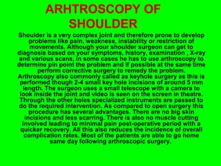
  • 1. ARHTROSCOPY OF SHOULDER Shoulder is a very complex joint and therefore prone to develop problems like pain, weakness, instability or restriction of movements. Although your shoulder surgeon can get to diagnosis based on your symptoms, history, examination , X-ray and various scans, in some cases he has to use arthroscopy to determine pin point the problem and if possible at the same time perform corrective surgery to remedy the problem. Arthroscopy also commonly called as keyhole surgery as this is performed though 2-4 small key hole incisions of around 5 mm length. The surgeon uses a small telescope with a camera to look inside the joint and video is seen on the screen in theatre. Through the other holes specialized instruments are passed to do the required intervention. As compared to open surgery this procedure has several advantages. There are no big skin incisions and less scarring. There is also no muscle cutting involved leading to minimal pain post-operative period with a quicker recovery. All this also reduces the incidence of overall complication rates. Most of the patients are able to go home same day following arthroscopic surgery.
  • 2. Biceps Problems • The famous Biceps tendon in front of your arm takes its origin in the shoulder. The pathologies affecting this tendon are one of the common cause of pain in the front of shoulder. The inflammation an degenerations of this tendon are called as biceps tendinitis. Ina advanced cases the tendon is torn from the shoulder with resultant bruising and a bulge in the mid arm causing weakness of the muscle and pain with cosmetic deformity. The treatment consists of rest, avoiding activities bringing on the pain and supervised physiotherapy. An injection in the tendon sheath also is of immense help. If all these non- operative measures fail to give relief then surgery provides excellent pain relief where your surgeon reattach the tendon on the top end of your arm bone, which can be performed arthroscopically ( key hole) or open surgery. PHOTO- anatomical, pappoyi and injection. Occasionally, in younger age group specially, this tendon gets pulled off from its far attachment in front of elbow causing pain, bruising and marked weakness in this area. This requires very early surgery to reattach this tendon to prevent permanent loss of function of this important muscle.
  • 3. Calcific Tendonitis • One of the common and most painful condition in 30-60 age group affecting the shoulder. This is due to intense reaction to the build up of calcium in the shoulder muscles/ tendons (rotator cuff). The exact cause of this calcium deposit is unknown but repetitive micro injury to the tendon or degeneration can be one of reasons .This calcium deposits triggers an extremely painful response resulting in swelling and pain. This reduces the space available in the shoulder joint to move. This can be diagnosed on clinical examination after taking careful history and confirmed on X-rays. The small amount of calcium not visible on xrays. can be seen on ultrasound scans which are very sensitive to presence of calcium and associated damage to shoulder tendon/muscle. PHOTOS OF CALCIUM ON XRAY.
  • 4. Fractured around the shoulder CLAVICLE (COLLAR BONE) FRACTURE & AC JOINT DISRUPTION- • A fractured collarbone and acromio-clavicular joint (ACJ) separation are common injuries involving all age groups who fall on the side of their shoulder or on the outstretched hand. Historically, these injuries were treated non surgically with slings or splints, but the long follow up in these cases have shown compromised functions at shoulder and persistence of pain in cases of severe displacement of clavicle fracture, resulting shortening of clavicle bone or in case of joint disruption. Results specially in elderly females are poor with non-operative measures. Therefore, severe displaced fractures of clavicle, bony spike right under skin threatening to come through the skin, soft tissue interposition, multi fragmented fracture ( comminuted) or acromio- clavicular joint separation should be treated with surgical repair to get the normal anatomy back and ensuring normal function of the shoulder joint. Injuries in this region specially sustained in high velocity accidents canalso involve associated injuries to chest, neurovascular structures ( subclavian artery or brachial plexus) which can be fatal.