Download to read offline


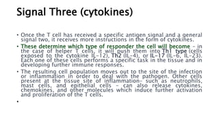
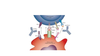
![3rd signal cytokine
production(Il12)
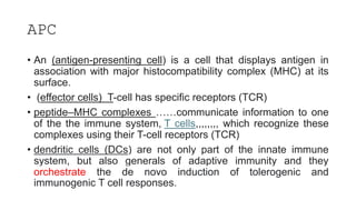
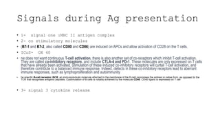
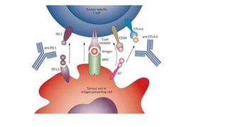
he third signal in vivo to show that the presence of signals
1 and 2 but the absence of IL-12 results in peripheral
tolerance in the CD8+ T-cell compartment [5].
CD8+ T cells are able to proliferate and to produce IFN-γ in
vivo in the absence of IL-12, but this cytokine production and
cytotoxic T-lymphocyte (CTL) activity are limited.
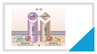
the important role of IL-12 in driving IFN-γ effector
function by T cells [6]. Further upstream, 7]. In this
regard, it is of interest that the minimum required
signals for CD154 (CD40L) expression by CD4+ T cells are
CD80/86 and CD54, even in the absence of signal 1 [](https://image.slidesharecdn.com/apcabdagpresentaion-240726193544-e2637e0b/85/APC-abd-Ag-presentaion-APC-abd-Ag-presentaion-37-320.jpg)
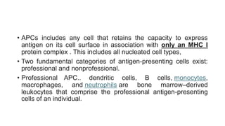
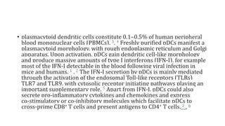
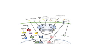
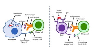
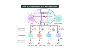
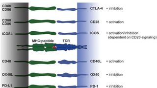
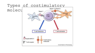
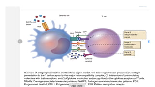
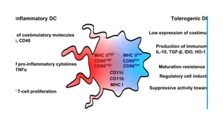
![• DCs undergo a series of phenotypic and functional changes upon exposure to
activation signals, leading to their maturation (10). This process is associated with
the following events: (1) downregulated antigen-capture activity, (2) increased
expression of surface MHC class II molecules and enhanced antigen processing and
presentation, (3) increased levels of chemokine receptors, e.g., CCR7, which allows
migration of the DC to lymphoid tissues; (4) increased expression of costimulatory
molecules associated with the capacity to stimulate or suppress T cells through
different signaling axes: CD80/CD86-CD28, CD40-CD40L, OX40L-OX40, ICOSL-ICOS
and galectin (GAL)9-TIM3, CD80-CTLA4, PDL1-PD1, PDL2-PD1, respectively (Figure
2); and (5) enhanced secretion of cytokines and chemokines, leading to the
development of an immune response T cell subtypes, e.g., CD4+ T cells such as TH1,
TH2 and Tregs
• 0. Dudek AM, Martin S, Garg AD, Agostinis P. Immature, semi-mature, and fully mature dendritic cells:
toward a DC-cancer cells interface that augments anticancer immunity. Front Immunol. (2013) 4:438.
10.3389/fimmu.2013.00438 [PMC free article] [PubMed] [CrossRef] [Google Scholar]](https://image.slidesharecdn.com/apcabdagpresentaion-240726193544-e2637e0b/85/APC-abd-Ag-presentaion-APC-abd-Ag-presentaion-39-320.jpg)


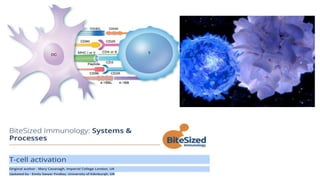
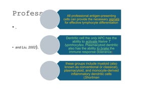
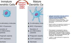
The document discusses antigen-presenting cells (APCs), highlighting their role in immune system activation, particularly T-cell responses. It explains the different types of APCs, including professional (dendritic cells, B cells, macrophages) and non-professional cells, and emphasizes the importance of co-stimulatory signals in T-cell activation. Additionally, it details dendritic cell differentiation and function, including their ability to mediate tolerance or immunity through cytokine release and the expression of inhibitory receptors.


![PERI-PROSTHETIC FRACTURE NAIL-PLATE CONSTRUCT [NPC].pptx](https://cdn.slidesharecdn.com/ss_thumbnails/drarunkumardrmohamedashrafperiprostheticfrasturenail-plateconstructnpc-260209164459-7e9d15a1-thumbnail.jpg?width=640&height=640&fit=bounds)


![CTEV [ clubfoot] DR ARUN LAL ,DR MOHAMED ASHRAF travancore medical college k...](https://cdn.slidesharecdn.com/ss_thumbnails/ctevclubfootdrarunlaldrmohamedashraftravancoremedicalcollegekollamkeralaindia-260208063247-18fc466c-thumbnail.jpg?width=640&height=640&fit=bounds)
