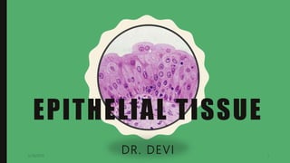
3. EPITHELIUM
• Basic tissue of the body
• Cells arranged as continuous sheets
• Single or multiple layers
• Covers the outer surface of the body or line the
luminal surface of tubular structures and cavities of
the body
5/28/2019 3
5. CHARACTERISTICS OF EPITHELIAL
TISSUE
• Epithelium consists of tightly packed cells- very
cellular with little intercellular space 20um
• Usually avascular- They lack a blood supply, instead
they receive nutrients by diffusion from capillaries in ct
• Epithelium readily divide, so they are continually
replaced = rapid healing- high capability to regenerate
5/28/2019 5
6. • Cells rest on basement membrane
• Cells shows polarity
• Cells may display surface
modification
• Supplied by nerves
• Secretory portion of glands and
cells lining the ducts are epithelial
in nature (glands are epithelial in
origin)5/28/2019 6
11. SIMPLE
SQUAMOUS
• Single layer of flattened cells
• Functions include diffusion,
filtration, and secretion
Locations include:
• air sacs of lungs (alveoli)
• lining of capillaries and the walls
of blood vessels, glomerulus of
kidneys, endocardium, internal
5/28/2019 11
12. S I M P L E
S Q UA M O U S
• Polygonal outlines
• Height less as compared with
width
• Thin layer
• Nucleus –oval/flat, Nuclei
forms bulging of cell surface
• Endothelium
• Mesothelium
5/28/2019 12
14. SIMPLE CUBOIDAL EPITHELIUM
• Height is same as that
of width
• Round nuclei
• Sectional view-
cuboidal in shape
• Surface view-
hexagonal in shape
5/28/2019 14
15. SIMPLE
CUBOIDAL
• Single layer of cube-shaped
cells
• Functions include secretion
and absorption
• Locations include
• Follicles of thyroid
• the lining and the ducts of
some glands
• Surface of ovary
• tubules within kidneys
5/28/2019 15
16. SIMPLE
COLUMNAR• Cells –taller
• Elongated nuclei-located at the
lower half
• All nuclei at same level
• Vertical section- rectangular ,
surface view- polygonal
• Cilia/ microvilli
5/28/2019 16
17. SIMPLE
COLUMNAR• Location: stomach & large
intestine, Brush border- gall
bladder, ciliated columnar
epithelium-respiratory tract,
uterus, central canal of spinal
cord, ventricles of brain
• Function: secretion, absorption
Goblet cells
5/28/2019 17
18. STRATIFIED EPITHELIUM
STRATIFIED SQUAMOUS
(NON-KERATINIZED)
• Many layers
• Basal layer-
cuboidal/round,
intermediate- polyhedral,
superficial layer- flattened
• Location- lining oral cavity,
tongue, part of epiglottis,
oesophagus , vagina
• Function- protection of
STRATIFIED SQUAMOUS
(KERATINIZED)
• Dead, dehydrated, non-
nucleated like scales
• Location- skin
• Function- protection,
prevents absorption of
water, prevents
dehydration of underlying
tissues
5/28/2019 18
21. STRATIFIED CUBOIDAL
• Many layers
• Ducts of sweat glands
• Provides passage to the
secretion and act as a
barrier
STRATIFIED COLUMNAR
• Many layers
• Epithelium lining large
ducts of glands
• Fornix of conjunctiva,
urethra
• Provides passage to the
secretion and acts as
barrier
5/28/2019 21
22. PSEUDOSTRATIFIED EPITHELIUM
• Not a true stratified epithelium
• Appears to be stratified
• All cells are attached to the basement membrane but
are of different heights
• hence not all reach the apical surface
• Nuclei at different levels
• Ciliated/non-ciliated , goblet cells
5/28/2019 22
23. Location :
• Non- ciliated – auditory tube, ductus deferens, male urethra
• Ciliated –trachea & large bronchi
• Pseu stra col epi with sterocilia- epididymis
Function:
• Tall columnary- Secretory
• Short, basal cells are stem cells- replace tall cells
• Cilia help in clearance of mucous
• Stereocilia help in absorption
5/28/2019 23
27. TRANSITIONAL EPITHELIUM
• Mutilayered 4-6 cells thick
• Differs from stratified squamous epithelium , surface
cells are not squamous
• Deepest cells- columnar/ cuboidal
• Middle layer-polyhedral/pear-shaped
• Surface layer- umbrella shaped
• Can be stretched without losing heir integrity-
becomes thinner and flattened
5/28/2019 27
28. • Location :
Renal pelvis, calyces, ureter, urinary bladder, part of
urethra
Urothelium
• Function :
The presence of occluding junctions and
intramembranous plaques forms an effective barrier,
prevents absorption of toxic substances of urine
Distention
5/28/2019 28
30. BASEMENT MEMBRANE
• thin supporting layer placed between basal surface of
epithelium and under line connective tissue
• Epithelium cells rest on the basement membrane
• Under EM – appears by layers , basal lamina and
reticular lamina
Functions: provides adhesion on one side to epithelial
cells , other side connective tissue ,
• Act as barrier of diffusion
• Influence regeneration of peripheral nerves
5/28/2019 30
32. SPECIALIZATION OF THE FREE
SURFACE OF THE CELL
a. Microvilli
b. Stereocilia
c. Cilia
5/28/2019 32
33. Cilia :
• Hair like projections
• Free part of the each cilia- shaft
• Region of attachment of shaft to cell- base
• Outer covering- extension of cell membrane, inner
core- microtubules
• Functions : wave-like movements on the surface of
tracheal and bronchial epithelium help to move the
mucous in one direction , movement of ova in oviduct
•
5/28/2019 33
34. Micro villi:
• Fine vertical striations / finger like projections
• Closely packed
• 1-2 um , 75-90 um
• Plasma membrane/ cytoplasmic core/ microfilaments/
actin filaments
• Brush border
• Function : non-motile process, increase the surface
area, active absorption
• Location : intestine, proximal convoluted tubule of
kidney5/28/2019 34
37. • Glands – specialized secretory cells
• Epithelial in origin
• Unicellular or multicellular
• Exocrine glands : epithelial layer
invaginates in deeper tissue to
form a diverticulum. The proximal
end of this forms duct while distal
end forms secretory units. These
type of glands remain in contact
with surface epithelium by the
duct and pour their secretions on
its surface5/28/2019 37
38. • Endocrine glands: some glands
lose contact with the epithelial
surface. They pour their
secretions directly in the blood.
(internally secreting glands/
duct less glands)
• Paracrine : similar to endocrine,
but secretion diffuses directly
into target cells eg : bronchus
5/28/2019 38