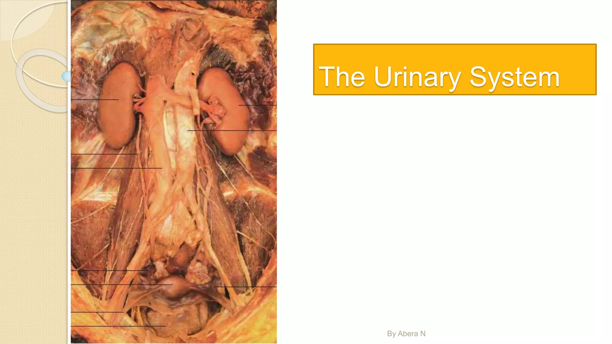
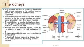
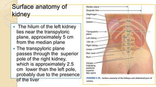
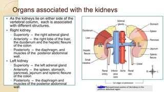
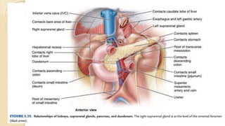
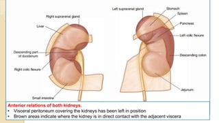
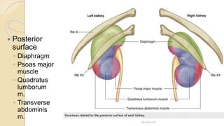
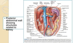
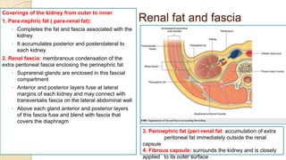
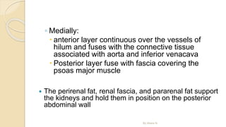
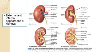
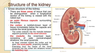
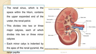
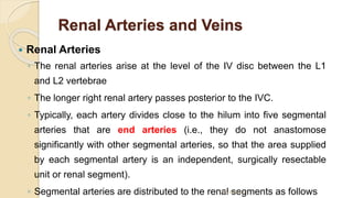
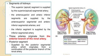
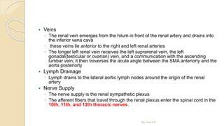
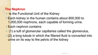
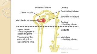
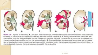
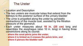
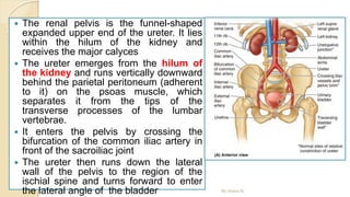
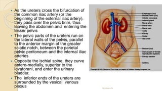
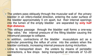
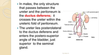
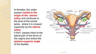
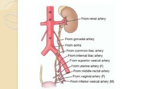
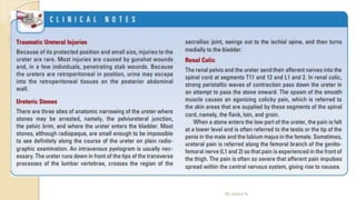
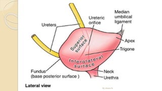
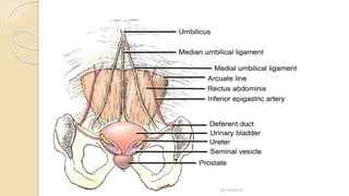
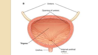
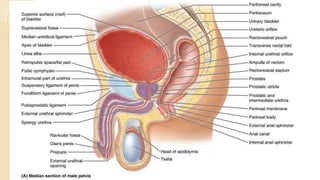
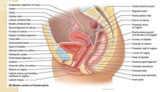
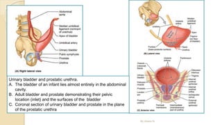
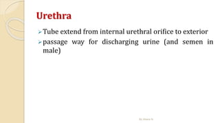
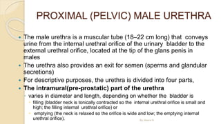
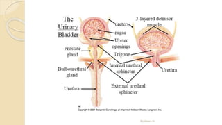
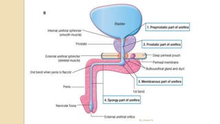
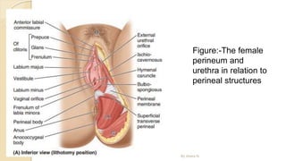
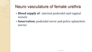
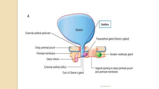
2. Overview of the urinary system
The urinary system is the main excretory
system and consists of the following structures:
◦ 2 kidneys, which secrete urine
◦ 2 ureters that convey the urine from the kidneys to the
urinary bladder
◦ the urinary bladder, which collects and stores urine
◦ the urethra through which urine leaves the body
By Abera N
3. overview of the urinary system
The urinary system plays a vital part in maintaining homeostasis of
water and electrolytes within the body
The kidneys produce urine that contains metabolic waste
products, including the nitrogenous compounds urea and uric acid,
excess ions and some drugs
The main functions of the kidneys are:
◦ formation of urine, maintaining water, electrolyte and acid–base balance
◦ excretion of waste products
◦ production and secretion of erythropoietin, the hormone that stimulates
formation of red blood cells
◦ production and secretion of renin, an important enzyme in the control of
blood pressure
◦ Urine is stored in the bladder and excreted by the process of micturition
By Abera N
4. The kidney
Learning outcomes
After studying this section, students should be able
to:
◦ Identify the organs associated with the kidneys
◦ Outline the gross structure of the kidneys
◦ describe the structure of a nephron
◦ Explain the processes involved in the formation of
urine
◦ Explain how body water and electrolyte balance is
maintained.
By Abera N
5. The kidneys
The kidneys lie on the posterior abdominal
wall, one on each side of the vertebral column,
behind the peritoneum and below the
diaphragm.
They extend from the level of the 12th thoracic
vertebra to the 3rd lumbar vertebra, receiving
some protection from the lower rib cage
The right kidney is usually slightly lower than
the left, probably because of the considerable
space occupied by the liver.
Kidneys are bean-shaped organs, about 11
cm long, 6 cm wide, 3 cm thick and weigh 150
g
They are embedded in, and held in position by,
a mass of fat
A sheath of fibrous connective tissue, the
renal fascia, encloses the kidney and the
renal fat.
By Abera N
6. Surface anatomy of
kidney
The hilum of the left kidney
lies near the transpyloric
plane, approximately 5 cm
from the median plane
The transpyloric plane
passes through the superior
pole of the right kidney,
which is approximately 2.5
cm lower than the left pole,
probably due to the presence
of the liver
By Abera N
7. Cont…
The levels of the kidneys change during respiration
and with changes in posture
Each kidney moves 2–3 cm in a vertical direction
during the movement of the diaphragm that occurs
with deep breathing.
the usual surgical approach to the kidneys is
through the posterior abdominal wall
the inferior pole of the right kidney is approximately
a finger’s breadth superior to the iliac crest
By Abera N
8. Organs associated with the kidneys
As the kidneys lie on either side of the
vertebral column, each is associated
with different structures.
Right kidney
◦ Superiorly – the right adrenal gland
◦ Anteriorly – the right lobe of the liver,
the duodenum and the hepatic flexure
of the colon
◦ Posteriorly – the diaphragm, and
muscles of the posterior abdominal
wall.
Left kidney
◦ Superiorly – the left adrenal gland
◦ Anteriorly – the spleen, stomach,
pancreas, jejunum and splenic flexure
of the colon
◦ Posteriorly – the diaphragm and
muscles of the posterior abdominal
wall.
By Abera N
10. By Abera N
Anterior relations of both kidneys.
• Visceral peritoneum covering the kidneys has been left in position
• Brown areas indicate where the kidney is in direct contact with the adjacent viscera
13. Renal fat and fascia
Coverings of the kidney from outer to inner
1. Para-nephric fat ( para-renal fat):
◦ Completes the fat and fascia associated with the
kidney
◦ It accumulates posterior and posterolateral to
each kidney
2. Renal fascia: membranous condensation of the
extra peritoneal fascia enclosing the perinephric fat
◦ Suprarenal glands are enclosed in this fascial
compartment
◦ Anterior and posterior layers fuse at lateral
margins of each kidney and may connect with
transversalis fascia on the lateral abdominal wall
◦ Above each gland anterior and posterior layers
of this fascia fuse and blend with fascia that
covers the diaphragm
By Abera N
3. Perinephric fat (peri-renal fat: accumulation of extra
peritoneal fat immediately outside the renal
capsule
4. Fibrous capsule: surrounds the kidney and is closely
applied to its outer surface
14. ◦ Medially:
anterior layer continuous over the vessels of
hilum and fuses with the connective tissue
associated with aorta and inferior venacava
Posterior layer fuse with fascia covering the
psoas major muscle
The perirenal fat, renal fascia, and pararenal fat support
the kidneys and hold them in position on the posterior
abdominal wall
By Abera N
16. Structure of the kidney
Gross structure of the kidney
◦ There are three areas of tissue that can
be distinguished when a longitudinal
section of the kidney is viewed with the
naked eye:
◦ an outer fibrous capsule: surrounding
the kidney
◦ the cortex: a reddish-brown layer of
tissue immediately below the capsule and
outside the renal pyramids
The cortex extends into the medulla between
adjacent pyramids as the renal columns
◦ the medulla: the innermost layer,
consisting of pale conical-shaped
striations, the renal pyramids
its base oriented toward the cortex and its
apex, the renal papilla, projecting medially
Extending from the bases of the renal
pyramids into the cortex are striations known
as medullary rays
By Abera N
17. The renal sinus, which is the
space within the hilum, contains
the upper expanded end of the
ureter, the renal pelvis
This divides into two or three
major calyces, each of which
divides into two or three minor
calyces
Each minor calyx is indented by
the apex of the renal pyramid, the
renal papilla By Abera N
18. Renal Arteries and Veins
Renal Arteries
◦ The renal arteries arise at the level of the IV disc between the L1
and L2 vertebrae
◦ The longer right renal artery passes posterior to the IVC.
◦ Typically, each artery divides close to the hilum into five segmental
arteries that are end arteries (i.e., they do not anastomose
significantly with other segmental arteries, so that the area supplied
by each segmental artery is an independent, surgically resectable
unit or renal segment).
◦ Segmental arteries are distributed to the renal segments as follows
By Abera N
19. Segments of kidneys
◦ The superior (apical) segment is supplied
by the superior(apical) segmental artery;
◦ the anterosuperior and antero inferior
segments are supplied by the
anterosuperior segmental and antero-
inferior segmental arteries; and
◦ the inferior segment is supplied by the
inferior segmental artery
These arteries originate from the
anterior branch of the renal artery.
Posterior segment
◦ Supplied by the posterior segmental
artery, which originates from a
continuation of the posterior branch of the
renal artery
By Abera N
20. Blood Supply, venous drainage, lymphatic drainage and
innervation
Arteries
The renal artery arises from the aorta at the level of the 2nd lumbar
vertebra.
Each renal artery usually divides into five segmental arteries that enter the
hilum of the kidney
They are distributed to different segments or areas of the kidney
◦ Lobar arteries arise from each segmental artery, one for each renal pyramid.
Before entering the renal substance, each lobar artery gives off two or three interlobar arteries
◦ The interlobar arteries run toward the cortex on each side of the renal pyramid.
At the junction of the cortex and the medulla, the interlobar arteries give off the arcuate
arteries, which arch over the bases of the pyramids
◦ The arcuate arteries give off several interlobular arteries that ascend in the cortex.
The afferent glomerular arterioles arise as branches of the interlobular
arteries
By Abera N
21. The renal artery enters the
kidney through the hilum and
then branches progressively to
form
◦ the interlobar arteries -- >
◦ arcuate arteries -- > interlobular
arteries (also called radial
arteries) --- >
◦ afferent arterioles -- > the
glomerular capillarie
The distal ends of the capillaries
of each glomerulus coalesce to
form --- > the efferent arteriole,
which leads to a second capillary
network, the peritubular
capillaries, that surrounds the
renal tubules.
By Abera N
22. About 22 % of the COP, or 1100 ml/min.
The renal circulation is unique in having two capillary
beds, the glomerular and peritubular capillaries
High hydrostatic pressure in the glomerular capillaries
(about 60 mm Hg) causes rapid fluid filtration,
whereas a much lower hydrostatic pressure in the
peritubular capillaries (about 13 mm Hg) permits rapid
fluid reabsorption.
By Abera N
23. Veins
◦ The renal vein emerges from the hilum in front of the renal artery and drains into
the inferior vena cava
◦ these veins lie anterior to the right and left renal arteries
◦ The longer left renal vein receives the left suprarenal vein, the left
gonadal(testicular or ovarian) vein, and a communication with the ascending
lumbar vein; it then traverses the acute angle between the SMA anteriorly and the
aorta posteriorly
Lymph Drainage
◦ Lymph drains to the lateral aortic lymph nodes around the origin of the renal
artery
Nerve Supply
◦ The nerve supply is the renal sympathetic plexus
◦ The afferent fibers that travel through the renal plexus enter the spinal cord in the
10th, 11th, and 12th thoracic nerves.
By Abera N
24. The Nephron
Is the Functional Unit of the Kidney
Each kidney in the human contains about 800,000 to
1,000,000 nephrons, each capable of forming urine.
Each nephron contains
(1) a tuft of glomerular capillaries called the glomerulus,
(2) a long tubule in which the filtered fluid is converted into
urine on its way to the pelvis of the kidney
By Abera N
28. The Ureter
Location and Description
The two ureters are muscular tubes that extend from the
kidneys to the posterior surface of the urinary bladder
The urine is propelled along the ureter by peristaltic
contractions of the muscle coat, assisted by the filtration
pressure of the glomeruli.
Each ureter measures about 10 in. (25 cm) long and
resembles the esophagus (also 10 in. long) in having three
constrictions along its course:
◦ where the renal pelvis joins the ureter,
◦ where it is kinked as it crosses the pelvic brim, and
◦ where it pierces the bladder wall
By Abera N
29. The renal pelvis is the funnel-shaped
expanded upper end of the ureter. It lies
within the hilum of the kidney and
receives the major calyces
The ureter emerges from the hilum of
the kidney and runs vertically downward
behind the parietal peritoneum (adherent
to it) on the psoas muscle, which
separates it from the tips of the
transverse processes of the lumbar
vertebrae.
It enters the pelvis by crossing the
bifurcation of the common iliac artery in
front of the sacroiliac joint
The ureter then runs down the lateral
wall of the pelvis to the region of the
ischial spine and turns forward to enter
the lateral angle of the bladder By Abera N
30. As the ureters cross the bifurcation of
the common iliac artery (or the
beginning of the external iliac artery),
they pass over the pelvic brim, thus
leaving the abdomen and entering the
lesser pelvis
The pelvic parts of the ureters run on
the lateral walls of the pelvis, parallel
to the anterior margin of the greater
sciatic notch, between the parietal
pelvic peritoneum and the internal iliac
arteries.
Opposite the ischial spine, they curve
antero-medially, superior to the
levatorani, and enter the urinary
bladder.
The inferior ends of the ureters are
surrounded by the vesical venous
plexus By Abera N
31. The ureters pass obliquely through the muscular wall of the urinary
bladder in an infero-medial direction, entering the outer surface of
the bladder approximately 5 cm apart, but their internal openings
into the lumen of the empty bladder are separated by only half
that distance.
This oblique passage through the bladder wall forms a one-way
“flap valve,” the internal pressure of the filling bladder causing the
intramural passage to collapse.
In addition, contractions of the bladder musculature act as a
sphincter preventing the reflux of urine into the ureters when the
bladder contracts, increasing internal pressure during micturition.
Urine is transported down the ureters by means of peristaltic
contractions, a few drops being transported at intervals of 12–20
sec By Abera N
32. In males, the only structure
that passes between the
ureter and the peritoneum is
the ductus deferens; it
crosses the ureter within the
ureteric fold of peritoneum.
The ureter lies posterolateral
to the ductus deferens and
enters the postero-superior
angle of the bladder, just
superior to the seminal
gland.
By Abera N
33. By Abera N
In females, the ureter
passes medial to the
origin of the uterine
artery and continues to
the level of the ischial
spine, where it is crossed
superiorly by the uterine
artery
It then passes close to the
lateral part of the fornix of
the vagina and enters the
postero-superior angle
of the bladder
34. Blood Supply
Arteries
◦ The arterial supply to the ureter is as follows: upper end, the renal artery;
middle portion, the testicular or ovarian artery; and in the pelvis, the superior
vesical artery.
Veins
◦ Venous blood drains into veins that correspond to the arteries.
Lymph Drainage
◦ The lymph drains to the lateral aortic nodes and the iliac nodes.
Nerve Supply
◦ The nerve supply is the renal, testicular (or ovarian), and hypogastric plexuses
(in the pelvis)
◦ Afferent fibers travel with the sympathetic nerves and enter the spinal cord in
the 1st and 2nd lumbar segments
By Abera N
37. URINARY BLADDER
The urinary bladder, a hollow viscus with strong muscular walls, is characterized by
its distensibility
The bladder is a temporary reservoir for urine, and varies in size, shape, position,
and relationships according to its content, and the state of neighboring viscera.
When empty, the adult urinary bladder is located in the lesser pelvis, lying partially
superior to and partially posterior to the pubic bones
It is separated from these bones by the potential retropubic space(of Retzius) and
lies mostly inferior to the peritoneum, resting on the pubic bones and pubic
symphysis anteriorly and the prostate (males) or anterior wall of the vagina
(females) posteriorly
The bladder is relatively free within the extra peritoneal subcutaneous fatty tissue,
except for its neck, which is held firmly by the lateral ligaments of bladder and the
tendinous arch of the pelvic fascia—especially its anterior component, the pubo-
prostatic ligament in males and the pubo-vesical ligament in females
In females, since the posterior aspect of the bladder rests directly upon the anterior
wall of the vagina, the lateral attachment of the vagina to the tendinous arch of the
pelvic fascia, the paracolpium, is an indirect but important factor in supporting the
urinary bladder By Abera N
38. In infants and young children, the urinary bladder is in the
abdomen even when empty
The bladder usually enters the greater pelvis by 6 years of
age; however, it is not located entirely within the lesser pelvis
until after puberty
An empty bladder in adults lies almost entirely in the lesser
pelvis, its superior surface level with the superior margin of
the pubic symphysis
As the bladder fills, it enters the greater pelvis as it ascends in
the extra-peritoneal fatty tissue of the anterior abdominal wall
In some individuals, a full bladder may ascend to the level of
the umbilicus
By Abera N
39. Urinary bladder
• a smooth, collapsible, muscular sac that
stores urine
• Position
shape, size and position vary with amount of
urine and age
In adults: empty bladder lies in pelvis minor
posterior and slightly superior to pubic bones.
In infants: found in abdomen
Enters pelvic major at about 6 years of age and
pelvic minor at puberty
When filled ascends to pelvic major and
abdomen
By Abera N
42. The bladder is very distensible and uniquely suited
for its function of urine storage
Empty its walls are thick and thrown into folds
(rugae)
As it expands it becomes pear shaped and rises in
the abdominal cavity
The bladder can store more than 300 ml of urine
without a significant increase in internal
pressure
A moderately full bladder holds approximately 500
ml and can about 1000 ml at capacity
In males, the bladder lies immediately anterior to
the rectum.
In females, the bladder is anterior to the vagina
and uterus.
By Abera N
44. Arterial supply
Branches of internal iliac areteries
Superior vesical artery: supply anterosuperior
parts
Inferior vesical artery and Vaginal artery: supply
fundus in male and female, respectively
Obturator and inferior gluteal arteries: small
branches
Venous drainage
Correspond to arteries and drain into internal iliac
vein
Innervation
Parasympathetic fibers from pelvic splanchnic
nerves (S2,S3,S4)
Motor to muscle and inhibitory to internal sphincter
Sympathetic fibers from T11,T12,L1 & L2
By Abera N
47. Urinary bladder and prostatic urethra.
A. The bladder of an infant lies almost entirely in the abdominal
cavity.
B. Adult bladder and prostate demonstrating their pelvic
location (inlet) and the surfaces of the bladder
C. Coronal section of urinary bladder and prostate in the plane
of the prostatic urethra
By Abera N
48. Urethra
Tube extend from internal urethral orifice to exterior
passage way for discharging urine (and semen in
male)
By Abera N
49. PROXIMAL (PELVIC) MALE URETHRA
The male urethra is a muscular tube (18–22 cm long) that conveys
urine from the internal urethral orifice of the urinary bladder to the
external urethral orifice, located at the tip of the glans penis in
males
The urethra also provides an exit for semen (sperms and glandular
secretions)
For descriptive purposes, the urethra is divided into four parts,
The intramural(pre-prostatic) part of the urethra
◦ varies in diameter and length, depending on whether the bladder is
filling (bladder neck is tonically contracted so the internal urethral orifice is small and
high; the filling internal urethral orifice) or
emptying (the neck is relaxed so the orifice is wide and low; the emptying internal
urethral orifice). By Abera N
51. prostatic urethra
about 3cm long
passes through prostate
widest and most dilatable part
ejaculatory ducts open on each side of prostatic utricle
By Abera N
52. Membranous urethra
shortest (1-2cm), thinnest and narrowest
traverse urogenital diaphragm
surrounded by sphincter urethrae muscle and
perineal membrane
posterolateral to it on each side is bulbourethral
gland
Spongy urethra
longest, about 15 cm
passes through bulb of penis and corpus
spongiosum and ends at external urethral orifice
ducts of bulbourethral glands opens into proximal
part
expanded in bulb and in glans penis
By Abera N
56. Blood supply
• Arterial supply
Prostatic urethra: prostatic branches of inferior
vesical and middle rectal arteries
Membranous and spongy urethra: internal
pudendal artery
• Venous drainage
veins accompany arteries and have similar names
inervation
Branches of pudendal nerve
Afferents run in pelvic splanchnic
nerves(parasympathetic)
By Abera N
57. FEMALE URETHRA
The female urethra is short (approximately 4 cm long and 6
mm in diameter) that makes females prone to UTI
It passes antero-inferiorly from the internal urethral orifice of the
urinary bladder, posterior and then inferior to the pubic
symphysis, to the external urethral orifice
The musculature surrounding the internal urethral orifice of the
female bladder is not organized into an internal sphincter
The female external urethral orifice is located in the vestibule of
the vagina, the cleft between the labia minora of the external
genitalia, directly anterior to the vaginal orifice
The urethra lies anterior to the vagina (forming an elevation in
the anterior vaginal wall)
its axis is parallel to that of the vagina
By Abera N
58. The urethra passes with the vagina through the
pelvic diaphragm, external urethral sphincter,
and perineal membrane.
Urethral glands are present, particularly in the
superior part of the urethra
the para urethral glands, are homologs to the
prostate
◦ Are glands located on each side of the urethra
◦ These glands have a common para-urethral duct, which
opens (one on each side) near the external urethral
orifice
By Abera N
60. Neuro vasculature of female urethra
Blood supply of : internal pudendal and vaginal
vessels
Innervation: pudendal nerve and pelvic splanchnic
nerves
By Abera N
62. Urethral Catheterization
Urethral catheterization is performed to remove urine from a
person who is unable to micturate
It is also performed to irrigate the bladder and to obtain an
uncontaminated sample of urine.
When inserting the catheters and urethral sounds (slightly
conical instruments for exploring and dilating a constricted
urethra), the curves of the male urethra must be considered
Dilation of Female Urethra
◦ The female urethra is distensible because it contains considerable
elastic tissue as well as smooth muscle.
◦ It can easily dilate without injury to it. Consequently, the passage of
catheters or cystoscopes in females is much easier than it is in males
By Abera N
The peritubular capillaries empty into the vessels of the venous system, which run parallel to the arteriolar vessels. The blood vessels of the venous system progressively form the interlobular vein, arcuate vein, interlobar vein, and renal vein, which leaves the kidney beside the renal artery and ureter.
The kidney cannot regenerate new nephrons. Therefore, with renal injury, disease, or normal aging, there is a gradual decrease in nephron number. After age 40, the number of functioning nephrons usually decreases about 10 percent every 10 years; thus, at age 80, many people have 40 percent fewer functioning nephrons than they did at age 40This loss is not life threatening because adaptive changes in the remaining nephrons allow them to excrete the proper amounts of water, electrolytes, and waste products,