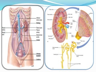
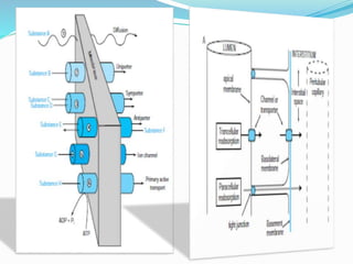
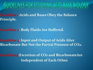
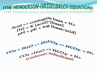
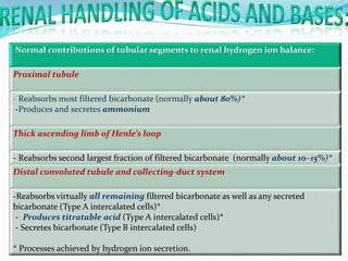
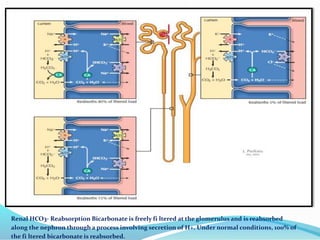
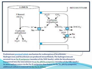
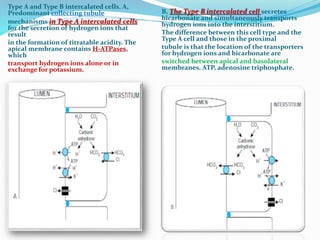
Guideline 1:Acids and Bases Obey the Balance
Principle.
Guideline 2: Body Fluids Are Buffered.
Guideline 3: Input and Output of Acids Alter
Bicarbonate But Not the Partial Pressure of CO2.
Guideline4:Excretion of CO2 and BicarbonateAre
Independent of Each Other.
 metabolism oF dietary protein.
 metabolism oF dietary weak acids.
 GI Secretions.
 Anaerobic metabolism of Carbohydrate and Fat.
Normal contributions of tubular segments to renal hydrogen ion balance:
Proximal tubule
- Reabsorbs most filtered bicarbonate (normally about 80%)*
-Produces and secretes ammonium
Thick ascending limb of Henle’s loop
- Reabsorbs second largest fraction of filtered bicarbonate (normally about 10–15%)*
Distal convoluted tubule and collecting-duct system
-Reabsorbs virtually all remaining filtered bicarbonate as well as any secreted
bicarbonate (Type A intercalated cells)*
- Produces titratable acid (Type A intercalated cells)*
- Secretes bicarbonate (Type B intercalated cells)
* Processes achieved by hydrogen ion secretion.
Renal HCO3- Reabsorption Bicarbonate is freely fi ltered at the glomerulus and is reabsorbed
along the nephron through a process involving secretion of H+. Under normal conditions, 100% of
the fi ltered bicarbonate is reabsorbed.
Predominant proximal tubule mechanism for reabsorption of bicarbonate.
Hydrogen ions and bicarbonate are produced intracellularly. The hydrogen ions are
secreted via an Na-H antiporter (member of the NHE family), while the bicarbonate is
transported into the interstitium via an Na-3HCO3 symporter (member of the NBC family).
As more sodium enters via the Na-H antiporter than leaves via the Na-3HCO3 symporter,
additional sodium is removed via the Na-K-ATPase.
Type A and Type B intercalated cells. A,
Predominant collecting tubule
mechanisms in Type A intercalated cells
for the secretion of hydrogen ions that
result
in the formation of titratable acidity. The
apical membrane contains H-ATPases,
which
transport hydrogen ions alone or in
exchange for potassium.
B, The Type B intercalated cell secretes
bicarbonate and simultaneously transports
hydrogen ions into the interstitium.
The difference between this cell type and the
Type A cell and those in the proximal
tubule is that the location of the transporters
for hydrogen ions and bicarbonate are
switched between apical and basolateral
membranes. ATP, adenosine triphosphate.
Excretion of hydrogen ions on
filtered phosphate. Divalent
phosphate (baseform) that has been
filtered and not reabsorbed reaches
the collecting tubule, where it
combines with secreted hydrogen
ions to form monovalent phosphate
(acid form) and is
then excreted in the urine. The
bicarbonate entering the blood is
new bicarbonate, not merely a
replacement for filtered
bicarbonate. ATP, adenosine
triphosphate.
Ammoniagenesis and excretion. A,
Ammonium production from
glutamine.
Glutamine is originally
synthesized in the liver from NH4
+ and bicarbonate. When
it reaches the proximal tubule
cells, it is converted back to NH4
+ and bicarbonate.There are more
biochemical steps in the
conversion of glutamine to
ammonium and bicarbonate
than indicated here; only the end
result is shown.
B, Ammonium reabsorption in the
thick ascending limb. Ammonium
reaches the thick ascending limb from
two sources. Most comes from
secretion in the proximal tubule.
Some also enters the thin limbs from
the medullary interstitium in the form
of neutral ammonia and is
subsequently reprotonated in the
lumen (ammonium recycling).
Ammonium is reabsorbed in the thick
ascending limb by several
mechanisms,the predominant one
being entrance
via the NKCC multiporter
(where ammonium substitutes for
potassium).
C, Ammonium secretion in the
inner medulla. Several
mechanisms are involved.
A prominent one involves uptake
and secretion of neutral
ammonia via specific
transporters in parallel with
hydrogen ion secretion, resulting
in reformation of ammonium in
the lumen. In the innermost
medulla, the high interstitial
ammonium concentration allows
ammonium to substitute for
potassium on the Na-K-ATPase.
Summary of renal ammonia
metabolism. The proximal
tubule produces ammonia, as
NH 4 + , from glutamine.
NH 4 + is then secreted
preferentially into the luminal
fluid, primarily by NHE-3. It is
then reabsorbed by the
TAL, primarily by NKCC2. This
results in ammonia delivery
to the distal nephron
accounting for ~20% of final
urinary ammonia; the
remaining ~80% is secreted in
the collecting duct through
parallel NH 3 and
H + transport.
Numbers in red indicate
proportion of total urinary
ammonia present at the
indicated sites under baseline
conditions
 Glutamine metabolism and NH4 excretion are
increased during acidosis and decreased during
alkalosis. The signal is unknown.
 Tubular hydrogen ion secretion is
-Increased by the increased blood Pco2 of
respiratory acidosis and decreased by the decreased
Pco2 of respiratory alkalosis.
-Increased, independently of changes in Pco2, by the
local effects of decreased extracellular pH on the
tubules; the opposite is true for increased
extracellular pH
-How much bicarbonate is excreted in the urine?
- How much new bicarbonate is contributed to the
plasma by secretion of hydrogen ions that combine in
the tubular lumen with non-bicarbonate urinary
buffers?
-How much new bicarbonate is returned to the plasma
by secretion of hydrogen ions that are excreted as
ammonium?
Summary of processes that acidify or alkalinize the blood
Nonrenal mechanisms of acidifying the blood
-Consumption and metabolism of protein (meat) containing acidic or sulfur-containing amino acids
-Consumption of acidic drugs
- Metabolism of substrate without complete oxidation (fat to ketones and carbohydrate to lactic
acid)
-GI tract secretion of bicarbonate (puts acid in blood)
Nonrenal mechanisms of alkalinizing the blood
-Consumption and metabolism of fruit and vegetables containing basic amino acids or the salts of
weak acids
- Consumption of antacids
- Infusion of lactated Ringer’s solution
-GI tract secretion of acid (puts bicarbonate in the blood)
Renal mechanisms of acidifying the blood
-Allow some filtered bicarbonate to pass into the urine
- Secrete bicarbonate (Type B intercalated cells)
Renal means of alkalinizing the blood
- Secrete protons that form urine titratable acidity (Type A intercalated cells)
-Excrete NH4
-synthesized from glutamine
High anion gap type
Ketoacidosis
Diabetic
Alcoholic
Starvation
Lactic acidosis
Type A
Type B
D-Lactic acidosis
Intoxications
Ethylene glycol
M ethanol
Salicylate
Pyroglutamic acidosis from acetaminophen
Advanced renal failure
Normal anion gap type
I. Gastrointestinal bicarbonate loss
A. Diarrhea
B. External pancreatic or small-bowel drainage
C. Ureterosigmoidostomy, jejunal loop, ileal loop
D. Drugs
1. Calcium chloride (acidifying agent)
2. Magnesium sulfate (diarrhea)
3. Cholestyramine (bile acid diarrhea)
III. Drug-induced hyperkalemia (with
renal insufficiency)
A. Potassium-sparing diuretics
(amiloride, triamterene, spironolactone)
B. Trimethoprim
C. Pentamidine
D. ACE-Is and ARBs
E. Nonsteroidal anti-inflammatory
drugs
F. Cyclosporine and tacrolimus
Renal acidosis
A. Hypokalemia
1. Proximal RTA (type 2)
Drug induced: acetazolamide, topiramate
2. Distal (classic) RTA (type 1)
Drug induced: amphotericin B, ifosfamide
B. Hyperkalemia
1. Generalized distal nephron dysfunction (typ4 RTA)
a. Mineralocorticoid deficiency
b. Mineralocorticoid resistance (autosomal
dominant PHA
c. Voltage defect (autosomal dominant PHA I and
PHA II)
d. Tubulointerstitial disease
IV. Other
A. Acid loads (ammonium chloride,
hyperalimentation)
B. Loss of potential bicarbonate: ketosis
with ketone excretion
C. Expansion acidosis (rapid saline
administration)
D. Hippurate
E. Cation exchange resins
Volume-depleted type
Gastric acid loss
Vomiting
Gastric suction
Renal chloride loss
Diuretics
Posthypercapnia
Cystic fibrosis
Volume-replete type
Mineralocorticoid excess
Hyperaldosteronism
Gitelman's syndrome
Bartter's syndrome
Cushing's syndrome
Licorice excess
Profound potassium depletion
I . A l k a l o s i s
A. Central nervous system stimulation D-Stimulation of chest receptors
1. Pain
2. Anxiety, psychosis
3. Fever
4. Cerebrovascular accident
5. Meningitis, encephalitis
6. Tumor
7. Trauma
1. Hemothorax
2. Flail chest
3. Cardiac failure
4. Pulmonary embolism
B. Hypoxemia or tissue hypoxia E. Miscellaneous
1. High altitude
2. Pneumonia, pulmonary edema
3. Aspiration
4. Severe anemia
1. Septicemia
2. Hepatic failure
3. Mechanical hyperventilation
4. Heat exposure
5. Recovery from metabolic acidosis
C. Drugs or hormones
1. Pregnancy, progesterone
2. Salicylates
3. Cardiac failure
A c u t e :
A i r w a y o b s t r u c i o n t
—aspiration of foreign body or vomitus , laryngospasm , generalized bronchospasm ,
obstructive sleep apnea.
Re s p i r a t o r y c e n te r d e p r e s s i o n
general anesthesia , sedative overdosage , cerebral trauma or infarction,central sleep apnea
C i r c u l a t o r y c a ta s t r o p h e s
— cardiac arrest , severe pulmonary edema.
N e u r o m u s c u l a r d e f e c t s
— high cervical cordotomy , botulism , tetanus , Guillain-Barrй syndrome , crisis in
myasthenia gravis , familial hypokalemic periodic paralysis , hypokalemic myopathy , toxic
drug agents
(eg , curare , succinylcholine , aminoglycosides , organophosphates ).
R e s t r i c t i v e d e f e c t s
— pneumothorax , hemothorax , fl ail chest , severe pneumonitis , hyaline membrane
Disease , adult respiratory distress syndrome.
P u l m o n a r y d i s o r d e r s
— pneumonia , massive pulmonary embolism , pulmonary edema , mechanical
Underventilation.
C h r o n i c :
A i r w a y o b s t r u c t i o n :
— chronic obstructive lung disease (bronchitis , emphysema) .
R e s p i ra to r y ce n te r d e p r e s s i o n :
— chronic sedative depression , primary alveolar hypoventilation , obesity
hypoventilation syndrome , brain tumor , bulbar poliomyelitis .
Ne u r o m u s c u l a r d e f e c t s :
— poliomyelitis , multiple sclerosis , muscular dystrophy , amyotrophic lateral
sclerosis , diaphragmatic paralysis , myxedema, myopathic disease (eg , polymyositis ,
acid maltase deficiency ) .
R e s t r i c t i ve d e f e c t s :
— kyphoscoliosis , spinal arthritis , fi brothorax , hydrothorax , interstitial fi brosis ,
decreased diaphragmatic movement (eg, ascites) , prolonged pneumonitis , obesity .
II. Acidosis
A. Central
Drugs (anesthetics, morphine, sedatives)
2. Stroke
3. Infectio
B. Airway
Obstruction
2. Asthma
Parenchyma
1. Emphysema
2. Pneumoconiosis
3. Bronchitis
4. Adult respiratory distress syndrome
5. Barotrauma
D. Neuromuscular
Poliomyelitis
2. Kyphoscoliosis
3. Myasthenia
4. Muscular dystrophies
E. Miscellaneous
1. Obesity
2. Hypoventilation
3. Permissive hypercapnia
Step 1: assess oxygenation status .
Step 2: Measure pH. This identifies acidemia or alkalemia.
Step 3: Determine partial pressure of CO2 .
Step 4: Determine The change in bicarbonate .
simple or mixed. Calculate the serum anion gap (AG)
Step 5: Determine the cause of the acid–base disorder from
the clinical setting and laboratory tests.
Treat the underlying disorder .
pH PaCO2 HCO3
-
normal 7.35 – 7.45 (35–45 mmHg) 22 – 26 mmol l-1
acidotic <7,35 >45 <22
Alkalotic >7,45 <35 >26
Expected PaO2 FiO2 * 5
*Metabolic acidosis with compensation
respiratory
*Respiratory acidosis with compensation
kidney
NMetabolic alkalosis
NRespiratory alkalosis


pH
N

PaCO2
Metabolic acidosis
Respiratory acidosis

N
HCO3
-
Metabolic and respiratory
alkalosis
Metabolic and respiratory
acidosis
 *
 *
pH


PaCO2
metabolic alkalosis with
compensation respirtory
Respiratory alkalosis with
compenasation kidney


HCO3
-
 56 F with vomiting and diarrhea 3 days ago her last
urine output was 12 hours ago .
 BP 80/60 HR 110 RR 28
 Poor skin turgor
NA 130 PH 7,30
K 2,5 P co2 30
CL 105 Hco3 15
BUN 42 Po2 90
CREA 2
 Pre-renal azotemia.
 Metabolic acidosis.
 Simple.
 NAG.
 Hydrate + Correct hypokalemia.
Acid base balance(ph)

Acid base balance(ph)

  • 5.
    Guideline 1:Acids andBases Obey the Balance Principle. Guideline 2: Body Fluids Are Buffered. Guideline 3: Input and Output of Acids Alter Bicarbonate But Not the Partial Pressure of CO2. Guideline4:Excretion of CO2 and BicarbonateAre Independent of Each Other.
  • 7.
     metabolism oFdietary protein.  metabolism oF dietary weak acids.  GI Secretions.  Anaerobic metabolism of Carbohydrate and Fat.
  • 12.
    Normal contributions oftubular segments to renal hydrogen ion balance: Proximal tubule - Reabsorbs most filtered bicarbonate (normally about 80%)* -Produces and secretes ammonium Thick ascending limb of Henle’s loop - Reabsorbs second largest fraction of filtered bicarbonate (normally about 10–15%)* Distal convoluted tubule and collecting-duct system -Reabsorbs virtually all remaining filtered bicarbonate as well as any secreted bicarbonate (Type A intercalated cells)* - Produces titratable acid (Type A intercalated cells)* - Secretes bicarbonate (Type B intercalated cells) * Processes achieved by hydrogen ion secretion.
  • 13.
    Renal HCO3- ReabsorptionBicarbonate is freely fi ltered at the glomerulus and is reabsorbed along the nephron through a process involving secretion of H+. Under normal conditions, 100% of the fi ltered bicarbonate is reabsorbed.
  • 14.
    Predominant proximal tubulemechanism for reabsorption of bicarbonate. Hydrogen ions and bicarbonate are produced intracellularly. The hydrogen ions are secreted via an Na-H antiporter (member of the NHE family), while the bicarbonate is transported into the interstitium via an Na-3HCO3 symporter (member of the NBC family). As more sodium enters via the Na-H antiporter than leaves via the Na-3HCO3 symporter, additional sodium is removed via the Na-K-ATPase.
  • 15.
    Type A andType B intercalated cells. A, Predominant collecting tubule mechanisms in Type A intercalated cells for the secretion of hydrogen ions that result in the formation of titratable acidity. The apical membrane contains H-ATPases, which transport hydrogen ions alone or in exchange for potassium. B, The Type B intercalated cell secretes bicarbonate and simultaneously transports hydrogen ions into the interstitium. The difference between this cell type and the Type A cell and those in the proximal tubule is that the location of the transporters for hydrogen ions and bicarbonate are switched between apical and basolateral membranes. ATP, adenosine triphosphate.
  • 16.
    Excretion of hydrogenions on filtered phosphate. Divalent phosphate (baseform) that has been filtered and not reabsorbed reaches the collecting tubule, where it combines with secreted hydrogen ions to form monovalent phosphate (acid form) and is then excreted in the urine. The bicarbonate entering the blood is new bicarbonate, not merely a replacement for filtered bicarbonate. ATP, adenosine triphosphate.
  • 17.
    Ammoniagenesis and excretion.A, Ammonium production from glutamine. Glutamine is originally synthesized in the liver from NH4 + and bicarbonate. When it reaches the proximal tubule cells, it is converted back to NH4 + and bicarbonate.There are more biochemical steps in the conversion of glutamine to ammonium and bicarbonate than indicated here; only the end result is shown.
  • 18.
    B, Ammonium reabsorptionin the thick ascending limb. Ammonium reaches the thick ascending limb from two sources. Most comes from secretion in the proximal tubule. Some also enters the thin limbs from the medullary interstitium in the form of neutral ammonia and is subsequently reprotonated in the lumen (ammonium recycling). Ammonium is reabsorbed in the thick ascending limb by several mechanisms,the predominant one being entrance via the NKCC multiporter (where ammonium substitutes for potassium).
  • 19.
    C, Ammonium secretionin the inner medulla. Several mechanisms are involved. A prominent one involves uptake and secretion of neutral ammonia via specific transporters in parallel with hydrogen ion secretion, resulting in reformation of ammonium in the lumen. In the innermost medulla, the high interstitial ammonium concentration allows ammonium to substitute for potassium on the Na-K-ATPase.
  • 20.
    Summary of renalammonia metabolism. The proximal tubule produces ammonia, as NH 4 + , from glutamine. NH 4 + is then secreted preferentially into the luminal fluid, primarily by NHE-3. It is then reabsorbed by the TAL, primarily by NKCC2. This results in ammonia delivery to the distal nephron accounting for ~20% of final urinary ammonia; the remaining ~80% is secreted in the collecting duct through parallel NH 3 and H + transport. Numbers in red indicate proportion of total urinary ammonia present at the indicated sites under baseline conditions
  • 21.
     Glutamine metabolismand NH4 excretion are increased during acidosis and decreased during alkalosis. The signal is unknown.  Tubular hydrogen ion secretion is -Increased by the increased blood Pco2 of respiratory acidosis and decreased by the decreased Pco2 of respiratory alkalosis. -Increased, independently of changes in Pco2, by the local effects of decreased extracellular pH on the tubules; the opposite is true for increased extracellular pH
  • 22.
    -How much bicarbonateis excreted in the urine? - How much new bicarbonate is contributed to the plasma by secretion of hydrogen ions that combine in the tubular lumen with non-bicarbonate urinary buffers? -How much new bicarbonate is returned to the plasma by secretion of hydrogen ions that are excreted as ammonium?
  • 23.
    Summary of processesthat acidify or alkalinize the blood Nonrenal mechanisms of acidifying the blood -Consumption and metabolism of protein (meat) containing acidic or sulfur-containing amino acids -Consumption of acidic drugs - Metabolism of substrate without complete oxidation (fat to ketones and carbohydrate to lactic acid) -GI tract secretion of bicarbonate (puts acid in blood) Nonrenal mechanisms of alkalinizing the blood -Consumption and metabolism of fruit and vegetables containing basic amino acids or the salts of weak acids - Consumption of antacids - Infusion of lactated Ringer’s solution -GI tract secretion of acid (puts bicarbonate in the blood) Renal mechanisms of acidifying the blood -Allow some filtered bicarbonate to pass into the urine - Secrete bicarbonate (Type B intercalated cells) Renal means of alkalinizing the blood - Secrete protons that form urine titratable acidity (Type A intercalated cells) -Excrete NH4 -synthesized from glutamine
  • 25.
    High anion gaptype Ketoacidosis Diabetic Alcoholic Starvation Lactic acidosis Type A Type B D-Lactic acidosis Intoxications Ethylene glycol M ethanol Salicylate Pyroglutamic acidosis from acetaminophen Advanced renal failure
  • 26.
    Normal anion gaptype I. Gastrointestinal bicarbonate loss A. Diarrhea B. External pancreatic or small-bowel drainage C. Ureterosigmoidostomy, jejunal loop, ileal loop D. Drugs 1. Calcium chloride (acidifying agent) 2. Magnesium sulfate (diarrhea) 3. Cholestyramine (bile acid diarrhea) III. Drug-induced hyperkalemia (with renal insufficiency) A. Potassium-sparing diuretics (amiloride, triamterene, spironolactone) B. Trimethoprim C. Pentamidine D. ACE-Is and ARBs E. Nonsteroidal anti-inflammatory drugs F. Cyclosporine and tacrolimus Renal acidosis A. Hypokalemia 1. Proximal RTA (type 2) Drug induced: acetazolamide, topiramate 2. Distal (classic) RTA (type 1) Drug induced: amphotericin B, ifosfamide B. Hyperkalemia 1. Generalized distal nephron dysfunction (typ4 RTA) a. Mineralocorticoid deficiency b. Mineralocorticoid resistance (autosomal dominant PHA c. Voltage defect (autosomal dominant PHA I and PHA II) d. Tubulointerstitial disease IV. Other A. Acid loads (ammonium chloride, hyperalimentation) B. Loss of potential bicarbonate: ketosis with ketone excretion C. Expansion acidosis (rapid saline administration) D. Hippurate E. Cation exchange resins
  • 27.
    Volume-depleted type Gastric acidloss Vomiting Gastric suction Renal chloride loss Diuretics Posthypercapnia Cystic fibrosis Volume-replete type Mineralocorticoid excess Hyperaldosteronism Gitelman's syndrome Bartter's syndrome Cushing's syndrome Licorice excess Profound potassium depletion
  • 28.
    I . Al k a l o s i s A. Central nervous system stimulation D-Stimulation of chest receptors 1. Pain 2. Anxiety, psychosis 3. Fever 4. Cerebrovascular accident 5. Meningitis, encephalitis 6. Tumor 7. Trauma 1. Hemothorax 2. Flail chest 3. Cardiac failure 4. Pulmonary embolism B. Hypoxemia or tissue hypoxia E. Miscellaneous 1. High altitude 2. Pneumonia, pulmonary edema 3. Aspiration 4. Severe anemia 1. Septicemia 2. Hepatic failure 3. Mechanical hyperventilation 4. Heat exposure 5. Recovery from metabolic acidosis C. Drugs or hormones 1. Pregnancy, progesterone 2. Salicylates 3. Cardiac failure
  • 29.
    A c ut e : A i r w a y o b s t r u c i o n t —aspiration of foreign body or vomitus , laryngospasm , generalized bronchospasm , obstructive sleep apnea. Re s p i r a t o r y c e n te r d e p r e s s i o n general anesthesia , sedative overdosage , cerebral trauma or infarction,central sleep apnea C i r c u l a t o r y c a ta s t r o p h e s — cardiac arrest , severe pulmonary edema. N e u r o m u s c u l a r d e f e c t s — high cervical cordotomy , botulism , tetanus , Guillain-Barrй syndrome , crisis in myasthenia gravis , familial hypokalemic periodic paralysis , hypokalemic myopathy , toxic drug agents (eg , curare , succinylcholine , aminoglycosides , organophosphates ). R e s t r i c t i v e d e f e c t s — pneumothorax , hemothorax , fl ail chest , severe pneumonitis , hyaline membrane Disease , adult respiratory distress syndrome. P u l m o n a r y d i s o r d e r s — pneumonia , massive pulmonary embolism , pulmonary edema , mechanical Underventilation.
  • 30.
    C h ro n i c : A i r w a y o b s t r u c t i o n : — chronic obstructive lung disease (bronchitis , emphysema) . R e s p i ra to r y ce n te r d e p r e s s i o n : — chronic sedative depression , primary alveolar hypoventilation , obesity hypoventilation syndrome , brain tumor , bulbar poliomyelitis . Ne u r o m u s c u l a r d e f e c t s : — poliomyelitis , multiple sclerosis , muscular dystrophy , amyotrophic lateral sclerosis , diaphragmatic paralysis , myxedema, myopathic disease (eg , polymyositis , acid maltase deficiency ) . R e s t r i c t i ve d e f e c t s : — kyphoscoliosis , spinal arthritis , fi brothorax , hydrothorax , interstitial fi brosis , decreased diaphragmatic movement (eg, ascites) , prolonged pneumonitis , obesity .
  • 31.
    II. Acidosis A. Central Drugs(anesthetics, morphine, sedatives) 2. Stroke 3. Infectio B. Airway Obstruction 2. Asthma Parenchyma 1. Emphysema 2. Pneumoconiosis 3. Bronchitis 4. Adult respiratory distress syndrome 5. Barotrauma D. Neuromuscular Poliomyelitis 2. Kyphoscoliosis 3. Myasthenia 4. Muscular dystrophies E. Miscellaneous 1. Obesity 2. Hypoventilation 3. Permissive hypercapnia
  • 32.
    Step 1: assessoxygenation status . Step 2: Measure pH. This identifies acidemia or alkalemia. Step 3: Determine partial pressure of CO2 . Step 4: Determine The change in bicarbonate . simple or mixed. Calculate the serum anion gap (AG) Step 5: Determine the cause of the acid–base disorder from the clinical setting and laboratory tests. Treat the underlying disorder .
  • 33.
    pH PaCO2 HCO3 - normal7.35 – 7.45 (35–45 mmHg) 22 – 26 mmol l-1 acidotic <7,35 >45 <22 Alkalotic >7,45 <35 >26 Expected PaO2 FiO2 * 5
  • 35.
    *Metabolic acidosis withcompensation respiratory *Respiratory acidosis with compensation kidney NMetabolic alkalosis NRespiratory alkalosis   pH N  PaCO2 Metabolic acidosis Respiratory acidosis  N HCO3 -
  • 36.
    Metabolic and respiratory alkalosis Metabolicand respiratory acidosis  *  * pH   PaCO2 metabolic alkalosis with compensation respirtory Respiratory alkalosis with compenasation kidney   HCO3 -
  • 37.
     56 Fwith vomiting and diarrhea 3 days ago her last urine output was 12 hours ago .  BP 80/60 HR 110 RR 28  Poor skin turgor NA 130 PH 7,30 K 2,5 P co2 30 CL 105 Hco3 15 BUN 42 Po2 90 CREA 2
  • 38.
     Pre-renal azotemia. Metabolic acidosis.  Simple.  NAG.  Hydrate + Correct hypokalemia.