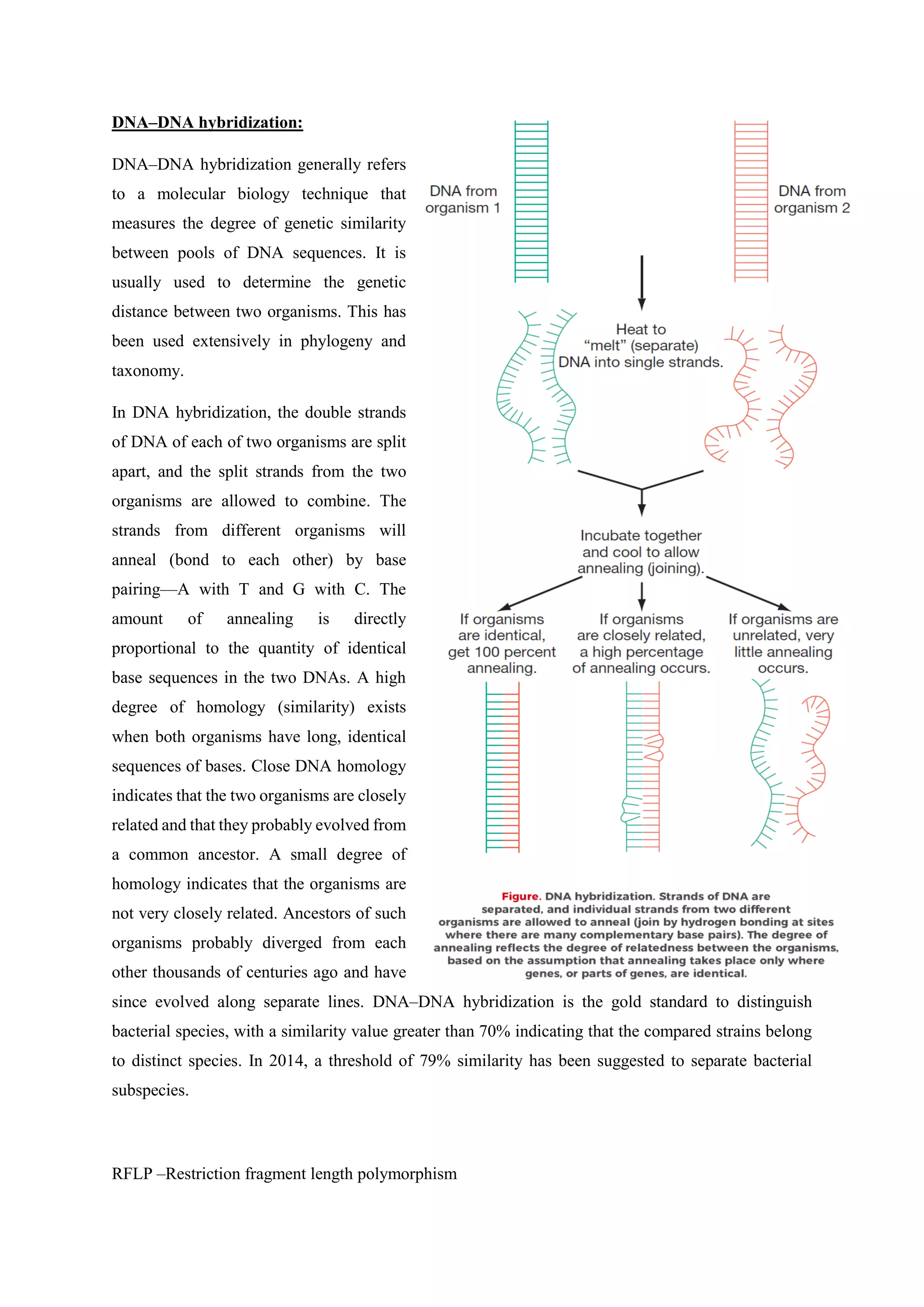
NAME OF THE ASSIGNMENT
“A lecture note on Microbial Growth and Nutrition, and Clones, Enzymes
and Informative Hybridizations”
Prepared by:
Md. Akram Hossain
B. Sc. in Food and Process Engineering, HSTU
M. Sc in Agro – Processing, BSMRAU
Bangabandhu Sheikh Mujibur Rahman Agricultural University,
Salna, Gazipur-1706
A lecture note on Microbial Growth and Nutrition, and Clones,
Enzymes and Informative Hybridizations.
Microbial growth:
In everyday language, growth refers to an increase in size. We are accustomed to seeing children, other
animals, and plants grow. Unicellular organisms also grow, but as soon as a cell, called the mother (or
parent) cell, has approximately doubled in size and duplicated its contents, it divides into two daughter
cells. Then the daughter cells grow, and subsequently they also divide. Because individual cells grow
larger only to divide into two new individuals, microbial growth is defined not in terms of cell size but
as the increase in the number of cells, which occurs by cell division.
So microbial growth refers to the growth of a population
(or an increase in the number of cells).
Cell division in bacteria, unlike cell division in eukaryotes,
usually occurs by binary fission or sometimes by
budding.
Binary Fission:
In binary fission, a cell duplicates its components and
divides into two cells. The daughter cells become
independent when a septum (partition) grows between
them and they separate. Unlike eukaryotic cells,
prokaryotic cells do not have a cell cycle with a specific
period of DNA synthesis. Instead, in continuously
dividing cells, DNA synthesis also is continuous and
replicates the bacterial chromosome shortly before the cell
divides.
Budding:
Cell division in yeast and a few bacteria occurs through
budding. In that process, a small, new cell develops from
the surface of an existing cell and subsequently separates
from the parent cell.
Phases of growth:
Consider a population of organisms introduced into a fresh, nutrient-rich medium (plural: media), a
mixture of substances on or in which
microorganisms grow. Such organisms
display four major phases of growth:
(1) the lag phase,
(2) the log (logarithmic) phase,
(3) the stationary phase, and
(4) the decline phase, or death phase.
These phases form the microbial growth
curve.
The lag Phase:
For a while, the number of cells changes very little because the cells do not immediately
reproduce in a new medium. This period of little or no cell division is called the lag phase, and
it can last for 1 hour or several days. During this time, however, the cells are not dormant. The
microbial population is undergoing a period of intense metabolic activity involving, in
particular, synthesis of enzymes and various molecules.
The log Phase:
Eventually, the cells begin to divide and enter a period of growth, or logarithmic increase, called
the log phase, or exponential growth phase. Cellular reproduction is most active during this
period, and generation time reaches a constant minimum. Because the generation time is
constant, a logarithmic plot of growth during the log phase is a straight line. The log phase is
the Bac1erilll populations follow a sequential series of growth phases; the lag, log. stationary.
and death phases. time when cells are most active metabolically and is preferred for industrial
purposes where, for example, a product needs to be produced efficiently.
The Stationary Phase:
If exponential growth continues unchecked, startlingly large numbers of cells could arise. For
example, a single bacterium (at a weight of9.5 X 10 - 13 g per cell) dividing every 20 minutes
for only 25.5 hours can theoretically produce a population equivalent in weight to that of an 80,
OOO-ton aircraft carrier. [n reality, this does not happen. Eventually, the growth rate slows, the
number of microbial deaths balances the number of new cells, and the population stabilizes.
This period of equilibrium is called the stationary phase. What causes exponential growth to
stop is not always clear. The exhaustion of nutrients, accumulation of waste products, and
harmful changes in pH may all play a role.
The Death Phase:
The number of deaths eventually exceeds the number of new cells formed, and the population
enters the death phase, or logarithmic decline phase. This phase continues until the population
is diminished to a tiny fraction of the number of cells in the previous phase or until the
population dies out entirely. Some species pass through the entire series of phases in only a few
days; others retain some surviving cells almost indefinitely.
Measuring bacterial growth:
• Growth can be measured by serial dilution, in which successive 1:10 dilutions of a liquid culture of
bacteria are made and transferred onto an agar plate; the colonies that arise are counted. Each colony
represents one live cell from the original sample.
• Growth also can be measured by direct microscopic counts, the most probable number (MPN)
technique, filtration, observing or measuring turbidity, measuring products of metabolism, and
obtaining the dry weight of cells.
Factors affecting bacterial growth:
Physical factors:
• Acidity and alkalinity of the medium affect growth, and most organisms have an optimum pH range
of no more than one pH unit.
• Temperature affects bacterial growth.
(1) Most bacteria can grow over a 30C temperature range.
(2) Bacteria can be classified according to growth temperature into three categories:
Psychrophiles, which grow at low temperatures (below 25C);
Mesophiles, which grow best at temperatures between 25C and 40C;
and thermophiles, which grow at high temperatures (above 40C).
(3) The temperature range of an organism is closely related to the temperature at which its
enzymes function best.
• The quantity of oxygen in the environment affects the growth of bacteria.
(1) Obligate aerobes require relatively large amounts of free molecular oxygen to grow.
(2) Obligate anaerobes are killed by free oxygen and must be grown in the absence of free
oxygen.
(3) Facultative anaerobes can metabolize substances aerobically if oxygen is available or
anaerobically if it is absent.
(4) Aerotolerant anaerobes metabolize substances anaerobically but are not harmed by free
oxygen.
(5) Microaerophiles must have only small amounts of oxygen to grow.
• Actively metabolizing bacteria require some water in their environment.
• Some bacteria, but no other living things, can withstand extreme hydrostatic pressures in deep valleys
in the ocean.
• Osmotic pressure affects bacterial growth, and water can be drawn into or out of cells according to the
relative osmotic pressure created by dissolved substances in the cell and the environment.
(1) Active transport minimizes the effects of high osmotic pressure in the environment.
(2) Bacteria called halophiles require moderate to large amounts of salt and are found in the
ocean and in exceptionally salty bodies of water.
Nutritional factors:
• All organisms require a carbon source:
(1) Autotrophs use CO2 as their carbon source and synthesize other substances they need.
(2) Heterotrophs require glucose or another organic carbon source from which they obtain
energy and intermediates for synthetic processes.
• Microorganisms require an organic or inorganic nitrogen source from which to synthesize proteins
and nucleic acids. They also require a source of other elements found within them, including sulfur,
phosphorus, potassium, iron, and many trace elements.
• Microorganisms that lack the enzymes to synthesize particular vitamins must obtain those vitamins
from their environment.
• The nutritional requirements of an organism are determined by the kind and number of its enzymes.
Nutritional complexity reflects a deficiency in biosynthetic enzymes.
• Bioassay techniques use metabolic properties of organisms to determine quantities of vitamins and
other compounds in foods and other materials.
• Most microorganisms move substances of low molecular weight across their cell membranes and
metabolize them internally. Some bacteria (and fungi) also produce exoenzymes that digest large
molecules outside the cell membrane of the organism.
• Microorganisms adjust to limited nutrient supplies by increasing the quantities of enzymes they
produce, by making enzymes to metabolize another available nutrient, or by adjusting their metabolic
activities to grow at a rate consistent with availability of nutrients.
Microbial nutrition:
Because human food sources are of plant and animal origin, it is important to understand the biological
principles of the microbial biota associated with plants and animals in their natural habitats and
respective roles. Although it sometimes appears that microorganisms are trying to ruin our food sources
by infecting and destroying plants and animals, including humans, this is by no means their primary
role in nature. In our present view of life on this planet, the primary function of microorganisms in
nature is self-perpetuation. During this process, the heterotrophs and autotrophs carry out the following
general reaction:
All organic matter
(carbohydrates, proteins, lipids, etc.)
↓
Energy + Inorganic compounds
(nitrates, sulfates, etc.)
This, of course, is essentially nothing more than the operation of the nitrogen cycle and the cycle of
other elements. The microbial spoilage of foods may be viewed simply as an attempt by the food biota
to carry out what appears to be their primary role in nature. This should not be taken in the teleological
sense. In spite of their simplicity when compared to higher forms, microorganisms are capable of
carrying out many complex chemical reactions essential to their perpetuation. To do this, they must
obtain nutrients from nature, some of which constitutes our food supply.
All organisms, including microbes, can be classified metabolically according to their nutritional pattern-
their source of energy and their source of carbon.
First considering the energy source, we can generally classify organisms as phototrophs or
chemotrophs.
Phototrophs use light as their primary energy source, whereas chemotrophs depend on oxidation-
reduction reactions of inorganic or organic compounds for energy.
For their principal carbon source, autotrophs (self-feeders) use carbon dioxide, and heterotrophs
(feeders on others) require an organic carbon source.
Autotrophs are also referred to as lithotrophs (rock eating), and heterotrophs are also referred to as
organotrophs.
If we combine the energy and carbon sources, we derive the following nutritional classification for
organisms: photoautotrophs, photoheterotrophs, chemoautotrophs, and chemoheterotrophs.
Chemoautotrophs (Chemolithotrophs) - use inorganic substrates as sources of energy and C02 as the
main source of carbon.
Chemoheterotrophs (Chemorganotrophs) - utilize organic substrates for both needs.
Photoautotrophs (Photolithotrophs) - use light as energy source and C02 as carbon source.
Photoheterotrophs (photoorganotrophs) - use light as energy source and organic carbon.
All fungi are all chemoheterotrophic microorganisms.
In nature fungi obtain their food either by infecting living organisms as biotrophs or by attacking dead
organic matter as saprotrophs.
Many also form symbiotic relationships with plants as in lichens and in mycorrhizae. The majority of
known fungi, whether normally parasitic or not, are capable of living on dead organic material, as shown
by their ability to grow artificially on synthetic media. Fungi that live on dead matter and are incapable
of infecting living organism we call obligate saprotrophs (obligate saprobes); those capable of causing
disease or of living on dead organic matter, according to circumstances,
Facultative parasite (or facultative saprobes); and those cannot live except on living protoplasm,
obligate parasites.
How do nutrients get into the microbial cell?
Having found a source of a given nutrient, a microorganism must:
• have some means of taking it up from the environment
• possess the appropriate enzyme systems to utilize it.
The plasma membrane represents a selective barrier, allowing into the cell only those substances it is
able to utilize. This selectivity is due in large part to the hydrophobic nature of the lipid bilayer. A
substance can be transported across the cell membrane in one of three ways, known as simple
diffusion, facilitated diffusion and active transport.
In simple diffusion, small molecules move across the membrane in response to a concentration gradient
(from high to low), until concentrations on either side of the membrane are in equilibrium. The ability
to do this depends on being small (H2O, Na+, Cl−) or soluble in the lipid component of the membrane
(non-polar gases such as O2 and CO2).
Larger polar molecules such as glucose and amino acids are unable to enter the cell unless assisted by
membrane-spanning transport proteins by the process of facilitated diffusion.
Like enzymes, these proteins are specific for a single/small number of related solutes; another parallel
is that they too can become saturated by too much ‘substrate’. As with simple diffusion, there is no
expenditure of cellular energy, and an inward concentration gradient is required. The transported
substance tends to be metabolized rapidly once inside the cell, thus maintaining the concentration
gradient from outside to inside.
Diffusion is only an effective method of internalizing substances when their concentrations are greater
outside the cell than inside. Generally, however, microorganisms find themselves in very dilute
environments; hence the concentration gradient runs in the other direction, and diffusion into the cell is
not possible. Active transport enables the cell to overcome this unfavorable gradient. Here, regardless
of the direction of the gradient, transport takes place in one direction only, into the cell.
Prokaryotic cells can carry out a specialized form of active transport called group translocation, whereby
the solute is chemically modified as it crosses the membrane, preventing its escape. A well-studied
example of this is the phosphorylation of glucose in E. coli by the phosphotransferase system. Glucose
present in very low concentrations outside the cell can be concentrated within it by this mechanism.
Glucose is unable to pass back across the membrane in its phosphorylated form (glucose-6-phosphate),
however it can be utilized in metabolic pathways in this form. Often it may be necessary to employ
extracellular enzymes to break down large molecules before any of these mechanisms can be used to
transport nutrients into the cell.
Growth media for the cultivation of bacteria:
A synthetic growth medium may be defined, that is, its exact chemical composition is known, or
undefined.
A defined growth medium may have few or many constituents, depending on the nutritional
requirements of the organism in question.
An undefined or complex medium may have a variable composition due to the inclusion of a component
such as blood, yeast extract or tap water.
Peptones are also commonly found in complex media; these are the products of partially digesting
protein sources such as beef or casein.
The exact composition of a complex medium is neither known nor critically important. A medium of
this type would generally be chosen for the cultivation of fastidious bacteria such as Neisseria
gonorrhoeae (the causative agent of gonorrhea); it is easier and less expensive to supply the many
nutrients required by such an organism in this form rather than supplying them all individually. Bacteria
whose specific nutrient requirements are not known are also grown on complex media.
Whilst media such as nutrient agar are used to support the growth of a wide range of organisms, others
are specifically designed for the isolation and identification of particular types.
Selective media such as bismuth sulphite medium preferentially support the growth of particular
bacteria. The bismuth ion inhibits the growth of Gram-positive organisms as well as many Gram-
negative types; this medium is used for the isolation of the pathogenic bacterium Salmonella typhi, one
of the few organisms that can tolerate the bismuth.
Specific media called differential media can be used to distinguish between organisms whose growth
they support, usually by means of a colored indicator. MacConkey agar contains lactose and a pH
indicator, allowing the differentiation between lactose fermenters (red colonies) and non-lactose
fermenters (white/pale pink colonies).
Many media act both selectively and differentially; MacConkey agar, for example, also contains bile
salts and the dye crystal violet, both of which serve to inhibit the growth of unwanted Gram-positive
bacteria. Mannitol salt agar is also both selective and differential. The high (7.5 per cent) salt content
suppresses growth of most bacteria, whilst a combination of mannitol and an indicator permits the
detection of mannitol fermenters in a similar fashion to that just described. Sometimes, it is desirable to
isolate an organism that is present in small numbers in a large mixed population (e.g. faeces or soil).
Enrichment media provide conditions that selectively encourage the growth of these organisms; the use
of blood agar in the isolation of streptococci provides an example of such a medium.
Blood agar can act as a differential medium, in allowing the user to distinguish between haemolytic and
nonhemolytic bacteria.
If we are to culture microorganisms successfully in the laboratory, we must provide appropriate physical
conditions as well as providing an appropriate nutrient medium.
A defined medium is one whose precise chemical composition is known.
An undefined or complex medium is one whose precise chemical composition is not known.
A selective medium is one that favors the growth of a particular organism or group of organisms, often
by suppressing the growth of others.
A differential medium allows colonies of a particular organism to be differentiated from others
growing in the same culture.
An enrichment culture uses a selective medium to encourage the growth of an organism present in
low numbers.
Clones:
Clones are organisms that are exact genetic copies. Every single bit of their DNA is identical.
Clones can happen naturally—identical twins are just one of many examples. Or they can be made in
the lab. Below, find out how natural identical twins are similar to and different from clones made
through modern cloning technologies.
There are two ways to make an exact genetic copy of an organism in a lab: artificial embryo twinning
and somatic cell nuclear transfer.
1. Artificial Embryo Twinning
Artificial embryo twinning is a relatively low-tech way to make clones. As the name suggests, this
technique mimics the natural process that creates identical twins.
In nature, twins form very early in development when the embryo splits in two. Twinning happens in
the first days after egg and sperm join, while the embryo is made of just a small number of
unspecialized cells. Each half of the embryo continues dividing on its own, ultimately developing into
separate, complete individuals. Since they developed from the same fertilized egg, the resulting
individuals are genetically identical.
Artificial embryo twinning uses the same approach, but it is carried out in a Petri dish instead of
inside the mother. A very early embryo is separated into individual cells, which are allowed to divide
and develop for a short time in the Petri dish. The embryos are then placed into a surrogate mother,
where they finish developing. Again, since all the embryos came from the same fertilized egg, they
are genetically identical.
2. Somatic Cell Nuclear Transfer
Somatic cell nuclear transfer (SCNT), also called nuclear transfer, uses a different approach than
artificial embryo twinning, but it produces the same result: an exact genetic copy, or clone, of an
individual. This was the method used to create Dolly the Sheep.
What does SCNT mean? Let's take it apart:
Somatic cell: A somatic cell is any cell in the body other than sperm and egg, the two types of
reproductive cells. Reproductive cells are also called germ cells. In mammals, every somatic cell has
two complete sets of chromosomes, whereas the germ cells have only one complete set.
Nuclear: The nucleus is a compartment that holds the cell's DNA. The DNA is divided into packages
called chromosomes, and it contains all the information needed to form an organism. It's small
differences in our DNA that make each of us unique.
Transfer: Moving an object from one place to another. To make Dolly, researchers isolated a somatic
cell from an adult female sheep. Next, they removed the nucleus and all of its DNA from an egg cell.
Then they transferred the nucleus from the somatic cell to the egg cell. After a couple of chemical
tweaks, the egg cell, with its new nucleus, was behaving just like a freshly fertilized egg. It developed
into an embryo, which was implanted into a surrogate mother and carried to term. (The transfer step is
most often done using an electrical current to fuse the membranes of the egg and the somatic cell.)
When scientists clone an organism, they are making an exact genetic copy of the whole organism, as
described above.
When scientists clone a gene, they isolate and make exact copies of just one of an organism's genes.
Cloning a gene usually involves copying the DNA sequence of that gene into a smaller, more easily
manipulated piece of DNA, such as a plasmid. This process makes it easier to study the function of
the individual gene in the laboratory.
Cloning of any DNA fragment essentially involves four steps:
fragmentation - breaking apart a strand of DNA
ligation - gluing together pieces of DNA in a desired sequence
transfection – inserting the newly formed pieces of DNA into cells
screening/selection – selecting out the cells that were successfully transfected with the new
DNA
Although these steps are invariable among cloning procedures a number of alternative routes can be
selected; these are summarized as a cloning strategy.
Initially, the DNA of interest needs to be isolated to provide a DNA segment of suitable size.
Subsequently, a ligation procedure is used where the amplified fragment is inserted into a vector
(piece of DNA). The vector (which is frequently circular) is linearized using restriction enzymes and
incubated with the fragment of interest under appropriate conditions with an enzyme called DNA
ligase. Following ligation, the vector with the insert of interest is transfected into cells. Finally, the
transfected cells are cultured. As the aforementioned procedures are of particularly low efficiency,
there is a need to identify the cells that have been successfully transfected with the vector construct
containing the desired insertion sequence in the required orientation. Modern cloning vectors include
selectable antibiotic resistance markers, which allow only cells in which the vector has been
transfected, to grow. Additionally, the cloning vectors may contain colour selection markers, which
provide blue/white screening (alpha-factor complementation) on X-gal medium. Nevertheless, these
selection steps do not absolutely guarantee that the DNA insert is present in the cells obtained. Further
investigation of the resulting colonies must be required to confirm that cloning was successful. This
may be accomplished by means of PCR, restriction fragment analysis and/or DNA sequencing.
DNA–DNA hybridization:
DNA–DNA hybridization generally refers
to a molecular biology technique that
measures the degree of genetic similarity
between pools of DNA sequences. It is
usually used to determine the genetic
distance between two organisms. This has
been used extensively in phylogeny and
taxonomy.
In DNA hybridization, the double strands
of DNA of each of two organisms are split
apart, and the split strands from the two
organisms are allowed to combine. The
strands from different organisms will
anneal (bond to each other) by base
pairing—A with T and G with C. The
amount of annealing is directly
proportional to the quantity of identical
base sequences in the two DNAs. A high
degree of homology (similarity) exists
when both organisms have long, identical
sequences of bases. Close DNA homology
indicates that the two organisms are closely
related and that they probably evolved from
a common ancestor. A small degree of
homology indicates that the organisms are
not very closely related. Ancestors of such
organisms probably diverged from each
other thousands of centuries ago and have
since evolved along separate lines. DNA–DNA hybridization is the gold standard to distinguish
bacterial species, with a similarity value greater than 70% indicating that the compared strains belong
to distinct species. In 2014, a threshold of 79% similarity has been suggested to separate bacterial
subspecies.
RFLP –Restriction fragment length polymorphism
Restriction Fragment Length Polymorphism; a molecular marker based on the differential hybridization
of cloned DNA to DNA fragments in a sample of restriction enzyme digested DNAs; the marker is
specific to a single clone/restriction enzyme combination
RFLP analysis is an application of the Southern hybridization procedure.
RFLP markers are defined by a specific enzyme-probe combination. The first step in the analysis is to
derive a set of clones that can be used to identify RFLPs. Genomic clones that represent sequences at
random are a poor choice as hybridization probes because plant genomes consist of a large percentage
of repeated sequences. Thus, many of the clones will contain repeated sequences, and hybridizations
with those clones containing repeated sequences generate many hybridization bands that are difficult to
analyze genetically.
The two primary sources of these clones for RFLP mapping of plants are cDNA clones and PstI-derived
genomic clones. These two clone sources are generally representing expressed genes which are in low
copy number. cDNA clones are DNA copies of expressed genes. PstI clones are based on the suggestion
that expressed genes are not methylated. As we saw earlier, GC and GXC methylation is the most
prominent form of methylation in plants. The enzyme PstI enzyme is C-methylation sensitive.
Therefore, the enzyme will only cut non-methylated sites. If a gene is expressed, then its sequence will
not be methylated and will be susceptible to PstI digestion. And because they probably contain
expressed sequences, these fragments would have a greater probability of being low copy number.
Once a series of clones are derived, DNA from potential parental genotypes is digested with a series of
enzymes and hybridized with the clones. Some of these hybridizations will generate fragments of only
one size and are not polymorphic. Other hybridizations will give a distinctive hybridization pattern for
each parent. These polymorphisms occur because the sequence of the probe is homologous to restriction
fragments of different sizes. Those genotypes that are highly polymorphic are candidates as parents
from which a mapping population can be derived
Advantages:
The three most common types of markers used today are RFLP, RAPD and isozymes. Of the three
marker types, RFLPs have been used the most extensively. RFLP markers have several advantages in
comparison with the RAPD and isozyme markers:
1. they are codominant and unaffected by the environment;
2. any source DNA can be used for the analysis; and
3. many markers can be mapped in a population that is not stressed by the effects of phenotypic
mutations.
Disadvantages:
1. require large amount of DNA
2.problem for biotrophic fungi
3.uses of radioactive substance
4.longer development time.
5.requires more equipment and technical expertise
Questions
1. Why do we need to know about microbial nutrition?
2. In which growth phase should we stop microbes and why?
3. What do you know about cloning?
4. What are the advantages of cloning?
5. Explain binary fission with schematic figure.
6. Explain microbial growth phases.
7. Explain classification of microbes based on their energy and carbon sources.
8. Differentiate between autotrophs and heterotrophs.
9. What is DNA-DNA hybridization?
10. Why hybridization is done?
References:
Black, Jacquelyn G. “Microbiology: principles and explorations / Jacquelyn G. Black.” John Wiley &
Sons, Inc. 7th ed. ISBN 978-0-470-10748-5
Stuart Hogg “Essential Microbiology / Stuart Hogg.” John Wiley & Sons, Inc. ISBN 0 471 49753 3
Tortora, Gerard J. “Microbiology: an introduction / Gerard J. Tortora, Berdell R. Funke, Christine L.
Case.” Pearson Education Inc. 10th
Ed. 2010. ISBN 10: 0-321-58202-0
http://learn.genetics.utah.edu/content/cloning/whatiscloning/
https://en.wikipedia.org/wiki/Cloning
https://www.cliffsnotes.com/study-guides/biology/biochemistry-ii/molecular-cloning-of-dna/dna-
hybridization
https://en.wikipedia.org/wiki/DNA%E2%80%93DNA_hybridization
https://www.ndsu.edu/pubweb/~mcclean/plsc731/mapping/mapping1.htm

A lecture note on Microbial Growth and Nutrition, and Clones, Enzymes and Informative Hybridizations

  • 1.
    NAME OF THEASSIGNMENT “A lecture note on Microbial Growth and Nutrition, and Clones, Enzymes and Informative Hybridizations” Prepared by: Md. Akram Hossain B. Sc. in Food and Process Engineering, HSTU M. Sc in Agro – Processing, BSMRAU Bangabandhu Sheikh Mujibur Rahman Agricultural University, Salna, Gazipur-1706
  • 2.
    A lecture noteon Microbial Growth and Nutrition, and Clones, Enzymes and Informative Hybridizations. Microbial growth: In everyday language, growth refers to an increase in size. We are accustomed to seeing children, other animals, and plants grow. Unicellular organisms also grow, but as soon as a cell, called the mother (or parent) cell, has approximately doubled in size and duplicated its contents, it divides into two daughter cells. Then the daughter cells grow, and subsequently they also divide. Because individual cells grow larger only to divide into two new individuals, microbial growth is defined not in terms of cell size but as the increase in the number of cells, which occurs by cell division. So microbial growth refers to the growth of a population (or an increase in the number of cells). Cell division in bacteria, unlike cell division in eukaryotes, usually occurs by binary fission or sometimes by budding. Binary Fission: In binary fission, a cell duplicates its components and divides into two cells. The daughter cells become independent when a septum (partition) grows between them and they separate. Unlike eukaryotic cells, prokaryotic cells do not have a cell cycle with a specific period of DNA synthesis. Instead, in continuously dividing cells, DNA synthesis also is continuous and replicates the bacterial chromosome shortly before the cell divides. Budding: Cell division in yeast and a few bacteria occurs through budding. In that process, a small, new cell develops from the surface of an existing cell and subsequently separates from the parent cell.
  • 3.
    Phases of growth: Considera population of organisms introduced into a fresh, nutrient-rich medium (plural: media), a mixture of substances on or in which microorganisms grow. Such organisms display four major phases of growth: (1) the lag phase, (2) the log (logarithmic) phase, (3) the stationary phase, and (4) the decline phase, or death phase. These phases form the microbial growth curve. The lag Phase: For a while, the number of cells changes very little because the cells do not immediately reproduce in a new medium. This period of little or no cell division is called the lag phase, and it can last for 1 hour or several days. During this time, however, the cells are not dormant. The microbial population is undergoing a period of intense metabolic activity involving, in particular, synthesis of enzymes and various molecules. The log Phase: Eventually, the cells begin to divide and enter a period of growth, or logarithmic increase, called the log phase, or exponential growth phase. Cellular reproduction is most active during this period, and generation time reaches a constant minimum. Because the generation time is constant, a logarithmic plot of growth during the log phase is a straight line. The log phase is the Bac1erilll populations follow a sequential series of growth phases; the lag, log. stationary. and death phases. time when cells are most active metabolically and is preferred for industrial purposes where, for example, a product needs to be produced efficiently. The Stationary Phase: If exponential growth continues unchecked, startlingly large numbers of cells could arise. For example, a single bacterium (at a weight of9.5 X 10 - 13 g per cell) dividing every 20 minutes for only 25.5 hours can theoretically produce a population equivalent in weight to that of an 80, OOO-ton aircraft carrier. [n reality, this does not happen. Eventually, the growth rate slows, the number of microbial deaths balances the number of new cells, and the population stabilizes.
  • 4.
    This period ofequilibrium is called the stationary phase. What causes exponential growth to stop is not always clear. The exhaustion of nutrients, accumulation of waste products, and harmful changes in pH may all play a role. The Death Phase: The number of deaths eventually exceeds the number of new cells formed, and the population enters the death phase, or logarithmic decline phase. This phase continues until the population is diminished to a tiny fraction of the number of cells in the previous phase or until the population dies out entirely. Some species pass through the entire series of phases in only a few days; others retain some surviving cells almost indefinitely. Measuring bacterial growth: • Growth can be measured by serial dilution, in which successive 1:10 dilutions of a liquid culture of bacteria are made and transferred onto an agar plate; the colonies that arise are counted. Each colony represents one live cell from the original sample. • Growth also can be measured by direct microscopic counts, the most probable number (MPN) technique, filtration, observing or measuring turbidity, measuring products of metabolism, and obtaining the dry weight of cells. Factors affecting bacterial growth: Physical factors: • Acidity and alkalinity of the medium affect growth, and most organisms have an optimum pH range of no more than one pH unit. • Temperature affects bacterial growth. (1) Most bacteria can grow over a 30C temperature range. (2) Bacteria can be classified according to growth temperature into three categories: Psychrophiles, which grow at low temperatures (below 25C); Mesophiles, which grow best at temperatures between 25C and 40C; and thermophiles, which grow at high temperatures (above 40C). (3) The temperature range of an organism is closely related to the temperature at which its enzymes function best. • The quantity of oxygen in the environment affects the growth of bacteria. (1) Obligate aerobes require relatively large amounts of free molecular oxygen to grow.
  • 5.
    (2) Obligate anaerobesare killed by free oxygen and must be grown in the absence of free oxygen. (3) Facultative anaerobes can metabolize substances aerobically if oxygen is available or anaerobically if it is absent. (4) Aerotolerant anaerobes metabolize substances anaerobically but are not harmed by free oxygen. (5) Microaerophiles must have only small amounts of oxygen to grow. • Actively metabolizing bacteria require some water in their environment. • Some bacteria, but no other living things, can withstand extreme hydrostatic pressures in deep valleys in the ocean. • Osmotic pressure affects bacterial growth, and water can be drawn into or out of cells according to the relative osmotic pressure created by dissolved substances in the cell and the environment. (1) Active transport minimizes the effects of high osmotic pressure in the environment. (2) Bacteria called halophiles require moderate to large amounts of salt and are found in the ocean and in exceptionally salty bodies of water. Nutritional factors: • All organisms require a carbon source: (1) Autotrophs use CO2 as their carbon source and synthesize other substances they need. (2) Heterotrophs require glucose or another organic carbon source from which they obtain energy and intermediates for synthetic processes. • Microorganisms require an organic or inorganic nitrogen source from which to synthesize proteins and nucleic acids. They also require a source of other elements found within them, including sulfur, phosphorus, potassium, iron, and many trace elements. • Microorganisms that lack the enzymes to synthesize particular vitamins must obtain those vitamins from their environment. • The nutritional requirements of an organism are determined by the kind and number of its enzymes. Nutritional complexity reflects a deficiency in biosynthetic enzymes. • Bioassay techniques use metabolic properties of organisms to determine quantities of vitamins and other compounds in foods and other materials.
  • 6.
    • Most microorganismsmove substances of low molecular weight across their cell membranes and metabolize them internally. Some bacteria (and fungi) also produce exoenzymes that digest large molecules outside the cell membrane of the organism. • Microorganisms adjust to limited nutrient supplies by increasing the quantities of enzymes they produce, by making enzymes to metabolize another available nutrient, or by adjusting their metabolic activities to grow at a rate consistent with availability of nutrients. Microbial nutrition: Because human food sources are of plant and animal origin, it is important to understand the biological principles of the microbial biota associated with plants and animals in their natural habitats and respective roles. Although it sometimes appears that microorganisms are trying to ruin our food sources by infecting and destroying plants and animals, including humans, this is by no means their primary role in nature. In our present view of life on this planet, the primary function of microorganisms in nature is self-perpetuation. During this process, the heterotrophs and autotrophs carry out the following general reaction: All organic matter (carbohydrates, proteins, lipids, etc.) ↓ Energy + Inorganic compounds (nitrates, sulfates, etc.) This, of course, is essentially nothing more than the operation of the nitrogen cycle and the cycle of other elements. The microbial spoilage of foods may be viewed simply as an attempt by the food biota to carry out what appears to be their primary role in nature. This should not be taken in the teleological sense. In spite of their simplicity when compared to higher forms, microorganisms are capable of carrying out many complex chemical reactions essential to their perpetuation. To do this, they must obtain nutrients from nature, some of which constitutes our food supply. All organisms, including microbes, can be classified metabolically according to their nutritional pattern- their source of energy and their source of carbon. First considering the energy source, we can generally classify organisms as phototrophs or chemotrophs. Phototrophs use light as their primary energy source, whereas chemotrophs depend on oxidation- reduction reactions of inorganic or organic compounds for energy.
  • 7.
    For their principalcarbon source, autotrophs (self-feeders) use carbon dioxide, and heterotrophs (feeders on others) require an organic carbon source. Autotrophs are also referred to as lithotrophs (rock eating), and heterotrophs are also referred to as organotrophs. If we combine the energy and carbon sources, we derive the following nutritional classification for organisms: photoautotrophs, photoheterotrophs, chemoautotrophs, and chemoheterotrophs. Chemoautotrophs (Chemolithotrophs) - use inorganic substrates as sources of energy and C02 as the main source of carbon. Chemoheterotrophs (Chemorganotrophs) - utilize organic substrates for both needs. Photoautotrophs (Photolithotrophs) - use light as energy source and C02 as carbon source. Photoheterotrophs (photoorganotrophs) - use light as energy source and organic carbon. All fungi are all chemoheterotrophic microorganisms. In nature fungi obtain their food either by infecting living organisms as biotrophs or by attacking dead organic matter as saprotrophs. Many also form symbiotic relationships with plants as in lichens and in mycorrhizae. The majority of known fungi, whether normally parasitic or not, are capable of living on dead organic material, as shown by their ability to grow artificially on synthetic media. Fungi that live on dead matter and are incapable
  • 8.
    of infecting livingorganism we call obligate saprotrophs (obligate saprobes); those capable of causing disease or of living on dead organic matter, according to circumstances, Facultative parasite (or facultative saprobes); and those cannot live except on living protoplasm, obligate parasites. How do nutrients get into the microbial cell? Having found a source of a given nutrient, a microorganism must: • have some means of taking it up from the environment • possess the appropriate enzyme systems to utilize it. The plasma membrane represents a selective barrier, allowing into the cell only those substances it is able to utilize. This selectivity is due in large part to the hydrophobic nature of the lipid bilayer. A substance can be transported across the cell membrane in one of three ways, known as simple diffusion, facilitated diffusion and active transport. In simple diffusion, small molecules move across the membrane in response to a concentration gradient (from high to low), until concentrations on either side of the membrane are in equilibrium. The ability to do this depends on being small (H2O, Na+, Cl−) or soluble in the lipid component of the membrane (non-polar gases such as O2 and CO2). Larger polar molecules such as glucose and amino acids are unable to enter the cell unless assisted by membrane-spanning transport proteins by the process of facilitated diffusion. Like enzymes, these proteins are specific for a single/small number of related solutes; another parallel is that they too can become saturated by too much ‘substrate’. As with simple diffusion, there is no expenditure of cellular energy, and an inward concentration gradient is required. The transported substance tends to be metabolized rapidly once inside the cell, thus maintaining the concentration gradient from outside to inside. Diffusion is only an effective method of internalizing substances when their concentrations are greater outside the cell than inside. Generally, however, microorganisms find themselves in very dilute environments; hence the concentration gradient runs in the other direction, and diffusion into the cell is not possible. Active transport enables the cell to overcome this unfavorable gradient. Here, regardless of the direction of the gradient, transport takes place in one direction only, into the cell.
  • 9.
    Prokaryotic cells cancarry out a specialized form of active transport called group translocation, whereby the solute is chemically modified as it crosses the membrane, preventing its escape. A well-studied example of this is the phosphorylation of glucose in E. coli by the phosphotransferase system. Glucose present in very low concentrations outside the cell can be concentrated within it by this mechanism. Glucose is unable to pass back across the membrane in its phosphorylated form (glucose-6-phosphate), however it can be utilized in metabolic pathways in this form. Often it may be necessary to employ extracellular enzymes to break down large molecules before any of these mechanisms can be used to transport nutrients into the cell. Growth media for the cultivation of bacteria: A synthetic growth medium may be defined, that is, its exact chemical composition is known, or undefined. A defined growth medium may have few or many constituents, depending on the nutritional requirements of the organism in question. An undefined or complex medium may have a variable composition due to the inclusion of a component such as blood, yeast extract or tap water. Peptones are also commonly found in complex media; these are the products of partially digesting protein sources such as beef or casein. The exact composition of a complex medium is neither known nor critically important. A medium of this type would generally be chosen for the cultivation of fastidious bacteria such as Neisseria gonorrhoeae (the causative agent of gonorrhea); it is easier and less expensive to supply the many nutrients required by such an organism in this form rather than supplying them all individually. Bacteria whose specific nutrient requirements are not known are also grown on complex media. Whilst media such as nutrient agar are used to support the growth of a wide range of organisms, others are specifically designed for the isolation and identification of particular types.
  • 10.
    Selective media suchas bismuth sulphite medium preferentially support the growth of particular bacteria. The bismuth ion inhibits the growth of Gram-positive organisms as well as many Gram- negative types; this medium is used for the isolation of the pathogenic bacterium Salmonella typhi, one of the few organisms that can tolerate the bismuth. Specific media called differential media can be used to distinguish between organisms whose growth they support, usually by means of a colored indicator. MacConkey agar contains lactose and a pH indicator, allowing the differentiation between lactose fermenters (red colonies) and non-lactose fermenters (white/pale pink colonies). Many media act both selectively and differentially; MacConkey agar, for example, also contains bile salts and the dye crystal violet, both of which serve to inhibit the growth of unwanted Gram-positive bacteria. Mannitol salt agar is also both selective and differential. The high (7.5 per cent) salt content suppresses growth of most bacteria, whilst a combination of mannitol and an indicator permits the detection of mannitol fermenters in a similar fashion to that just described. Sometimes, it is desirable to isolate an organism that is present in small numbers in a large mixed population (e.g. faeces or soil). Enrichment media provide conditions that selectively encourage the growth of these organisms; the use of blood agar in the isolation of streptococci provides an example of such a medium. Blood agar can act as a differential medium, in allowing the user to distinguish between haemolytic and nonhemolytic bacteria. If we are to culture microorganisms successfully in the laboratory, we must provide appropriate physical conditions as well as providing an appropriate nutrient medium. A defined medium is one whose precise chemical composition is known. An undefined or complex medium is one whose precise chemical composition is not known. A selective medium is one that favors the growth of a particular organism or group of organisms, often by suppressing the growth of others. A differential medium allows colonies of a particular organism to be differentiated from others growing in the same culture. An enrichment culture uses a selective medium to encourage the growth of an organism present in low numbers.
  • 11.
    Clones: Clones are organismsthat are exact genetic copies. Every single bit of their DNA is identical. Clones can happen naturally—identical twins are just one of many examples. Or they can be made in the lab. Below, find out how natural identical twins are similar to and different from clones made through modern cloning technologies. There are two ways to make an exact genetic copy of an organism in a lab: artificial embryo twinning and somatic cell nuclear transfer. 1. Artificial Embryo Twinning Artificial embryo twinning is a relatively low-tech way to make clones. As the name suggests, this technique mimics the natural process that creates identical twins. In nature, twins form very early in development when the embryo splits in two. Twinning happens in the first days after egg and sperm join, while the embryo is made of just a small number of unspecialized cells. Each half of the embryo continues dividing on its own, ultimately developing into separate, complete individuals. Since they developed from the same fertilized egg, the resulting individuals are genetically identical. Artificial embryo twinning uses the same approach, but it is carried out in a Petri dish instead of inside the mother. A very early embryo is separated into individual cells, which are allowed to divide and develop for a short time in the Petri dish. The embryos are then placed into a surrogate mother, where they finish developing. Again, since all the embryos came from the same fertilized egg, they are genetically identical. 2. Somatic Cell Nuclear Transfer Somatic cell nuclear transfer (SCNT), also called nuclear transfer, uses a different approach than artificial embryo twinning, but it produces the same result: an exact genetic copy, or clone, of an individual. This was the method used to create Dolly the Sheep. What does SCNT mean? Let's take it apart: Somatic cell: A somatic cell is any cell in the body other than sperm and egg, the two types of reproductive cells. Reproductive cells are also called germ cells. In mammals, every somatic cell has two complete sets of chromosomes, whereas the germ cells have only one complete set. Nuclear: The nucleus is a compartment that holds the cell's DNA. The DNA is divided into packages called chromosomes, and it contains all the information needed to form an organism. It's small differences in our DNA that make each of us unique.
  • 12.
    Transfer: Moving anobject from one place to another. To make Dolly, researchers isolated a somatic cell from an adult female sheep. Next, they removed the nucleus and all of its DNA from an egg cell. Then they transferred the nucleus from the somatic cell to the egg cell. After a couple of chemical tweaks, the egg cell, with its new nucleus, was behaving just like a freshly fertilized egg. It developed into an embryo, which was implanted into a surrogate mother and carried to term. (The transfer step is most often done using an electrical current to fuse the membranes of the egg and the somatic cell.) When scientists clone an organism, they are making an exact genetic copy of the whole organism, as described above. When scientists clone a gene, they isolate and make exact copies of just one of an organism's genes. Cloning a gene usually involves copying the DNA sequence of that gene into a smaller, more easily manipulated piece of DNA, such as a plasmid. This process makes it easier to study the function of the individual gene in the laboratory. Cloning of any DNA fragment essentially involves four steps: fragmentation - breaking apart a strand of DNA ligation - gluing together pieces of DNA in a desired sequence transfection – inserting the newly formed pieces of DNA into cells screening/selection – selecting out the cells that were successfully transfected with the new DNA Although these steps are invariable among cloning procedures a number of alternative routes can be selected; these are summarized as a cloning strategy. Initially, the DNA of interest needs to be isolated to provide a DNA segment of suitable size. Subsequently, a ligation procedure is used where the amplified fragment is inserted into a vector (piece of DNA). The vector (which is frequently circular) is linearized using restriction enzymes and incubated with the fragment of interest under appropriate conditions with an enzyme called DNA ligase. Following ligation, the vector with the insert of interest is transfected into cells. Finally, the transfected cells are cultured. As the aforementioned procedures are of particularly low efficiency, there is a need to identify the cells that have been successfully transfected with the vector construct containing the desired insertion sequence in the required orientation. Modern cloning vectors include selectable antibiotic resistance markers, which allow only cells in which the vector has been transfected, to grow. Additionally, the cloning vectors may contain colour selection markers, which provide blue/white screening (alpha-factor complementation) on X-gal medium. Nevertheless, these selection steps do not absolutely guarantee that the DNA insert is present in the cells obtained. Further investigation of the resulting colonies must be required to confirm that cloning was successful. This may be accomplished by means of PCR, restriction fragment analysis and/or DNA sequencing.
  • 13.
    DNA–DNA hybridization: DNA–DNA hybridizationgenerally refers to a molecular biology technique that measures the degree of genetic similarity between pools of DNA sequences. It is usually used to determine the genetic distance between two organisms. This has been used extensively in phylogeny and taxonomy. In DNA hybridization, the double strands of DNA of each of two organisms are split apart, and the split strands from the two organisms are allowed to combine. The strands from different organisms will anneal (bond to each other) by base pairing—A with T and G with C. The amount of annealing is directly proportional to the quantity of identical base sequences in the two DNAs. A high degree of homology (similarity) exists when both organisms have long, identical sequences of bases. Close DNA homology indicates that the two organisms are closely related and that they probably evolved from a common ancestor. A small degree of homology indicates that the organisms are not very closely related. Ancestors of such organisms probably diverged from each other thousands of centuries ago and have since evolved along separate lines. DNA–DNA hybridization is the gold standard to distinguish bacterial species, with a similarity value greater than 70% indicating that the compared strains belong to distinct species. In 2014, a threshold of 79% similarity has been suggested to separate bacterial subspecies. RFLP –Restriction fragment length polymorphism
  • 14.
    Restriction Fragment LengthPolymorphism; a molecular marker based on the differential hybridization of cloned DNA to DNA fragments in a sample of restriction enzyme digested DNAs; the marker is specific to a single clone/restriction enzyme combination RFLP analysis is an application of the Southern hybridization procedure. RFLP markers are defined by a specific enzyme-probe combination. The first step in the analysis is to derive a set of clones that can be used to identify RFLPs. Genomic clones that represent sequences at random are a poor choice as hybridization probes because plant genomes consist of a large percentage of repeated sequences. Thus, many of the clones will contain repeated sequences, and hybridizations with those clones containing repeated sequences generate many hybridization bands that are difficult to analyze genetically. The two primary sources of these clones for RFLP mapping of plants are cDNA clones and PstI-derived genomic clones. These two clone sources are generally representing expressed genes which are in low copy number. cDNA clones are DNA copies of expressed genes. PstI clones are based on the suggestion that expressed genes are not methylated. As we saw earlier, GC and GXC methylation is the most prominent form of methylation in plants. The enzyme PstI enzyme is C-methylation sensitive. Therefore, the enzyme will only cut non-methylated sites. If a gene is expressed, then its sequence will not be methylated and will be susceptible to PstI digestion. And because they probably contain expressed sequences, these fragments would have a greater probability of being low copy number. Once a series of clones are derived, DNA from potential parental genotypes is digested with a series of enzymes and hybridized with the clones. Some of these hybridizations will generate fragments of only one size and are not polymorphic. Other hybridizations will give a distinctive hybridization pattern for each parent. These polymorphisms occur because the sequence of the probe is homologous to restriction fragments of different sizes. Those genotypes that are highly polymorphic are candidates as parents from which a mapping population can be derived Advantages: The three most common types of markers used today are RFLP, RAPD and isozymes. Of the three marker types, RFLPs have been used the most extensively. RFLP markers have several advantages in comparison with the RAPD and isozyme markers: 1. they are codominant and unaffected by the environment; 2. any source DNA can be used for the analysis; and 3. many markers can be mapped in a population that is not stressed by the effects of phenotypic mutations.
  • 15.
    Disadvantages: 1. require largeamount of DNA 2.problem for biotrophic fungi 3.uses of radioactive substance 4.longer development time. 5.requires more equipment and technical expertise Questions 1. Why do we need to know about microbial nutrition? 2. In which growth phase should we stop microbes and why? 3. What do you know about cloning? 4. What are the advantages of cloning? 5. Explain binary fission with schematic figure. 6. Explain microbial growth phases. 7. Explain classification of microbes based on their energy and carbon sources. 8. Differentiate between autotrophs and heterotrophs. 9. What is DNA-DNA hybridization? 10. Why hybridization is done? References: Black, Jacquelyn G. “Microbiology: principles and explorations / Jacquelyn G. Black.” John Wiley & Sons, Inc. 7th ed. ISBN 978-0-470-10748-5 Stuart Hogg “Essential Microbiology / Stuart Hogg.” John Wiley & Sons, Inc. ISBN 0 471 49753 3 Tortora, Gerard J. “Microbiology: an introduction / Gerard J. Tortora, Berdell R. Funke, Christine L. Case.” Pearson Education Inc. 10th Ed. 2010. ISBN 10: 0-321-58202-0 http://learn.genetics.utah.edu/content/cloning/whatiscloning/ https://en.wikipedia.org/wiki/Cloning https://www.cliffsnotes.com/study-guides/biology/biochemistry-ii/molecular-cloning-of-dna/dna- hybridization https://en.wikipedia.org/wiki/DNA%E2%80%93DNA_hybridization https://www.ndsu.edu/pubweb/~mcclean/plsc731/mapping/mapping1.htm