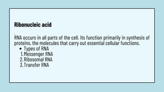
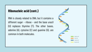
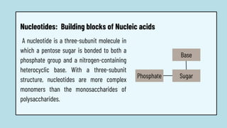
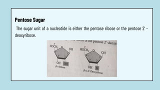
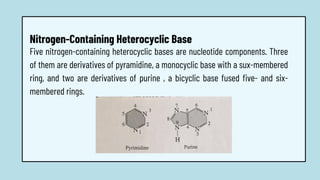
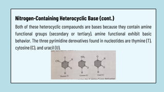
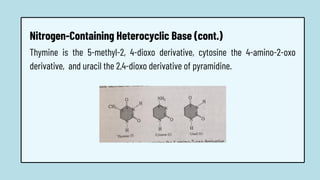
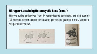
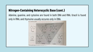
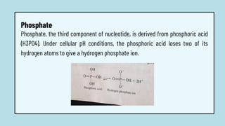
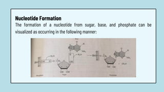
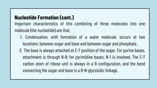
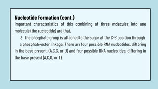
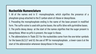
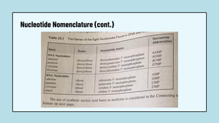
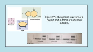
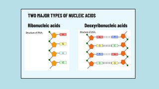
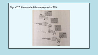
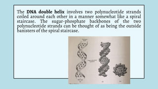
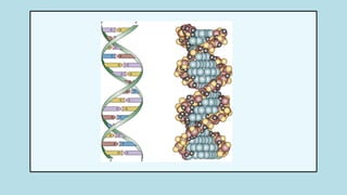
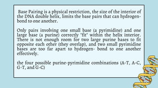
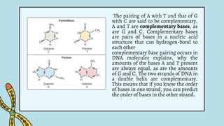
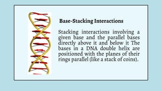
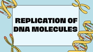
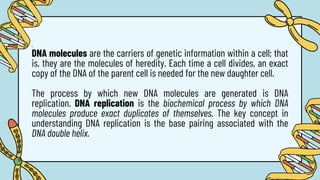
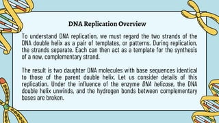
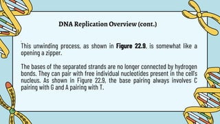
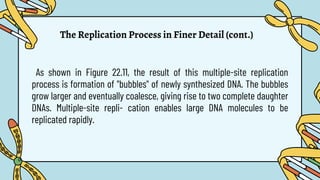
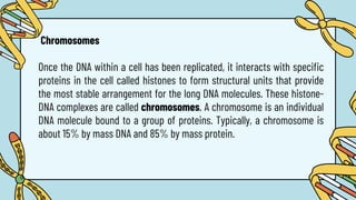
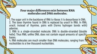
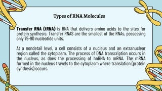
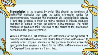
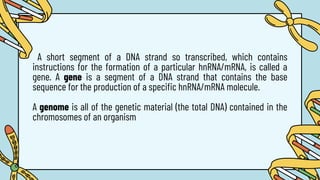
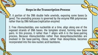
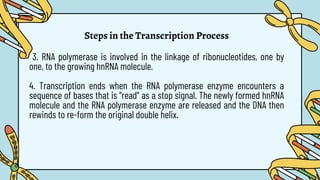
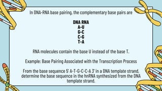
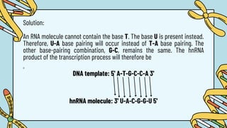
Nucleic
Acid
Group Members:
Andales, Shannel V.
Cortas, Ernalyn
Jordan, Rona
Maramot, Alexa Jahara S.
TYPES OF NUCLEIC
ACIDS
1.
Two types of nucleic acids are found within cells of higher organisms:
deoxyribonucleic acid (DNA) and ribonucleic acid (RNA). Nearly found
within the cell nucleus. Its primary function is the storage and transfer of
genetic information .
This information is used in (indirectly) to control many functions of living
cell. In addition, DNA is passed from existing cells to new cells during cell
division.
Deoxyribonucleic Acid
1.
Additionally, DNA does more than specify the structure and function
of living things — it also serves as the primary unit of heredity in
organisms of all types. In other words, whenever organisms
reproduce, a portion of their DNA is passed along to their offspring.
This transmission of all or part of an organism's DNA helps ensure a
certain level of continuity from one generation to the next, while
still allowing for slight changes that contribute to the diversity of
life.
Deoxyribonucleic Acid (cont.)
Types of RNA
Messenger RNA
Ribosomal RNA
Transfer RNA
1.
RNA occurs in all parts of the cell. Its function primarily in synthesis of
proteins, the molecules that carry out essential cellular functions.
1.
2.
3.
Ribonucleic acid
Ribonucleic acid (cont.)
RNA is closely related to DNA, but it contains a
different sugar – ribose – and the base uracil
(U) replaces thymine (T). The other bases,
adenine (A), cytosine (C) and guanine (G), are
common in both molecules.
A nucleotide is a three-subunit molecule in
which a pentose sugar is bonded to both a
phosphate group and a nitrogen-containing
heterocyclic base. With a three-subunit
structure, nucleotides are more complex
monomers than the monosaccharides of
polysaccharides.
Phosphate
Nucleotides: Building blocks of Nucleic acids
Sugar
Base
The sugar unit of a nucleotide is either the pentose ribose or the pentose 2' -
deoxyribose.
Pentose Sugar
Structurally, the only difference these two sugars occurs at carbon 2'. The -OH
group present on this carbon in ribose becomes a -H atom in 2' -deoxyribose.
RNA and DNA differ in the identity of the sugars unit in their nucleotides. In RNA
the sugar unit is ribose -hence the R in RNA. In DNA the sugar unit 2' -
deoxyribose -hence the D in DNA.
Pentose Sugar (cont.)
Five nitrogen-containing heterocyclic bases are nucleotide components. Three
of them are derivatives of pyramidine, a monocyclic base with a sux-membered
ring, and two are derivatives of purine , a bicyclic base fused five- and six-
membered rings.
Nitrogen-Containing Heterocyclic Base
Both of these heterocyclic compaounds are bases because they contain amine
functional groups (secondary or tertiary), amine functional exhibit basic
behavior. The three pyrimidine derevatives found in nucleotides are thymine (T),
cytosine (C), and uracil (U).
Nitrogen-Containing Heterocyclic Base (cont.)
Thymine is the 5-methyl-2, 4-dioxo derivative, cytosine the 4-amino-2-oxo
derivative, and uracil the 2,4-dioxo derivative of pyramidine.
Nitrogen-Containing Heterocyclic Base (cont.)
The two purine derivatives found in nucleotides re adenine (A) and and guanine
(G). Adenine is the 6-amino derivative of purine and guanine is the 2-amino-6-
oxo purine derivative.
Nitrogen-Containing Heterocyclic Base (cont.)
Adenine, guanine, and cytosine are found in both DNA and RNA. Uracil is found
only in RNA, and thymuine usually occures only in DNA.
Nitrogen-Containing Heterocyclic Base (cont.)
Phosphate, the third component of nucleotide, is derived from phosphoric acid
(H3PO4). Under cellular pH conditions, the phosphoric acid loses two of its
hydrogen atoms to give a hydrogen phosphate ion.
Phosphate
The formation of a nucleotide from sugar, base, and phosphate can be
visualized as occurring in the following manner:
Nucleotide Formation
Condensation, with formation of a water molecule, occurs at two
locations: between sugar and base and between sugar and phosphate.
The base is always attached at C-1' position of the sugar. For purine bases,
attachment is through N-9; for pyrimidine bases, N-1 is involved. The C-1'
carbon atom of ribose unit is always in a B configuration, and the bond
connecting the sugar and base is a B-N-glycosidic linkage.
Important characteristics of this combining of three molecules into one
molecule (the nucleotide) are that,
1.
2.
Nucleotide Formation (cont.)
Important characteristics of this combining of three molecules into one
molecule (the nucleotide) are that,
3. The phosphate group is attached to the sugar at the C-5' position through
a phosphate-ester linkage. There are four possible RNA nucleotides, differing
in the base present, (A,C,G, or U) and four possible DNA nucleotides, differing in
the base present (A,C,G, or T).
Nucleotide Formation (cont.)
1. All of the names end in 5' -monophosphate, which signifies the presence of a
phosphate group attached to the 5' carbon atom of ribose or deoxyribose.
2. Preceding the monophosphate ending is the name of the base present in modified
form. The suffix-osine is used eith purine bases, the suffix idine with pyrimidine bases.
3. The prefix deoxy deoxy- at the start of the names signifies that the sugar present is
deoxyribose. When no prfix is present, the sugar is ribose.
4. The abbreviations in Tbale 22.1 for the nucleotides come from the one-letter symbols
for the bases (A,C,G,T and U), the use of MP for monosphosphate , a lower-case d at the
start of the abbreviation whenever deoxyribose in the sugar.
Nucleotide Numenclature
Nucleotide Nomenclature (cont.)
PRIMARY NUCLEIC
ACID STRUCTURE
1.
Nucleic acids are polymers in which the repeating units, the monomers,
are nucleotides . The nucleotide units within a nucleic acid molecule are
linked to each other through sugar-phosphate bonds. The resulting
molecular structure .
The resulting molecular structure (Figure 22.3) involves a chain of
alternating sugar and phosphate groups with a base group protruding
from the chain at regular intervals. Nucleotides are related to nucleic
acids in the same way that amino acids are related to proteins.
1.
Figure 22.3 The general structure of a
nucleic acid in terms of nucleotide
subunits.
1.
TWO MAJOR TYPES OF NUCLEIC ACIDS
Ribonucleic acids Deoxyribonucleic acids
Ribonucleic Acid (RNA) is a nucleotide polymer in which each of
the monomers contains ribose, a phosphate group, and one of the
heterocyclic bases adenine, cytosine, guanine, or uracil. Two
changes to this definition generate the deoxyribonucleic acid
definition; deoxyribose replaces ribose and thymine replaces uracil
Deoxyribonucleic Acid (DNA) is a nucleotide polymer in which each
of the monomers contains deoxyribose, a phosphate group, and one
of the heterocyclic bases adenine, cytosine, guanine, or thymine.
The backbone of a nucleic acid structure is always an alternating
sequence of phosphate and sugar groups.
The alternating sugar-phosphate chain in a nucleic acid structure is often called
the nucleic acid backbone. This backbone is constant throughout the entire
nucleic acid structure. For DNA molecules,
Primary nucleic acid structure is the sequence in which nucleotides are linked
together in c nucleic acid. Because the sugar-phosphate backbone of a given
nucleic acid does not vary, the primary nucleic acid structure can be obtained by
considering the detailed four-nucleotide segment of a DNA structure of the nucleic
acid depends only on the sequence of bases present.
the order of nucleotide bases determines the primary structure of a nucleic
acid.
The following list describes some important points about nucleic acid
structure that are illustrated in Figure 22.5.
1. Each nonterminal phosphate group of the sugar-phosphate backbone is bonded
to two sugar molecules through a 3',5'-phosphodiester linkage. There is a
phosphoester bond to the 5' carbon of one sugar unit and a phosphoester bond to
the 3' carbon of the other sugar.
2. A nucleotide chain has directionality. One end of the nucleotide chain, the 5' end,
normally carries a free phosphate group attached to the 5' carbon atom. The other
end of the nucleotide chain, the 3' end, normally has a free hydroxyl group
attached to the 3' carbon atom. By convention, the sequence of bases of a nucleic
acid strand is read from the 5' end to the 3' end.
the order of nucleotide bases determines the primary structure of a nucleic
acid.
The following list describes some important points about nucleic acid
structure that are illustrated in Figure 22.5.
13. Each nonterminal phosphate group in the backbone of a nucleic acid carries a
-1 charge. The par- ent phosphoric acid molecule from which the phosphate was
derived originally had three -OH groups (Section 22.2). Two of these become
involved in the 3',5'-phosphodiester linkage. The re- maining-OH group is free to
exhibit acidic behavior—that is, to produce a H+ ion.
Figure 22.5 A four-nucleotide-long segment of DNA
Three parallels between primary nucleic acid structure and
primary protein structure (Section 20.9) are worth noting.
1. DNAS, RNAs, and proteins all have backbones that do not
vary in structure (see Figure 22.6).
2. The sequence of attachments to the backbones (nitrogen
bases in nucleic acids and amino acid R groups in proteins)
distinguishes one DNA from another, one RNA from another,
and one protein from another (see Figure 22.6)
3. Both nucleic acid polymer chains and protein polymer
chains have directionality; for nucleic acids there is a 5' end and
a 3' end, and for proteins there is an N-terminal end and a C-
terminal end.
THE DNA
DOUBLE HELIX
The DNA double helix involves two polynucleotide strands
coiled around each other in a manner somewhat like a spiral
staircase. The sugar-phosphate backbones of the two
polynucleotide strands can be thought of as being the outside
banisters of the spiral staircase.
Base Pairing is a physical restriction, the size of the interior of
the DNA double helix, limits the base pairs that can hydrogen-
bond to one another.
Only pairs involving one small base (a pyrimidine) and one
large base (a purine) correctly "fit" within the helix interior.
There is not enough room for two large purine bases to fit
opposite each other (they overlap), and two small pyrimidine
bases are too far apart to hydrogen- bond to one another
effectively.
the four possible purine-pyrimidine combinations (A-T, A-C,
G-T, and G-C)
The pairing of A with T and that of G
with C are said to be complementary.
A and T are complementary bases, as
are G and C. Complementary bases
are pairs of bases in a nucleic acid
structure that can hydrogen-bond to
each other
complementary base pairing occurs in
DNA molecules explains, why the
amounts of the bases A and T present
are always equal, as are the amounts
of G and C. The two strands of DNA in
a double helix are complementary.
This means that if you know the order
of bases in one strand, you can predict
the order of bases in the other strand.
The two strands of DNA in a double helix are not identical-they are
Complementary DNA strands are strands of DNA in a double helix with
base pairing such that each base is located opposite its complementary
base.
Example 22.1 Predicting Base Sequence in a Complementary DNA Strand
Predict the sequence of bases in the DNA strand that is complementary to
the single DNA strand shown.
Solution
5' C-G-A-A-T-C-C-T-A 3' end
Given: 5' C-G-A-A-T-C-C-T-A 3'
Complementary strand: 3' G-C-T-T-A-G-G-A-T 5'
Hydrogen bonding between base pairs is an important
factor in stabilizing the DNA double helix structure.
Although hydrogen bonds are relatively weak forces, each
DNA molecule has so many base pairs that collectively these
hydrogen bonds are a force of significant strength. In
addition to hydrogen bonding, base-stacking interactions
also contribute to DNA double-helix stabilization.
Hydrogen bonding is responsible for the secondary
structure (double helix) of DNA. Hydrogen bonding is also
responsible for secondary structure in proteins
Base-Stacking Interactions
Stacking interactions involving a
given base and the parallel bases
directly above it and below it The
bases in a DNA double helix are
positioned with the planes of their
rings parallel (like a stack of coins).
Use of the Term "DNA Molecule"
The term DNA molecule is actually a misnomer,
even though general usage of the term is common
in news reports, in textbooks, and even in the
vocabulary of scientists. It is technically a
misnomer for two reasons.
Use of the Term "DNA Molecule"
1. Cellular solutions have pH values such that the
phosphate groups present in the DNA backbone
structure are negatively charged. This means DNA is
actually a multicharged ionic species rather than a
neutral molecule.
2. The two strands of DNA in a double-helix structure
are not held together by covalent bonds but rather by
hydrogen bonds, which are noncovalent interactions.
Thus, double-helix DNA is an entity that involves two
intertwined ionic species rather than a single molecule.
REPLICATION OF
DNA MOLECULES
1.
DNA molecules are the carriers of genetic information within a cell; that
is, they are the molecules of heredity. Each time a cell divides, an exact
copy of the DNA of the parent cell is needed for the new daughter cell.
The process by which new DNA molecules are generated is DNA
replication. DNA replication is the biochemical process by which DNA
molecules produce exact duplicates of themselves. The key concept in
understanding DNA replication is the base pairing associated with the
DNA double helix.
DNA Replication Overview
1.
To understand DNA replication, we must regard the two strands of the
DNA double helix as a pair of templates, or patterns. During replication,
the strands separate. Each can then act as a template for the synthesis
of a new, complementary strand.
The result is two daughter DNA molecules with base sequences identical
to those of the parent double helix. Let us consider details of this
replication. Under the influence of the enzyme DNA helicase, the DNA
double helix unwinds, and the hydrogen bonds between complementary
bases are broken.
DNA Replication Overview (cont.)
1.
This unwinding process, as shown in Figure 22.9, is somewhat like a
opening a zipper.
The bases of the separated strands are no longer connected by hydrogen
bonds. They can pair with free individual nucleotides present in the cell's
nucleus. As shown in Figure 22.9, the base pairing always involves C
pairing with G and A pairing with T.
DNA Replication Overview (cont.)
The pairing process occurs
one nucleotide at a time.
After a free nucleotide has
formed hydrogen bonds with
a base of the old strand ( the
templatea) , the enzymes DNA
polymerase verifies that the
base pairing is correct and
then catalyzes the formation
The Replication Process in Finer Detail
Though simple in principle, the DNA replication process has many
intricacies.
1. The enzyme DNA polymerase can operate on a forming DNA daughter strand
only in the 5'-to-3' direction. Because the two strands of parent DNA run in
opposite directions (one is 5' to 3' and the other 3' to 5'; Section 22.4), only one
strand can grow continuously in the 5'-to-3' direction. The other strand must be
formed in short segments, called Okazaki fragments (after their discoverer, Reiji
Okazaki), as the DNA unwinds (see Figure 22.10).
The Replication Process in Finer Detail (cont.)
1.
The breaks or gaps in this daughter strand are called nicks. To complete
the formation of this strand, the Okazaki fragments are connected by
action of the enzyme DNA ligase.
2. The process of DNA unwinding does not have to begin at an end of the
DNA molecule. It may occur at any location within the molecule. Indeed,
studies show that unwinding usually occurs at several interior locations
simultaneously and that DNA replication is bidirectional for these
locations; that is, it proceeds in both directions from the unwinding sites.
The Replication Process in Finer Detail (cont.)
As shown in Figure 22.11, the result of this multiple-site replication
process is formation of "bubbles" of newly synthesized DNA. The bubbles
grow larger and eventually coalesce, giving rise to two complete daughter
DNAs. Multiple-site repli- cation enables large DNA molecules to be
replicated rapidly.
Chromosomes
Once the DNA within a cell has been replicated, it interacts with specific
proteins in the cell called histones to form structural units that provide
the most stable arrangement for the long DNA molecules. These histone-
DNA complexes are called chromosomes. A chromosome is an individual
DNA molecule bound to a group of proteins. Typically, a chromosome is
about 15% by mass DNA and 85% by mass protein.
Cells from différent kinds of organisms have different numbers of
chromosomes. A normal human has 46 chromosomes per cell, a mosquito
6, a frog 26, a dog 78, and a turkey 82.
Chromosomes occur in matched (homologous) pairs. The 46
chromosomes of a human cell constitute 23 homologous pairs. One
member of each homologous pair is derived from a chromosome
inherited from the father, and the other is a copy of one of the
chromosomes inherited from the mother.
Homologous chromosomes have similar, but not identical, DNA base
sequences; both code for the same traits but for different forms of the
trait (for example, blue eyes versus brown eyes). Offspring are like their
parents, but they are different as well; part of their DNA came from one
parent and part from the other parent. Occasionally, identical twins are
born (see Figure 22.12). Such twins have received identical DNA from their
parents.
OVERVIEW OF
PROTEIN
SYNTHESIS
t
Overview of Protein Synthesis
We saw in the previous section how the replication of DNA makes it
possible for a new cell to contain the same genetic information as its
parent cell. We will now consider how genetic information contained in a
cell is expressed in cell operation. This brings us to the topic of DNA
protein synthesis. The synthesis of proteins (skin, hair, enzymes,
hormones, and so on) is under the direction of DNA molecules. It is this role
of DNA that establishes the similarities between parent and offspring that
we regard as hereditary characteristics.
Overview of Protein Synthesis
Chromosomes are nucleoproteins. They are a combination of nucleic acid
(DNA) and various proteins.
We can divide the overall process of protein synthesis into two phases.
The first phase is called transcription and the second translation. The
following diagram summarizes the relationship between transcription
and translation.
DNA RNA PROTEIN
Transcription Translation
Ribonucleic
Acid
Four major differences exist between RNA
molecules and DNA molecules.
1. The sugar unit in the backbone of RNA is ribose; it is deoxyribose in DNA.
2. The base thymine found in DNA is replaced by uracil in RNA. In RNA,
uracil, instead of thymine, pairs with (forms hydrogen bonds with)
adenine.
3. RNA is a single-stranded molecule; DNA is double-stranded (double
helix). Thus RNA, unlike DNA, does not contain equal amounts of specific
bases.
4. RNA molecules are much smaller than DNA molecules, ranging from 75
nucleotides to a few thousand nucleotides.
Types of RNA Molecules
RNA molecules found in human cells are categorized into five major types,
distinguished function. These five RNA types are heterogeneous nuclear
RNA (hnRNA), messenger small nuclear RNA (snRNA), ribosomal RNA (rRNA),
and transfer RNA (tRNA). Heterogeneous nuclear RNA (hnRNA) also goes by
the name primary transcript RNA (ptRNA).
Heterogeneous nuclear RNA (hnRNA) is RNA formed directly by DNA
transcription. Post-transcription processing converts the heterogeneous
nuclear RNA to messenger RNA.
Types of RNA Molecules
Messenger RNA (mRNA) is RNA that carries instructions for protein
synthesis (genetic information) to the sites for protein synthesis. The
molecular mass of messenger RNA varies with the length of the protein
whose synthesis it will direct.
Small nuclear RNA (snRNA) is RNA that facilitates the conversion of
heterogeneous nuclear RNA to messenger RNA. It contains from 100 to 200
nucleotides.
Ribosomal RNA (rRNA) is RNA that combines with specific proteins to
form ribosomes, the physical sites for protein synthesis. Ribosomes have
molecular masses on the order of 3 million The rRNA present in ribosomes
has no informational function.
Types of RNA Molecules
Transfer RNA (tRNA) is RNA that delivers amino acids to the sites for
protein synthesis. Transfer RNAS are the smallest of the RNAs, possessing
only 75-90 nucleotide units.
At a nondetail level, a cell consists of a nucleus and an extranuclear
region called the cytoplasm. The process of DNA transcription occurs in
the nucleus, as does the processing of hnRNA to mRNA. The mRNA
formed in the nucleus travels to the cytoplasm where translation (protein
synthesis) occurs.
Transcription:
RNA Synthesis
Transcription is the process by which DNA directs the synthesis of
hnRNA/RNA molecules that carry the coded information needed for
protein synthesis. Messenger RNA production via transcription is actually
a "two-step" process in which an hnRNA molecule is initially produced
and then is "edited" to yield the desired mRNA molecule. The mRNA
molecule so produced then functions as the carrier of the information
needed to direct protein synthesis
Within a strand of a DNA molecule are instructions for the synthesis of
numerous hnRNA/mRNA molecules. During transcription, a DNA molecule
unwinds, under enzyme influence, at the particular location where the
appropriate base sequence is found for the hnRNA/mRNA of concern, and
the "exposed" base sequence is transcribed.
A short segment of a DNA strand so transcribed, which contains
instructions for the formation of a particular hnRNA/mRNA, is called a
gene. A gene is a segment of a DNA strand that contains the base
sequence for the production of a specific hnRNA/mRNA molecule.
A genome is all of the genetic material (the total DNA) contained in the
chromosomes of an organism
Steps in the Transcription Process
1. A portion of the DNA double helix unwinds, exposing some bases (a
gene). The unwinding process is governed by the enzyme RNA polymerase
rather than by DNA helicase (replication enzyme)
2. Free ribonucleotides, one nucleotide at a time, align along one of the
exposed strands of DNA bases, the template strand, forming new base
pairs. In this process, U rather than T aligns with A in the base-pairing
process. Because ribonucleotides rather than deoxyribonucleotides are
involved in the base-pairing, ribose, rather than deoxyribose, becomes
incorporated into the new nucleic acid backbone.
Steps in the Transcription Process
3. RNA polymerase is involved in the linkage of ribonucleotides, one by
one, to the growing hnRNA molecule.
4. Transcription ends when the RNA polymerase enzyme encounters a
sequence of bases that is "read" as a stop signal. The newly formed hnRNA
molecule and the RNA polymerase enzyme are released and the DNA then
rewinds to re-form the original double helix.
In DNA-RNA base pairing, the complementary base pairs are
DNA RNA
A-U
G-C
C-G
T-A
RNA molecules contain the base U instead of the base T.
Example: Base Pairing Associated with the Transcription Process
From the base sequence 5' A-T-G-C-C-A 3' in a DNA template strand,
determine the base sequence in the hnRNA synthesized from the DNA
template strand.
Solution:
An RNA molecule cannot contain the base T. The base U is present instead.
Therefore, U-A base pairing will occur instead of T-A base pairing. The
other base-pairing combination, G-C, remains the same. The hnRNA
product of the transcription process will therefore be
D
DNA template: 5' A-T-G-C-C-A 3'
hnRNA molecule: 3' U-A-C-G-G-U 5'
Post-Transcription Processing: Formation of
mRNA
The RNA produced from a gene through transcription is hnRNA, the
precursor for mRNA. The conversion of hnRNA to mRNA involves post-
transcription processing of the hnRNA. In this processing, certain portions
of the hnRNA are deleted and the retained parts are then spliced together.
This process leads us to the concepts of exons and introns.
It is now known that not all bases in a gene convey genetic information.
Instead, a gene is segmented; it has portions called exons that contain
genetic information and portions called introns that do not convey genetic
information.
Post-Transcription Processing: Formation of
mRNA
An exon is a gene segment that conveys (codes for) genetic information.
Exons are DNA segments that help express a genetic message. An intron is
a gene segment that does not convey (code for) genetic information.
Introns are DNA segments that interrupt a genetic message. A gene
consists of alternating exon and intron segments.
The splicing process involves snRNA molecules, the most recent of
Splicing is the process of removing introns from an hnRNA molecule and
joining the remaining exons the RNA types to be discovered.
An snRNA molecule is always found complexed with proteins in particles
called small nuclear ribonucleoprotein particles, which are usually called
snRNPs (pronounced "snurps"). A small nuclear ribonucleoprotein particle
is a complex formed from an snRNA molecule and several proteins.
"Snurps" always further collect together into larger complexes called
spliceosomes. A spliceosome is a large assembly of snRNA molecules and
proteins involved in the conversion of hnRNA molecules to mRNA
molecules.
Alternative Splicing
Alternative splicing is a process by which several different proteins that
are variations of a basic structural motif can be produced from a single
gene. In alternative splicing, an hnRNA molecule with multiple exons
present is spliced in several different ways
THANK YOU
FOR
LISTENING!

43ABF31E-080A-4577-BF72-518647F76D5C.pdf

  • 1.
    Nucleic Acid Group Members: Andales, ShannelV. Cortas, Ernalyn Jordan, Rona Maramot, Alexa Jahara S.
  • 2.
  • 3.
    1. Two types ofnucleic acids are found within cells of higher organisms: deoxyribonucleic acid (DNA) and ribonucleic acid (RNA). Nearly found within the cell nucleus. Its primary function is the storage and transfer of genetic information . This information is used in (indirectly) to control many functions of living cell. In addition, DNA is passed from existing cells to new cells during cell division. Deoxyribonucleic Acid
  • 4.
    1. Additionally, DNA doesmore than specify the structure and function of living things — it also serves as the primary unit of heredity in organisms of all types. In other words, whenever organisms reproduce, a portion of their DNA is passed along to their offspring. This transmission of all or part of an organism's DNA helps ensure a certain level of continuity from one generation to the next, while still allowing for slight changes that contribute to the diversity of life. Deoxyribonucleic Acid (cont.)
  • 5.
    Types of RNA MessengerRNA Ribosomal RNA Transfer RNA 1. RNA occurs in all parts of the cell. Its function primarily in synthesis of proteins, the molecules that carry out essential cellular functions. 1. 2. 3. Ribonucleic acid
  • 6.
    Ribonucleic acid (cont.) RNAis closely related to DNA, but it contains a different sugar – ribose – and the base uracil (U) replaces thymine (T). The other bases, adenine (A), cytosine (C) and guanine (G), are common in both molecules.
  • 7.
    A nucleotide isa three-subunit molecule in which a pentose sugar is bonded to both a phosphate group and a nitrogen-containing heterocyclic base. With a three-subunit structure, nucleotides are more complex monomers than the monosaccharides of polysaccharides. Phosphate Nucleotides: Building blocks of Nucleic acids Sugar Base
  • 8.
    The sugar unitof a nucleotide is either the pentose ribose or the pentose 2' - deoxyribose. Pentose Sugar
  • 9.
    Structurally, the onlydifference these two sugars occurs at carbon 2'. The -OH group present on this carbon in ribose becomes a -H atom in 2' -deoxyribose. RNA and DNA differ in the identity of the sugars unit in their nucleotides. In RNA the sugar unit is ribose -hence the R in RNA. In DNA the sugar unit 2' - deoxyribose -hence the D in DNA. Pentose Sugar (cont.)
  • 10.
    Five nitrogen-containing heterocyclicbases are nucleotide components. Three of them are derivatives of pyramidine, a monocyclic base with a sux-membered ring, and two are derivatives of purine , a bicyclic base fused five- and six- membered rings. Nitrogen-Containing Heterocyclic Base
  • 11.
    Both of theseheterocyclic compaounds are bases because they contain amine functional groups (secondary or tertiary), amine functional exhibit basic behavior. The three pyrimidine derevatives found in nucleotides are thymine (T), cytosine (C), and uracil (U). Nitrogen-Containing Heterocyclic Base (cont.)
  • 12.
    Thymine is the5-methyl-2, 4-dioxo derivative, cytosine the 4-amino-2-oxo derivative, and uracil the 2,4-dioxo derivative of pyramidine. Nitrogen-Containing Heterocyclic Base (cont.)
  • 13.
    The two purinederivatives found in nucleotides re adenine (A) and and guanine (G). Adenine is the 6-amino derivative of purine and guanine is the 2-amino-6- oxo purine derivative. Nitrogen-Containing Heterocyclic Base (cont.)
  • 14.
    Adenine, guanine, andcytosine are found in both DNA and RNA. Uracil is found only in RNA, and thymuine usually occures only in DNA. Nitrogen-Containing Heterocyclic Base (cont.)
  • 15.
    Phosphate, the thirdcomponent of nucleotide, is derived from phosphoric acid (H3PO4). Under cellular pH conditions, the phosphoric acid loses two of its hydrogen atoms to give a hydrogen phosphate ion. Phosphate
  • 16.
    The formation ofa nucleotide from sugar, base, and phosphate can be visualized as occurring in the following manner: Nucleotide Formation
  • 17.
    Condensation, with formationof a water molecule, occurs at two locations: between sugar and base and between sugar and phosphate. The base is always attached at C-1' position of the sugar. For purine bases, attachment is through N-9; for pyrimidine bases, N-1 is involved. The C-1' carbon atom of ribose unit is always in a B configuration, and the bond connecting the sugar and base is a B-N-glycosidic linkage. Important characteristics of this combining of three molecules into one molecule (the nucleotide) are that, 1. 2. Nucleotide Formation (cont.)
  • 18.
    Important characteristics ofthis combining of three molecules into one molecule (the nucleotide) are that, 3. The phosphate group is attached to the sugar at the C-5' position through a phosphate-ester linkage. There are four possible RNA nucleotides, differing in the base present, (A,C,G, or U) and four possible DNA nucleotides, differing in the base present (A,C,G, or T). Nucleotide Formation (cont.)
  • 19.
    1. All ofthe names end in 5' -monophosphate, which signifies the presence of a phosphate group attached to the 5' carbon atom of ribose or deoxyribose. 2. Preceding the monophosphate ending is the name of the base present in modified form. The suffix-osine is used eith purine bases, the suffix idine with pyrimidine bases. 3. The prefix deoxy deoxy- at the start of the names signifies that the sugar present is deoxyribose. When no prfix is present, the sugar is ribose. 4. The abbreviations in Tbale 22.1 for the nucleotides come from the one-letter symbols for the bases (A,C,G,T and U), the use of MP for monosphosphate , a lower-case d at the start of the abbreviation whenever deoxyribose in the sugar. Nucleotide Numenclature
  • 20.
  • 21.
  • 22.
    1. Nucleic acids arepolymers in which the repeating units, the monomers, are nucleotides . The nucleotide units within a nucleic acid molecule are linked to each other through sugar-phosphate bonds. The resulting molecular structure . The resulting molecular structure (Figure 22.3) involves a chain of alternating sugar and phosphate groups with a base group protruding from the chain at regular intervals. Nucleotides are related to nucleic acids in the same way that amino acids are related to proteins.
  • 23.
    1. Figure 22.3 Thegeneral structure of a nucleic acid in terms of nucleotide subunits.
  • 24.
    1. TWO MAJOR TYPESOF NUCLEIC ACIDS Ribonucleic acids Deoxyribonucleic acids
  • 25.
    Ribonucleic Acid (RNA)is a nucleotide polymer in which each of the monomers contains ribose, a phosphate group, and one of the heterocyclic bases adenine, cytosine, guanine, or uracil. Two changes to this definition generate the deoxyribonucleic acid definition; deoxyribose replaces ribose and thymine replaces uracil Deoxyribonucleic Acid (DNA) is a nucleotide polymer in which each of the monomers contains deoxyribose, a phosphate group, and one of the heterocyclic bases adenine, cytosine, guanine, or thymine. The backbone of a nucleic acid structure is always an alternating sequence of phosphate and sugar groups.
  • 26.
    The alternating sugar-phosphatechain in a nucleic acid structure is often called the nucleic acid backbone. This backbone is constant throughout the entire nucleic acid structure. For DNA molecules,
  • 27.
    Primary nucleic acidstructure is the sequence in which nucleotides are linked together in c nucleic acid. Because the sugar-phosphate backbone of a given nucleic acid does not vary, the primary nucleic acid structure can be obtained by considering the detailed four-nucleotide segment of a DNA structure of the nucleic acid depends only on the sequence of bases present.
  • 28.
    the order ofnucleotide bases determines the primary structure of a nucleic acid. The following list describes some important points about nucleic acid structure that are illustrated in Figure 22.5. 1. Each nonterminal phosphate group of the sugar-phosphate backbone is bonded to two sugar molecules through a 3',5'-phosphodiester linkage. There is a phosphoester bond to the 5' carbon of one sugar unit and a phosphoester bond to the 3' carbon of the other sugar. 2. A nucleotide chain has directionality. One end of the nucleotide chain, the 5' end, normally carries a free phosphate group attached to the 5' carbon atom. The other end of the nucleotide chain, the 3' end, normally has a free hydroxyl group attached to the 3' carbon atom. By convention, the sequence of bases of a nucleic acid strand is read from the 5' end to the 3' end.
  • 29.
    the order ofnucleotide bases determines the primary structure of a nucleic acid. The following list describes some important points about nucleic acid structure that are illustrated in Figure 22.5. 13. Each nonterminal phosphate group in the backbone of a nucleic acid carries a -1 charge. The par- ent phosphoric acid molecule from which the phosphate was derived originally had three -OH groups (Section 22.2). Two of these become involved in the 3',5'-phosphodiester linkage. The re- maining-OH group is free to exhibit acidic behavior—that is, to produce a H+ ion.
  • 30.
    the order ofnucleotide bases determines the primary structure of a nucleic acid. The following list describes some important points about nucleic acid structure that are illustrated in Figure 22.5. 13. Each nonterminal phosphate group in the backbone of a nucleic acid carries a -1 charge. The par- ent phosphoric acid molecule from which the phosphate was derived originally had three -OH groups (Section 22.2). Two of these become involved in the 3',5'-phosphodiester linkage. The re- maining-OH group is free to exhibit acidic behavior—that is, to produce a H+ ion.
  • 31.
    Figure 22.5 Afour-nucleotide-long segment of DNA
  • 32.
    Three parallels betweenprimary nucleic acid structure and primary protein structure (Section 20.9) are worth noting. 1. DNAS, RNAs, and proteins all have backbones that do not vary in structure (see Figure 22.6). 2. The sequence of attachments to the backbones (nitrogen bases in nucleic acids and amino acid R groups in proteins) distinguishes one DNA from another, one RNA from another, and one protein from another (see Figure 22.6) 3. Both nucleic acid polymer chains and protein polymer chains have directionality; for nucleic acids there is a 5' end and a 3' end, and for proteins there is an N-terminal end and a C- terminal end.
  • 33.
  • 34.
    The DNA doublehelix involves two polynucleotide strands coiled around each other in a manner somewhat like a spiral staircase. The sugar-phosphate backbones of the two polynucleotide strands can be thought of as being the outside banisters of the spiral staircase.
  • 36.
    Base Pairing isa physical restriction, the size of the interior of the DNA double helix, limits the base pairs that can hydrogen- bond to one another. Only pairs involving one small base (a pyrimidine) and one large base (a purine) correctly "fit" within the helix interior. There is not enough room for two large purine bases to fit opposite each other (they overlap), and two small pyrimidine bases are too far apart to hydrogen- bond to one another effectively. the four possible purine-pyrimidine combinations (A-T, A-C, G-T, and G-C)
  • 37.
    The pairing ofA with T and that of G with C are said to be complementary. A and T are complementary bases, as are G and C. Complementary bases are pairs of bases in a nucleic acid structure that can hydrogen-bond to each other complementary base pairing occurs in DNA molecules explains, why the amounts of the bases A and T present are always equal, as are the amounts of G and C. The two strands of DNA in a double helix are complementary. This means that if you know the order of bases in one strand, you can predict the order of bases in the other strand.
  • 38.
    The pairing ofA with T and that of G with C are said to be complementary. A and T are complementary bases, as are G and C. Complementary bases are pairs of bases in a nucleic acid structure that can hydrogen-bond to each other complementary base pairing occurs in DNA molecules explains, why the amounts of the bases A and T present are always equal, as are the amounts of G and C. The two strands of DNA in a double helix are complementary. This means that if you know the order of bases in one strand, you can predict the order of bases in the other strand.
  • 39.
    The two strandsof DNA in a double helix are not identical-they are Complementary DNA strands are strands of DNA in a double helix with base pairing such that each base is located opposite its complementary base. Example 22.1 Predicting Base Sequence in a Complementary DNA Strand Predict the sequence of bases in the DNA strand that is complementary to the single DNA strand shown. Solution 5' C-G-A-A-T-C-C-T-A 3' end Given: 5' C-G-A-A-T-C-C-T-A 3' Complementary strand: 3' G-C-T-T-A-G-G-A-T 5'
  • 40.
    Hydrogen bonding betweenbase pairs is an important factor in stabilizing the DNA double helix structure. Although hydrogen bonds are relatively weak forces, each DNA molecule has so many base pairs that collectively these hydrogen bonds are a force of significant strength. In addition to hydrogen bonding, base-stacking interactions also contribute to DNA double-helix stabilization. Hydrogen bonding is responsible for the secondary structure (double helix) of DNA. Hydrogen bonding is also responsible for secondary structure in proteins
  • 41.
    Base-Stacking Interactions Stacking interactionsinvolving a given base and the parallel bases directly above it and below it The bases in a DNA double helix are positioned with the planes of their rings parallel (like a stack of coins).
  • 42.
    Use of theTerm "DNA Molecule" The term DNA molecule is actually a misnomer, even though general usage of the term is common in news reports, in textbooks, and even in the vocabulary of scientists. It is technically a misnomer for two reasons.
  • 43.
    Use of theTerm "DNA Molecule" 1. Cellular solutions have pH values such that the phosphate groups present in the DNA backbone structure are negatively charged. This means DNA is actually a multicharged ionic species rather than a neutral molecule. 2. The two strands of DNA in a double-helix structure are not held together by covalent bonds but rather by hydrogen bonds, which are noncovalent interactions. Thus, double-helix DNA is an entity that involves two intertwined ionic species rather than a single molecule.
  • 44.
  • 45.
    1. DNA molecules arethe carriers of genetic information within a cell; that is, they are the molecules of heredity. Each time a cell divides, an exact copy of the DNA of the parent cell is needed for the new daughter cell. The process by which new DNA molecules are generated is DNA replication. DNA replication is the biochemical process by which DNA molecules produce exact duplicates of themselves. The key concept in understanding DNA replication is the base pairing associated with the DNA double helix.
  • 47.
    DNA Replication Overview 1. Tounderstand DNA replication, we must regard the two strands of the DNA double helix as a pair of templates, or patterns. During replication, the strands separate. Each can then act as a template for the synthesis of a new, complementary strand. The result is two daughter DNA molecules with base sequences identical to those of the parent double helix. Let us consider details of this replication. Under the influence of the enzyme DNA helicase, the DNA double helix unwinds, and the hydrogen bonds between complementary bases are broken.
  • 48.
    DNA Replication Overview(cont.) 1. This unwinding process, as shown in Figure 22.9, is somewhat like a opening a zipper. The bases of the separated strands are no longer connected by hydrogen bonds. They can pair with free individual nucleotides present in the cell's nucleus. As shown in Figure 22.9, the base pairing always involves C pairing with G and A pairing with T.
  • 49.
    DNA Replication Overview(cont.) The pairing process occurs one nucleotide at a time. After a free nucleotide has formed hydrogen bonds with a base of the old strand ( the templatea) , the enzymes DNA polymerase verifies that the base pairing is correct and then catalyzes the formation
  • 50.
    The Replication Processin Finer Detail Though simple in principle, the DNA replication process has many intricacies. 1. The enzyme DNA polymerase can operate on a forming DNA daughter strand only in the 5'-to-3' direction. Because the two strands of parent DNA run in opposite directions (one is 5' to 3' and the other 3' to 5'; Section 22.4), only one strand can grow continuously in the 5'-to-3' direction. The other strand must be formed in short segments, called Okazaki fragments (after their discoverer, Reiji Okazaki), as the DNA unwinds (see Figure 22.10).
  • 51.
    The Replication Processin Finer Detail (cont.) 1. The breaks or gaps in this daughter strand are called nicks. To complete the formation of this strand, the Okazaki fragments are connected by action of the enzyme DNA ligase. 2. The process of DNA unwinding does not have to begin at an end of the DNA molecule. It may occur at any location within the molecule. Indeed, studies show that unwinding usually occurs at several interior locations simultaneously and that DNA replication is bidirectional for these locations; that is, it proceeds in both directions from the unwinding sites.
  • 53.
    The Replication Processin Finer Detail (cont.) As shown in Figure 22.11, the result of this multiple-site replication process is formation of "bubbles" of newly synthesized DNA. The bubbles grow larger and eventually coalesce, giving rise to two complete daughter DNAs. Multiple-site repli- cation enables large DNA molecules to be replicated rapidly.
  • 54.
    Chromosomes Once the DNAwithin a cell has been replicated, it interacts with specific proteins in the cell called histones to form structural units that provide the most stable arrangement for the long DNA molecules. These histone- DNA complexes are called chromosomes. A chromosome is an individual DNA molecule bound to a group of proteins. Typically, a chromosome is about 15% by mass DNA and 85% by mass protein.
  • 55.
    Cells from différentkinds of organisms have different numbers of chromosomes. A normal human has 46 chromosomes per cell, a mosquito 6, a frog 26, a dog 78, and a turkey 82. Chromosomes occur in matched (homologous) pairs. The 46 chromosomes of a human cell constitute 23 homologous pairs. One member of each homologous pair is derived from a chromosome inherited from the father, and the other is a copy of one of the chromosomes inherited from the mother.
  • 56.
    Homologous chromosomes havesimilar, but not identical, DNA base sequences; both code for the same traits but for different forms of the trait (for example, blue eyes versus brown eyes). Offspring are like their parents, but they are different as well; part of their DNA came from one parent and part from the other parent. Occasionally, identical twins are born (see Figure 22.12). Such twins have received identical DNA from their parents.
  • 57.
  • 58.
    Overview of ProteinSynthesis We saw in the previous section how the replication of DNA makes it possible for a new cell to contain the same genetic information as its parent cell. We will now consider how genetic information contained in a cell is expressed in cell operation. This brings us to the topic of DNA protein synthesis. The synthesis of proteins (skin, hair, enzymes, hormones, and so on) is under the direction of DNA molecules. It is this role of DNA that establishes the similarities between parent and offspring that we regard as hereditary characteristics.
  • 59.
    Overview of ProteinSynthesis Chromosomes are nucleoproteins. They are a combination of nucleic acid (DNA) and various proteins. We can divide the overall process of protein synthesis into two phases. The first phase is called transcription and the second translation. The following diagram summarizes the relationship between transcription and translation.
  • 60.
  • 61.
  • 62.
    Four major differencesexist between RNA molecules and DNA molecules. 1. The sugar unit in the backbone of RNA is ribose; it is deoxyribose in DNA. 2. The base thymine found in DNA is replaced by uracil in RNA. In RNA, uracil, instead of thymine, pairs with (forms hydrogen bonds with) adenine. 3. RNA is a single-stranded molecule; DNA is double-stranded (double helix). Thus RNA, unlike DNA, does not contain equal amounts of specific bases. 4. RNA molecules are much smaller than DNA molecules, ranging from 75 nucleotides to a few thousand nucleotides.
  • 63.
    Types of RNAMolecules RNA molecules found in human cells are categorized into five major types, distinguished function. These five RNA types are heterogeneous nuclear RNA (hnRNA), messenger small nuclear RNA (snRNA), ribosomal RNA (rRNA), and transfer RNA (tRNA). Heterogeneous nuclear RNA (hnRNA) also goes by the name primary transcript RNA (ptRNA). Heterogeneous nuclear RNA (hnRNA) is RNA formed directly by DNA transcription. Post-transcription processing converts the heterogeneous nuclear RNA to messenger RNA.
  • 64.
    Types of RNAMolecules Messenger RNA (mRNA) is RNA that carries instructions for protein synthesis (genetic information) to the sites for protein synthesis. The molecular mass of messenger RNA varies with the length of the protein whose synthesis it will direct. Small nuclear RNA (snRNA) is RNA that facilitates the conversion of heterogeneous nuclear RNA to messenger RNA. It contains from 100 to 200 nucleotides. Ribosomal RNA (rRNA) is RNA that combines with specific proteins to form ribosomes, the physical sites for protein synthesis. Ribosomes have molecular masses on the order of 3 million The rRNA present in ribosomes has no informational function.
  • 65.
    Types of RNAMolecules Transfer RNA (tRNA) is RNA that delivers amino acids to the sites for protein synthesis. Transfer RNAS are the smallest of the RNAs, possessing only 75-90 nucleotide units. At a nondetail level, a cell consists of a nucleus and an extranuclear region called the cytoplasm. The process of DNA transcription occurs in the nucleus, as does the processing of hnRNA to mRNA. The mRNA formed in the nucleus travels to the cytoplasm where translation (protein synthesis) occurs.
  • 66.
  • 67.
    Transcription is theprocess by which DNA directs the synthesis of hnRNA/RNA molecules that carry the coded information needed for protein synthesis. Messenger RNA production via transcription is actually a "two-step" process in which an hnRNA molecule is initially produced and then is "edited" to yield the desired mRNA molecule. The mRNA molecule so produced then functions as the carrier of the information needed to direct protein synthesis Within a strand of a DNA molecule are instructions for the synthesis of numerous hnRNA/mRNA molecules. During transcription, a DNA molecule unwinds, under enzyme influence, at the particular location where the appropriate base sequence is found for the hnRNA/mRNA of concern, and the "exposed" base sequence is transcribed.
  • 68.
    A short segmentof a DNA strand so transcribed, which contains instructions for the formation of a particular hnRNA/mRNA, is called a gene. A gene is a segment of a DNA strand that contains the base sequence for the production of a specific hnRNA/mRNA molecule. A genome is all of the genetic material (the total DNA) contained in the chromosomes of an organism
  • 69.
    Steps in theTranscription Process 1. A portion of the DNA double helix unwinds, exposing some bases (a gene). The unwinding process is governed by the enzyme RNA polymerase rather than by DNA helicase (replication enzyme) 2. Free ribonucleotides, one nucleotide at a time, align along one of the exposed strands of DNA bases, the template strand, forming new base pairs. In this process, U rather than T aligns with A in the base-pairing process. Because ribonucleotides rather than deoxyribonucleotides are involved in the base-pairing, ribose, rather than deoxyribose, becomes incorporated into the new nucleic acid backbone.
  • 70.
    Steps in theTranscription Process 3. RNA polymerase is involved in the linkage of ribonucleotides, one by one, to the growing hnRNA molecule. 4. Transcription ends when the RNA polymerase enzyme encounters a sequence of bases that is "read" as a stop signal. The newly formed hnRNA molecule and the RNA polymerase enzyme are released and the DNA then rewinds to re-form the original double helix.
  • 71.
    In DNA-RNA basepairing, the complementary base pairs are DNA RNA A-U G-C C-G T-A RNA molecules contain the base U instead of the base T. Example: Base Pairing Associated with the Transcription Process From the base sequence 5' A-T-G-C-C-A 3' in a DNA template strand, determine the base sequence in the hnRNA synthesized from the DNA template strand.
  • 72.
    Solution: An RNA moleculecannot contain the base T. The base U is present instead. Therefore, U-A base pairing will occur instead of T-A base pairing. The other base-pairing combination, G-C, remains the same. The hnRNA product of the transcription process will therefore be D DNA template: 5' A-T-G-C-C-A 3' hnRNA molecule: 3' U-A-C-G-G-U 5'
  • 73.
    Post-Transcription Processing: Formationof mRNA The RNA produced from a gene through transcription is hnRNA, the precursor for mRNA. The conversion of hnRNA to mRNA involves post- transcription processing of the hnRNA. In this processing, certain portions of the hnRNA are deleted and the retained parts are then spliced together. This process leads us to the concepts of exons and introns. It is now known that not all bases in a gene convey genetic information. Instead, a gene is segmented; it has portions called exons that contain genetic information and portions called introns that do not convey genetic information.
  • 74.
    Post-Transcription Processing: Formationof mRNA An exon is a gene segment that conveys (codes for) genetic information. Exons are DNA segments that help express a genetic message. An intron is a gene segment that does not convey (code for) genetic information. Introns are DNA segments that interrupt a genetic message. A gene consists of alternating exon and intron segments.
  • 75.
    The splicing processinvolves snRNA molecules, the most recent of Splicing is the process of removing introns from an hnRNA molecule and joining the remaining exons the RNA types to be discovered. An snRNA molecule is always found complexed with proteins in particles called small nuclear ribonucleoprotein particles, which are usually called snRNPs (pronounced "snurps"). A small nuclear ribonucleoprotein particle is a complex formed from an snRNA molecule and several proteins. "Snurps" always further collect together into larger complexes called spliceosomes. A spliceosome is a large assembly of snRNA molecules and proteins involved in the conversion of hnRNA molecules to mRNA molecules.
  • 76.
    Alternative Splicing Alternative splicingis a process by which several different proteins that are variations of a basic structural motif can be produced from a single gene. In alternative splicing, an hnRNA molecule with multiple exons present is spliced in several different ways
  • 77.