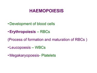
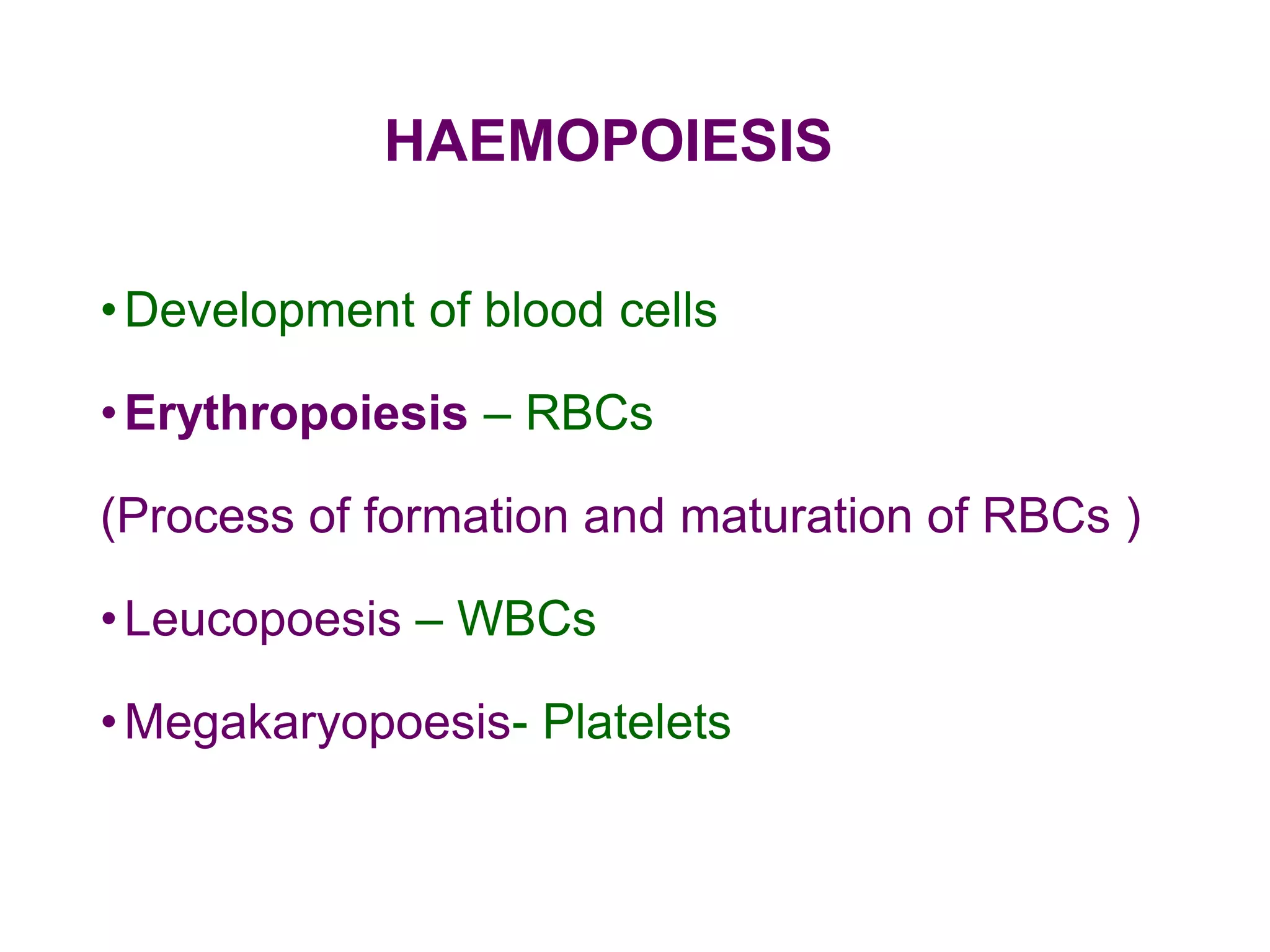
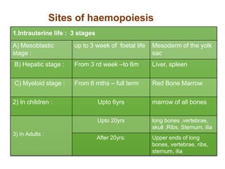
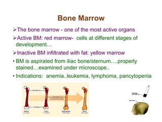
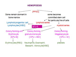
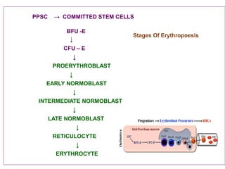
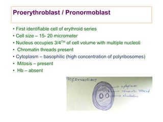
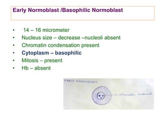
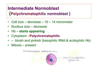
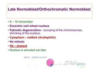
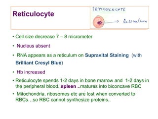
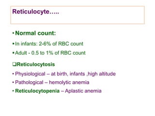
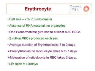
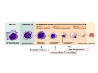
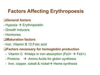
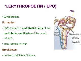
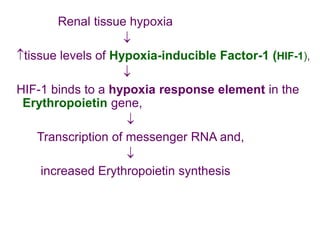
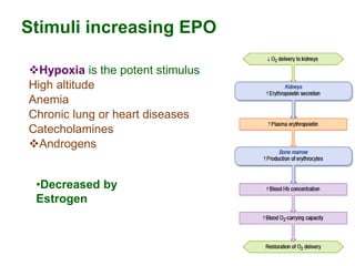
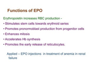
HAEMOPOIESIS
•Development of blood cells
•Erythropoiesis – RBCs
(Process of formation and maturation of RBCs )
•Leucopoesis – WBCs
•Megakaryopoesis- Platelets
Sites of haemopoiesis
1.Intrauterine life : 3 stages
A) Mesoblastic
stage :
up to 3 week of foetal life Mesoderm of the yolk
sac
B) Hepatic stage : From 3 rd week –to 6m Liver, spleen
C) Myeloid stage : From 6 mths – full term Red Bone Marrow
2) In children : Upto 6yrs marrow of all bones
3) In Adults :
Upto 20yrs long bones ,vertebrae,
skull ,Ribs, Sternum, ilia
After 20yrs: Upper ends of long
bones, vertebrae, ribs,
sternum, ilia
Bone Marrow
The bone marrow - one of the most active organs
Active BM: red marrow- cells at different stages of
development…
Inactive BM infiltrated with fat: yellow marrow
• BM is aspirated from iliac bone/sternum….properly
stained…examined under microscope..
• Indications: anemia..leukemia, lymphoma, pancytopenia
BFU-E
HEMOPOESIS
Bone Marrow….
• 75% of the cells in the marrow belong to the myeloid
series ( for WBC & Platelets)
• Only 25% - red cells.
• Normal myeloid- erythroid ratio is 3:1
• In circulation RBC count is 500 times more than WBC
count…because of longer life span of RBCs…
PPSC → COMMITTED STEM CELLS
BFU -E
↓
CFU – E
↓
PROERYTHROBLAST
↓
EARLY NORMOBLAST
↓
INTERMEDIATE NORMOBLAST
↓
LATE NORMOBLAST
↓
RETICULOCYTE
↓
ERYTHROCYTE
Stages Of Erythropoesis
Proerythroblast / Pronormoblast
• First identifiable cell of erythroid series
• Cell size – 15- 20 micrometer
• Nucleus occupies 3/4TH of cell volume with multiple nucleoli
• Chromatin threads present
• Cytoplasm – basophilic (high concentration of polyribosomes)
• Mitosis – present
• Hb – absent
Early Normoblast /Basophilic Normoblast
• 14 – 16 micrometer
• Nucleus size – decrease –nucleoli absent
• Chromatin condensation present
• Cytoplasm – basophilic
• Mitosis – present
• Hb – absent
Intermediate Normoblast
(Polychromatophilic normoblast )
• Cell size – decrease – 10 – 14 micrometer
• Nucleus size – decrease
• Hb – starts appearing
• Cytoplasm –Polychromatophilic
• – bluish and pinkish (basophilic RNA & acidophilic Hb)
• Mitosis – present
Late Normoblast/Orthochromatic Normoblast
• 8 – 10 micrometer
• Eccentric cart wheel nucleus
• Pyknotic degeneration- clumping of the chromosomes,
shrinking of the nucleus.
• Cytoplasm - reddish (Acidophilic)
• No mitosis
• Hb – present
• Nucleus is extruded out later
Reticulocyte
• Cell size decrease 7 – 8 micrometer
• Nucleus absent
• RNA appears as a reticulum on Supravital Staining (with
Brilliant Cresyl Blue)
• Hb increased
• Reticulocyte spends 1-2 days in bone marrow and 1-2 days in
the peripheral blood..spleen ..matures into biconcave RBC
• Mitochondria, ribosomes etc are lost when converted to
RBCs…so RBC cannot synthesize proteins..
Reticulocyte…..
•Normal count:
In infants: 2-6% of RBC count
Adult - 0.5 to 1% of RBC count
Reticulocytosis
• Physiological – at birth, infants ,high altitude
• Pathological – hemolytic anemia
• Reticulocytopenia – Aplastic anemia
Erythrocyte
• Cell size – 7.2- 7.5 micrometer
• Absence of RNA material, no organelles
• One Pronormoblast give rise to at least 8-10 RBCs
• 2 million RBCs produced each sec
• Average duration of Erythropoiesis: 7 to 9 days
Proerythroblast to reticulocyte takes 5 to 7 days
Maturation of reticulocyte to RBC takes 2 days..
• Life span = 120days
Factors Affecting Erythropoesis
General factors
- Hypoxia  Erythropoietin
- Growth inducers
- Hormones
Maturation factors
- Iron, Vitamin B 12,Folic acid
Factors necessary for hemoglobin production
- Vitamin C Helps in iron absorption (Fe3+  Fe2+)
- Proteins  Amino Acids for globin synthesis
- Iron, copper, cobalt & nickel Heme synthesis
1.ERYTHROPOETIN ( EPO)
• Glycoprotein.
Formation
• 85% formed in endothelial cells of the
peritubular capillaries of the renal
tubules.
• 15% formed in liver
Breakdown
 In liver. Half life is 5 hours
PTC
Renal tissue hypoxia

tissue levels of Hypoxia-inducible Factor-1 (HIF-1),

HIF-1 binds to a hypoxia response element in the
Erythropoietin gene,

Transcription of messenger RNA and,

increased Erythropoietin synthesis
Stimuli increasing EPO
Hypoxia is the potent stimulus
High altitude
Anemia
Chronic lung or heart diseases
Catecholamines
Androgens
•Decreased by
Estrogen
Functions of EPO
Erythropoietin increases RBC production -
• Stimulates stem cells towards erythroid series
• Promotes pronormoblast production from progenitor cells
• Enhances mitosis
• Accelerates Hb synthesis
• Promotes the early release of reticulocytes.
Applied – EPO injections in treatment of anemia in renal
failure
2.Growth inducers/ Differentiation inducers
• Interleukin 1, 3, 5 (IL-3 is a growth inducer for all cell lines )
Acts on stem cells to convert them to progenitor cells
• Colony stimulating factors – produced from T cells,
endothelial cells, fibroblasts stimulates production of
committed stem cells .
3. Hormones
•Androgens  & Estrogen  Erythropoesis
•. Thyroxine , GH, Cortisol –stimulates Erythropoesis
Maturation factors
Role of iron ,folic acid & vitamin B12.
• Iron- needed for Hb synthesis
• Folic acid & Vit B12- required for the synthesis of DNA and
maturation of nucleus and cell.
• Milk ,meat ,liver ,Green leafy vegetables
 If deficiency occurs… failure of maturation of nucleus.
 There occurs reduction in the cell division.
Cells remain large (megaloblasts) and become more
fragile.
• Intrinsic factor – needed for Vit B12 absorption
Other dietary factors
•Vitamin C Helps in iron absorption (Fe3+ 
Fe2+)
•Proteins  Amino Acids for globin synthesis
•Copper, cobalt & nickel.  Heme synthesis
(Copper- incorporation of Fe into Porphyrin Ring )

4. Erythropoesis - dental.pptx

  • 1.
    HAEMOPOIESIS •Development of bloodcells •Erythropoiesis – RBCs (Process of formation and maturation of RBCs ) •Leucopoesis – WBCs •Megakaryopoesis- Platelets
  • 2.
    Sites of haemopoiesis 1.Intrauterinelife : 3 stages A) Mesoblastic stage : up to 3 week of foetal life Mesoderm of the yolk sac B) Hepatic stage : From 3 rd week –to 6m Liver, spleen C) Myeloid stage : From 6 mths – full term Red Bone Marrow 2) In children : Upto 6yrs marrow of all bones 3) In Adults : Upto 20yrs long bones ,vertebrae, skull ,Ribs, Sternum, ilia After 20yrs: Upper ends of long bones, vertebrae, ribs, sternum, ilia
  • 3.
    Bone Marrow The bonemarrow - one of the most active organs Active BM: red marrow- cells at different stages of development… Inactive BM infiltrated with fat: yellow marrow • BM is aspirated from iliac bone/sternum….properly stained…examined under microscope.. • Indications: anemia..leukemia, lymphoma, pancytopenia
  • 4.
  • 5.
    Bone Marrow…. • 75%of the cells in the marrow belong to the myeloid series ( for WBC & Platelets) • Only 25% - red cells. • Normal myeloid- erythroid ratio is 3:1 • In circulation RBC count is 500 times more than WBC count…because of longer life span of RBCs…
  • 6.
    PPSC → COMMITTEDSTEM CELLS BFU -E ↓ CFU – E ↓ PROERYTHROBLAST ↓ EARLY NORMOBLAST ↓ INTERMEDIATE NORMOBLAST ↓ LATE NORMOBLAST ↓ RETICULOCYTE ↓ ERYTHROCYTE Stages Of Erythropoesis
  • 7.
    Proerythroblast / Pronormoblast •First identifiable cell of erythroid series • Cell size – 15- 20 micrometer • Nucleus occupies 3/4TH of cell volume with multiple nucleoli • Chromatin threads present • Cytoplasm – basophilic (high concentration of polyribosomes) • Mitosis – present • Hb – absent
  • 8.
    Early Normoblast /BasophilicNormoblast • 14 – 16 micrometer • Nucleus size – decrease –nucleoli absent • Chromatin condensation present • Cytoplasm – basophilic • Mitosis – present • Hb – absent
  • 9.
    Intermediate Normoblast (Polychromatophilic normoblast) • Cell size – decrease – 10 – 14 micrometer • Nucleus size – decrease • Hb – starts appearing • Cytoplasm –Polychromatophilic • – bluish and pinkish (basophilic RNA & acidophilic Hb) • Mitosis – present
  • 10.
    Late Normoblast/Orthochromatic Normoblast •8 – 10 micrometer • Eccentric cart wheel nucleus • Pyknotic degeneration- clumping of the chromosomes, shrinking of the nucleus. • Cytoplasm - reddish (Acidophilic) • No mitosis • Hb – present • Nucleus is extruded out later
  • 11.
    Reticulocyte • Cell sizedecrease 7 – 8 micrometer • Nucleus absent • RNA appears as a reticulum on Supravital Staining (with Brilliant Cresyl Blue) • Hb increased • Reticulocyte spends 1-2 days in bone marrow and 1-2 days in the peripheral blood..spleen ..matures into biconcave RBC • Mitochondria, ribosomes etc are lost when converted to RBCs…so RBC cannot synthesize proteins..
  • 12.
    Reticulocyte….. •Normal count: In infants:2-6% of RBC count Adult - 0.5 to 1% of RBC count Reticulocytosis • Physiological – at birth, infants ,high altitude • Pathological – hemolytic anemia • Reticulocytopenia – Aplastic anemia
  • 13.
    Erythrocyte • Cell size– 7.2- 7.5 micrometer • Absence of RNA material, no organelles • One Pronormoblast give rise to at least 8-10 RBCs • 2 million RBCs produced each sec • Average duration of Erythropoiesis: 7 to 9 days Proerythroblast to reticulocyte takes 5 to 7 days Maturation of reticulocyte to RBC takes 2 days.. • Life span = 120days
  • 15.
    Factors Affecting Erythropoesis Generalfactors - Hypoxia  Erythropoietin - Growth inducers - Hormones Maturation factors - Iron, Vitamin B 12,Folic acid Factors necessary for hemoglobin production - Vitamin C Helps in iron absorption (Fe3+  Fe2+) - Proteins  Amino Acids for globin synthesis - Iron, copper, cobalt & nickel Heme synthesis
  • 16.
    1.ERYTHROPOETIN ( EPO) •Glycoprotein. Formation • 85% formed in endothelial cells of the peritubular capillaries of the renal tubules. • 15% formed in liver Breakdown  In liver. Half life is 5 hours PTC
  • 17.
    Renal tissue hypoxia  tissuelevels of Hypoxia-inducible Factor-1 (HIF-1),  HIF-1 binds to a hypoxia response element in the Erythropoietin gene,  Transcription of messenger RNA and,  increased Erythropoietin synthesis
  • 18.
    Stimuli increasing EPO Hypoxiais the potent stimulus High altitude Anemia Chronic lung or heart diseases Catecholamines Androgens •Decreased by Estrogen
  • 19.
    Functions of EPO Erythropoietinincreases RBC production - • Stimulates stem cells towards erythroid series • Promotes pronormoblast production from progenitor cells • Enhances mitosis • Accelerates Hb synthesis • Promotes the early release of reticulocytes. Applied – EPO injections in treatment of anemia in renal failure
  • 20.
    2.Growth inducers/ Differentiationinducers • Interleukin 1, 3, 5 (IL-3 is a growth inducer for all cell lines ) Acts on stem cells to convert them to progenitor cells • Colony stimulating factors – produced from T cells, endothelial cells, fibroblasts stimulates production of committed stem cells . 3. Hormones •Androgens  & Estrogen  Erythropoesis •. Thyroxine , GH, Cortisol –stimulates Erythropoesis
  • 21.
    Maturation factors Role ofiron ,folic acid & vitamin B12. • Iron- needed for Hb synthesis • Folic acid & Vit B12- required for the synthesis of DNA and maturation of nucleus and cell. • Milk ,meat ,liver ,Green leafy vegetables  If deficiency occurs… failure of maturation of nucleus.  There occurs reduction in the cell division. Cells remain large (megaloblasts) and become more fragile. • Intrinsic factor – needed for Vit B12 absorption
  • 22.
    Other dietary factors •VitaminC Helps in iron absorption (Fe3+  Fe2+) •Proteins  Amino Acids for globin synthesis •Copper, cobalt & nickel.  Heme synthesis (Copper- incorporation of Fe into Porphyrin Ring )