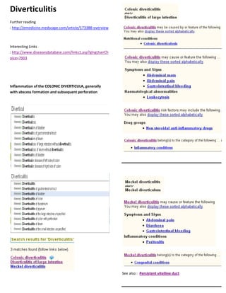
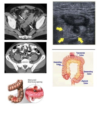
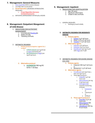
Diverticulitis is inflammation of diverticula in the colon. It can range from mild to severe, with complications including colonic perforation, abscesses, and peritonitis. Symptoms include abdominal pain, fever, and tenderness. Treatment involves antibiotics, clear liquid diets, and sometimes surgery for recurrent or complicated cases.
Dyslipidemia refers to abnormalities in serum lipid levels, including high or low levels of total cholesterol, triglycerides, LDL cholesterol, and HDL cholesterol. These abnormalities can be inherited, acquired, or secondary to other primary conditions. Dyslipidemias are classified based on the pattern of lipoproteins in electrophoresis or ultracentrifugation testing.
Tetrodotoxin is a potent neurotoxin found in several marine animals like pufferfish and blue-ringed octopus that blocks nerve impulses by binding to sodium channels. It produces paralysis by preventing action potentials, though bacteria are its actual source. While lethal in large doses, tetrodotoxin has helped scientists study sodium channel function and may lead to new pain treatments if a human antidote can be developed.
1. Progressive bulbar paralysis is a subtype of amyotrophic lateral sclerosis characterized by atrophy of motor cranial nerve nuclei in the brainstem.
2. It causes weakness of the muscles involved in swallowing and speech leading to drooling, difficulty chewing and talking, and risk of aspiration pneumonia.
3. Symptoms start around age 50-70 and progressively worsen over 1 to 3 years ultimately resulting in respiratory failure and death.
Acalculous cholecystitis is inflammation of the gallbladder wall that occurs in the absence of gallstones. It is most commonly observed in very ill patients, those with severe injuries or burns, or those who have been on total parenteral nutrition for more than 3 months. Imaging findings include a distended gallbladder with thickened walls and potentially pericholecystic fluid without evidence of gallstones. Prompt treatment with broad-spectrum antibiotics and cholecystectomy or cholecystostomy is recommended to prevent complications like perforation or gangrene.
1. Irritable bowel syndrome (IBS) is a common functional gastrointestinal disorder that affects 10-22% of the population. It is characterized by abdominal pain and changes in bowel habits without any underlying structural or biochemical abnormalities.
2. Management of IBS focuses on lifestyle modifications, diet changes, stress reduction techniques, and symptom-targeted medications. Dietary triggers like caffeine, alcohol, dairy, and fatty or gas-producing foods should be avoided. Bulk-forming fibers can help constipation, while antidiarrheals may relieve diarrhea.
3. Evaluation involves a careful history, physical exam, and initial labs as warranted. Colonoscopy is recommended if red flags are present. While there are no
This document provides an overview of the gastrointestinal system including its structure and function. It discusses several common gastrointestinal disorders such as parotitis, appendicitis, liver cirrhosis, pancreatitis, cholecystitis, and cholelithiasis. For each disorder, it describes the etiology, signs and symptoms, diagnostics, and nursing management. The document is authored by Mark Fredderick Abejo RN, MAN and intended to educate nurses on gastrointestinal disorders and their treatment.
Biliary colic is a painful condition caused by gallstones obstructing the cystic duct or ampulla of Vater. This leads to distention of the gallbladder or biliary tree. Pain is relieved when the gallstone passes through or moves locations. The main symptom is intermittent abdominal pain in the right upper quadrant that may radiate to the right shoulder. Diagnostic tests include ultrasound and bloodwork. Treatment options range from pain medications and watchful waiting to laparoscopic cholecystectomy to permanently remove the gallbladder in symptomatic patients.
Casts are used to immobilize and support injured bones and joints. Potential complications include pressure ulcers, cast syndrome, infection, and impaired circulation. To prevent complications, casts must not be too tight and the patient's neurovascular status should be frequently monitored. Any signs of complications such as pain, odor, or skin changes require evaluation and treatment which may include cast removal or modification.
This document discusses Meniere's disease, including its history, symptoms, pathophysiology, variants, and diagnostic criteria. Some key points:
- Meniere's disease causes episodes of vertigo, hearing loss, tinnitus, and a feeling of fullness in the ear. It is associated with endolymphatic hydrops (fluid buildup) in the inner ear.
- The cause is multifactorial but may involve abnormalities in endolymphatic fluid production, absorption, or circulation within the inner ear.
- Diagnosis requires recurrent episodes of vertigo, hearing loss, and tinnitus. Hearing loss must be sensorineural and involve low frequencies.
-
This document discusses several congenital laryngeal disorders classified by location in the larynx. Laryngomalacia, the most common cause of congenital stridor, involves soft, flabby laryngeal tissues that prolapse inward during inspiration. Laryngoceles are air-filled dilations of the laryngeal saccule that can be internal, external, or combined. They may cause respiratory distress or neck swelling. Laryngeal webs are failures of complete laryngeal canalization, most commonly involving the vocal cords. They can cause weak crying, recurrent croup, or inspiratory stridor. Flexible laryngoscopy is used to diagnose these conditions, while management depends on severity and may include observation,
This document discusses several congenital laryngeal disorders classified by location in the larynx. Laryngomalacia, the most common cause of congenital stridor, involves soft, flabby laryngeal tissues that prolapse inward during inspiration. Laryngoceles are air-filled dilations of the laryngeal saccule that can be internal, external, or combined. They may cause respiratory distress or neck swelling. Laryngeal webs are failures of complete laryngeal canalization, most commonly involving the vocal cords. They can cause weak crying, recurrent croup, or inspiratory stridor. Flexible laryngoscopy aids diagnosis while temporary tracheostomy or endoscopic procedures may help treat severe cases.
Bowel obstruction, also known as intestinal obstruction, is a mechanical or functional obstruction of the intestines which prevents the normal movement of the products of digestion. Either the small bowel or large bowel may be affected. Signs and symptoms include abdominal pain, vomiting, bloating and not passing gas.
The document defines and classifies periodontal pockets, describing their pathogenesis, clinical features, histopathology, contents, and measurement. Periodontal pockets are classified as gingival, suprabonny, or infrabony depending on their location relative to the alveolar bone crest. Treatment involves non-surgical approaches like scaling and root planing or surgical procedures such as gingivectomy, flap surgery, and bone grafting to reduce pocket depth. Accurately measuring and classifying pocket types is important for determining periodontal treatment.
This document discusses different types of periodontal abscesses and diseases. It begins by defining an acute periodontal abscess and listing potential causes. There are four main types of abscesses of the periodontium: gingival abscess, periodontal abscess, periapical abscess, and pericoronal abscess. Necrotizing periodontal disease is also discussed, which can present as necrotizing gingivitis, necrotizing periodontitis, or necrotizing stomatitis depending on the extent of necrosis. Endo-perio lesions are addressed, which occur when there is communication between the pulp and periodontium. Differential diagnosis and treatment approaches are provided for
Inflammatory bowel disease is caused by inappropriate activation of the immune system in response to normal gut bacteria. The two main types are Crohn's disease and ulcerative colitis. Crohn's disease can affect any part of the gastrointestinal tract and often involves transmural inflammation. Ulcerative colitis only involves the colon and rectum in a continuous manner. Both have a genetic component and involve defects in the epithelial barrier and immune response to gut microbiota. Common symptoms include abdominal pain, diarrhea, and weight loss.
This document discusses inflammatory bowel disease (IBD), specifically ulcerative colitis and Crohn's disease. It provides definitions and comparisons of the key features of each condition.
Ulcerative colitis causes continuous mucosal inflammation of the colon that extends proximally from the rectum. Its clinical features include bloody diarrhea, tenesmus, and abdominal tenderness. Crohn's disease causes skip areas of transmural inflammation that can occur anywhere along the gastrointestinal tract from mouth to anus. It can cause diarrhea, abdominal pain, strictures, and fistulas.
Both conditions can lead to extra-intestinal manifestations affecting the eyes, skin, joints, and primary sclerosing cholangitis
Periodontal pockets can form when the gingival sulcus deepens through movement of the gingival margin or displacement of the gingival attachment. Pockets are classified based on their morphology, relationship to crestal bone, number of tooth surfaces involved, and nature of the soft tissue wall. The pathogenesis of pockets involves inflammatory changes that lead to degradation of collagen fibers and destruction of connective tissue and bone. Clinically, pockets present with signs like bleeding and suppuration. Microscopic examination reveals areas of bacterial accumulation, leukocyte emergence and interaction, and epithelial desquamation. Pockets contain debris, microorganisms, and inflammatory products. Probing is used to detect and measure pocket depth.
This document provides information on corrosive poisoning, including types, mechanisms of injury, clinical features, investigations, management, and treatment. It discusses three main types of corrosives - acids, alkalis, and oxidants. Alkalis are noted as the most dangerous due to their ability to rapidly cause liquifactive necrosis and injury. Clinical features involve the gastrointestinal tract, respiratory system, eyes and skin. Investigations include endoscopy, imaging, and labs. Management focuses on airway protection, dilution, antibiotics, and monitoring. Long term complications like stricture formation may require repeated dilations.
Inflammatory bowel disease (IBD) refers to two conditions - Crohn's disease and ulcerative colitis. Crohn's disease causes transmural inflammation that can affect any part of the gastrointestinal tract, typically causing thickening of the bowel wall and formation of skip lesions. Ulcerative colitis only involves the large intestine and causes superficial ulcers and inflammation of the colonic mucosa in a continuous pattern. Both conditions are chronic, relapsing inflammatory disorders of unknown cause that are likely due to genetic and immunological factors. They can lead to complications like malnutrition, fistulas, strictures, and an increased risk of colon cancer.
This document summarizes amoebiasis, caused by the protozoan Entamoeba histolytica. It is prevalent in warm climates with unsanitary areas and is acquired by swallowing cysts. The cysts can survive a few days outside the body and pass into the intestines, where they may invade tissues and form liver abscesses. Symptoms include diarrhea, abdominal pain, and tenderness. Treatment involves antibiotics like metronidazole and prevention focuses on sanitation, water purification, and hygiene.
cornealulcers diagnosis treatment and other factorsMurali Krishna
This document discusses various types of keratitis including infective, allergic, trophic, and those associated with other diseases. It provides detailed information on bacterial corneal ulcers including causative organisms, stages of progression and treatment. It also describes herpes simplex keratitis and recurrent herpes infections of the cornea, noting the different clinical presentations and treatments for primary and recurrent infections.
1. Inflammation is defined as the local response to injury characterized by fluid and leukocyte movement into tissues. Acute inflammation has a rapid onset and short duration dominated by neutrophils, while chronic inflammation has a long duration dominated by mononuclear cells.
2. The classical signs of acute inflammation are redness, heat, swelling, pain, and loss of function. Systemic effects include fever, leukocytosis, lymphadenopathy, and acute phase protein production by the liver.
3. Exudative inflammation can be serous, lymphoplasmocytic, purulent, fibrinous, or gangrenous depending on the characteristics and cellular composition of the exudate and tendency for
56 Establishing A Bedside Diagnosis Of Hypovolemiakdiwavvou
This document summarizes a literature review on physical exam findings that can help diagnose hypovolemia. The review found that a large increase in pulse (over 30 beats per minute) when moving from lying to standing, or severe dizziness preventing standing, best indicate hypovolemia related to blood loss. However, these findings may be absent with moderate blood loss. Few physical exam findings reliably diagnose hypovolemia due to diarrhea, vomiting or low fluid intake. Prolonged capillary refill time and poor skin turgor did not prove useful. The authors recommend lab tests if hypovolemia is suspected.
This document summarizes three medications used to treat hyperaldosteronism: Canrenone, Spironolactone, and Eplerenone.
Canrenone and Spironolactone are aldosterone antagonists with diuretic effects that act to counteract aldosterone and promote excretion of sodium. Eplerenone selectively blocks aldosterone receptors in the kidneys and cardiovascular system.
The document provides information on indications, contraindications, side effects and dosing for each medication. It also notes that periodic monitoring of potassium levels is needed when using these aldosterone antagonists due to the risk of hyperkalemia.
The respiratory rate and pattern are determined by the respiratory control center in the brainstem. It receives feedback from peripheral chemoreceptors in the carotid bodies and central chemoreceptors in the brainstem to regulate ventilation and maintain normal blood gases. The respiratory rate, tidal volume, and use of accessory muscles are observed during a physical exam to detect any abnormalities. Changes in rate or tidal volume have different effects on gas exchange depending on whether the dead space or alveolar volume is altered.
The document summarizes essential thrombocytosis, a rare chronic blood disorder characterized by overproduction of platelets. It is one of four myeloproliferative disorders. The summary describes the epidemiology, pathophysiology involving abnormal megakaryocytes and platelet function, clinical features such as bleeding, thrombosis, and splenomegaly. Diagnostic criteria include persistent thrombocytosis over 600x109/L and exclusion of other causes, with some cases associated with a JAK2 kinase mutation. Treatment aims to reduce platelet count and risk of thrombosis.
Standing electrolyte replacement protocols are available for use in adult patients admitted to Orlando Regional Healthcare hospitals. These include protocols for calcium chloride or calcium gluconate, magnesium sulfate, potassium chloride, and potassium phosphate replacement. The protocols provide guidance on administration methods, dosage, rates of infusion, and monitoring based on current serum electrolyte levels. All electrolyte replacements must be administered via infusion pump with appropriate dilution and monitoring by medical staff.
Here are the key points about ionized calcium levels:
- Ionized calcium is the biologically active form of calcium and provides a more accurate assessment of calcium status compared to total calcium levels.
- Low ionized calcium levels are common in critically ill patients and those with conditions affecting calcium homeostasis like renal failure.
- Ionized calcium levels below 2.8 mg/dL increase the risk of cardiac arrest, so calcium replacement therapy is generally started once levels fall below this threshold.
- Measurement of ionized calcium is particularly important for monitoring unconscious or anesthetized patients where changes in calcium levels may not produce early warning signs.
- Ionized calcium can also be useful for evaluating conditions like neonatal hypocal
1. The Frederickson classification system outlines 5 types of hyperlipidemia based on elevated lipid levels and underlying genetic defects.
2. Type I is characterized by increased chylomicrons due to LPL deficiency. Type IIa is caused by LDL receptor deficiency leading to high LDL. Type IIb involves high LDL and VLDL due to LDL receptor and ApoB defects. Type III stems from ApoE defects causing elevated cholesterol and triglycerides. Type IV results from increased VLDL production and decreased elimination. Type V involves increased VLDL and chylomicron production coupled with low LPL.
Dyslipidemia refers to abnormalities in serum lipid levels, including high or low levels of cholesterol, triglycerides, and lipoproteins. Dyslipidemias can be inherited, acquired, primary, or secondary. They are classified based on the pattern of lipoproteins seen on electrophoresis or ultracentrifugation. Causes include genetic factors, endocrine conditions, drugs, and lifestyle factors like smoking. Symptoms are often nonspecific but may include obesity.
The document provides descriptions of various cardiac rhythms, conduction abnormalities, myocardial infarctions and other cardiac conditions as assessed by electrocardiogram findings. Key items summarized include descriptions of flutter, fibrillation, supraventricular and atrial tachycardias, bundle branch and fascicular blocks, atrioventricular blocks, preexcitation syndromes, myocardial infarction in various territories, athlete's heart, electrolyte abnormalities, drug effects and various cardiac pathologies.
The 11-step method provides a systematic approach to reading EKGs:
1. Gather data such as heart rate, intervals, and axis.
2. Diagnose rhythm, conduction blocks, enlargement, and infarction by applying specific criteria.
3. Potential diagnoses are identified through disturbances of rhythm, conduction, hypertrophy, and ischemia. The relationship between P waves and QRS complexes helps determine block types.
Hypertension, or high blood pressure, is a major risk factor for coronary artery disease and cerebrovascular accidents. The risk of these conditions increases as blood pressure rises. For those over age 60, pulse pressure is the best predictor of outcomes from hypertension. Essential or primary hypertension, which has no identifiable cause, accounts for 80% of hypertension cases. It is defined as a diastolic blood pressure of 90-104 mmHg. Isolated systolic hypertension, affecting those over age 75, occurs when systolic pressure is over 160 mmHg and diastolic is under 90 mmHg.
This document discusses vitamin A, including its sources, forms, and functions. It notes that vitamin A is found primarily in animal foods as retinol, retinal, and retinoic acid. These forms are essential for growth, tissue integrity, and vision. Deficiencies can occur rarely due to absorption or storage issues, though stores usually last over 2 years. Toxicity risks exist from excessive supplementation, with symptoms taking long to resolve as stores are depleted slowly. The document recommends vitamin A supplementation only for deficiencies, pregnancy, or lactation, as normal diets provide sufficient amounts.
The document discusses a clinical case of a 30-year-old woman experiencing intermittent right upper quadrant pain. Her lab tests and ultrasound were normal. The key signs and symptoms suggest a diagnosis of biliary dyskinesia. There is no standardized test for this condition, but HIDA scanning is commonly used to assess gallbladder ejection fraction, with values under 35-40% indicating dysfunction. However, the test protocol can vary between providers and affect results. The most reliable approach may be one where CCK is administered at 30 and 60 minutes to better evaluate gallbladder motility.
Hiranandani Hospital in Powai, Mumbai, is a premier healthcare institution that has been serving the community with exceptional medical care since its establishment. As a part of the renowned Hiranandani Group, the hospital is committed to delivering world-class healthcare services across a wide range of specialties, including kidney transplantation. With its state-of-the-art facilities, advanced medical technology, and a team of highly skilled healthcare professionals, Hiranandani Hospital has earned a reputation as a trusted name in the healthcare industry. The hospital's patient-centric approach, coupled with its focus on innovation and excellence, ensures that patients receive the highest standard of care in a compassionate and supportive environment.
Promoting Wellbeing - Applied Social Psychology - Psychology SuperNotesPsychoTech Services
A proprietary approach developed by bringing together the best of learning theories from Psychology, design principles from the world of visualization, and pedagogical methods from over a decade of training experience, that enables you to: Learn better, faster!
Cell Therapy Expansion and Challenges in Autoimmune DiseaseHealth Advances
There is increasing confidence that cell therapies will soon play a role in the treatment of autoimmune disorders, but the extent of this impact remains to be seen. Early readouts on autologous CAR-Ts in lupus are encouraging, but manufacturing and cost limitations are likely to restrict access to highly refractory patients. Allogeneic CAR-Ts have the potential to broaden access to earlier lines of treatment due to their inherent cost benefits, however they will need to demonstrate comparable or improved efficacy to established modalities.
In addition to infrastructure and capacity constraints, CAR-Ts face a very different risk-benefit dynamic in autoimmune compared to oncology, highlighting the need for tolerable therapies with low adverse event risk. CAR-NK and Treg-based therapies are also being developed in certain autoimmune disorders and may demonstrate favorable safety profiles. Several novel non-cell therapies such as bispecific antibodies, nanobodies, and RNAi drugs, may also offer future alternative competitive solutions with variable value propositions.
Widespread adoption of cell therapies will not only require strong efficacy and safety data, but also adapted pricing and access strategies. At oncology-based price points, CAR-Ts are unlikely to achieve broad market access in autoimmune disorders, with eligible patient populations that are potentially orders of magnitude greater than the number of currently addressable cancer patients. Developers have made strides towards reducing cell therapy COGS while improving manufacturing efficiency, but payors will inevitably restrict access until more sustainable pricing is achieved.
Despite these headwinds, industry leaders and investors remain confident that cell therapies are poised to address significant unmet need in patients suffering from autoimmune disorders. However, the extent of this impact on the treatment landscape remains to be seen, as the industry rapidly approaches an inflection point.
One health condition that is becoming more common day by day is diabetes.
According to research conducted by the National Family Health Survey of India, diabetic cases show a projection which might increase to 10.4% by 2030.
Osteoporosis - Definition , Evaluation and Management .pdfJim Jacob Roy
Osteoporosis is an increasing cause of morbidity among the elderly.
In this document , a brief outline of osteoporosis is given , including the risk factors of osteoporosis fractures , the indications for testing bone mineral density and the management of osteoporosis
5. Diverticulitis
4. Signs
1. See Also 1. Fever
1. Diverticulosis 2. Tenderness over left lower quadrant
3. GUARDING AND REBOUND TENDERNESS MAY BE
PRESENT
2. Pathophysiology
1. COMPLICATES 5% OF DIVERTICULOSIS
5. Labs
2. DISTRIBUTION 1. COMPLETE BLOOD COUNT
1. MOST OFTEN AFFECTS SIGMOID COLON 1. Leukocytosis (>68% of cases)
2. Right Diverticular Disease in age <60 and
asians
2. URINALYSIS
1. DYSURIA AND URINARY
FREQUENCY MAY OCCUR
3. INFLAMMATION OF COLONIC DIVERTICULA
1. Impacted with fecal material
(fecalith)
6. Radiology
1. ABDOMINAL FLAT AND UPRIGHT
ABDOMEN
2. Colon Perforation 1. Observe for abdominal free air
1. Microperforation 2. Small Bowel Obstruction
(Simple Diverticulitis)
1. Peridiverticulitis with
localized phlegmon
2. Infection walled off by
pericolic fat
2. ABDOMINAL CT WITH CONTRAST
1. Best test to confirm Diverticulitis
2. Macroperforation
(Complicated Diverticulitis) 2. Best test to identify complications
1. Pericolic abscess or (perforation)
2. Free perforation with
generalized peritonitis
3. Fistulas may form
between adjacent
3. Findings suggestive of perforation
structures
1. Pericolic fat infiltration
2. Fascial thickening and muscle
hypertrophy
3. Arrowhead sign
1. Localized bowel wall
thickening
2. Bowel lumen
resembles arrow
3. Symptoms shape at diverticulum
1. Mild anorexia
2. Nausea or Vomiting
3. Chills
4. Diarrhea or cobstipation
5. Abdominal Pain: Acute constant pain 3. AVOID COLONOSCOPY IN ACUTE DISEASE
1. Initial: Hypogastric pain 1. Risk of worsening perforation
2. LATER: LEFT LOWER 4. AVOID BARIUM ENEMA IN ACUTE DISEASE
QUADRANT ABDOMINAL PAIN 1. Risk of extravasation if perforation
(>92%)
6. 7. Management: General Measures
1. Clear Liquid Diet (NPO in severe disease)
2. Low fiber diet in acute phase 9. Management: Inpatient
3. Avoid Narcotics (INCREASES INTRACOLONIC 1. INDICATIONS FOR HOSPITALIZATION
PRESSURE)
1. Age >85 years
1. Except Meperidine (decreases
2. Significant inflammation
intraluminal pressure) 3. Unable to take oral fluids
4. ANTICIPATE IMPROVEMENT WITHIN 48-72 HOURS
2. GENERAL MEASURES
8. Management: Outpatient Mangement 1. Nothing by mouth initially
of mild disease
1. INDICATIONS FOR OUTPATIENT
MANAGEMENT
1. Uncomplicated Diverticulitis 3. ANTIBIOTIC REGIMEN FOR MODERATE
2. Stable clinically DISEASE
3. Tolerating oral fluids 1. Primary agents
1. Unasyn 3 g IV q6 hours
2. Zosyn 3.375 g IV q6 hours
3. Timentin 3.1 g IV q6 hours
2. Alternative agents
2. ANTIBIOTIC REGIMEN 1. Cefoxitin 2 g IV q8 hours
1. Primary protocol (requires 2 agents for 7-
2. Cefotetan 2 g IV q12 hours
10 days)
3. Ciprofloxacin 400 mg IV q12h
1. CIPROFLOXACIN 500 mg PO bid
with Flagyl 500 IV q6h
or Septra DS PO bid and
2. METRONIDAZOLE (Flagyl) 500
mg PO q6 hours
4. ANTIBIOTIC REGIMEN FOR SEVERE DISEASE
(E.G. ICU)
2. Alternative protocol 1. Primary agents
1. AUGMENTIN 500 mg PO 1. Imipenem 500 mg IV q6 hours
or
tid for 7-10 days 2. Merepenem 1 g IV q8 hours
2. Alternative agents
1. Trovafloxacin 300 mg IV day 1,
then 200 mg IV qd or
2. Three agent protocol 1
1. Ampicillin 2 g IV q6
hours and
2. Metronidazole 500 mg
IV q6 hours and
3. Aminoglycoside
(requires monitoring
of levels)
1. Gentamicin
or
2. Tobramycin
or
3. Amikacin
3. Three agent protocol 2
1. Ampicillin 2 g IV q6
hours and
2. Metronidazole 500
mg IV q6 hours
3. Ciprofloxacin 400 mg
IV q12 hours
7. 10.Course
1. Improves on antibiotics within 48 to 72
hours
11.Follow-up
1. COLONOSCOPY 6 WEEKS AFTER
DIVERTICULITIS EPISODE
1. Define extent of Diverticulosis
2. Evaluate for Colon Cancer
3. Barium Enema may be used as alternative
option
2. SURGICAL INDICATIONS
1. Recurrent Diverticulitis (more than
1 episode)
12.Complications
1. Colonic perforation
2. Colonic abscess
3. Generalized peritonitis
4. Colonic fistula
13.Prevention
1. High fiber diet (except in acute phase - see above)
2. Maintain adequate hydration
14.Prognosis
1. After first episode, recurs in 20-30% of cases
2. After second episode, recurs in 50% of cases
15. References
1. Gilbert (2002) Sanford Guide to
Antimicrobials, p. 14
2. Simmang in Feldman (1998)
Gastrointestinal, p. 1793-7
3. Salzman (2005) Am Fam Physician 72:1229