Downloaded 124 times

![SMALL AND LARGE BOWEL . . . CONT.
Common Clinical Syndromes
❏ Food Poisoning
• brief explosive diarrhea following exposure to food contaminated with bacteria or bacterial toxins
• 90% due to 4 bacteria: Salmonella > S. aureus > C. perfringens > B. cereus
• spontaneously resolves within 24-48 hours
❏ Traveller’s Diarrhea
• 3 unformed stools in 24 hours +/– nausea, vomiting, abdominal pain, tenesmus, blood/mucus in stool
• up to 50% of travelers to developing countries affected in first 2 weeks and 10-20% after returning home
• etiology - 80% bacterial, E. coli most common
• enterotoxigenic E. coli, other E. coli, Campylobacter, Shigella, Salmonella, Vibrio (non-cholera)
• viral - Norwalk and Rotavirus accounting for about 10%
• rarely protozoal (Giardiasis, Amebiasis)
• treatment and prophylaxis
• can use bismuth subsalicylate (Pepto-Bismol), empiric quinolone such as ciprofloxacin or
TMP/SMX prophylaxis for travelers who cannot tolerate inactivity, have underlying medical
condition (DM, AIDS, FBD, ESRD), or past history of traveler’s diarrhea
• if diarrhea persists after returning home, think of Giardia, Entamoeba histolytica, post-infections IBS
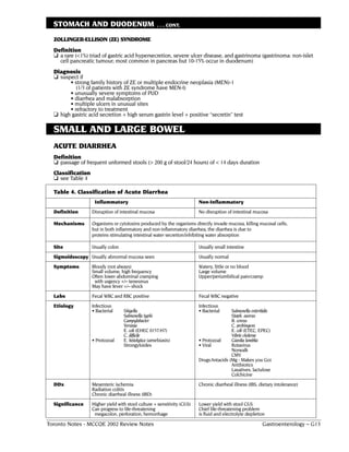
CHRONIC DIARRHEA
Definition
❏ passage of frequent unformed stools (> 200 mL of stool water/24 hours) of > 14 days duration
Etiology / Classification
❏ see Table 7
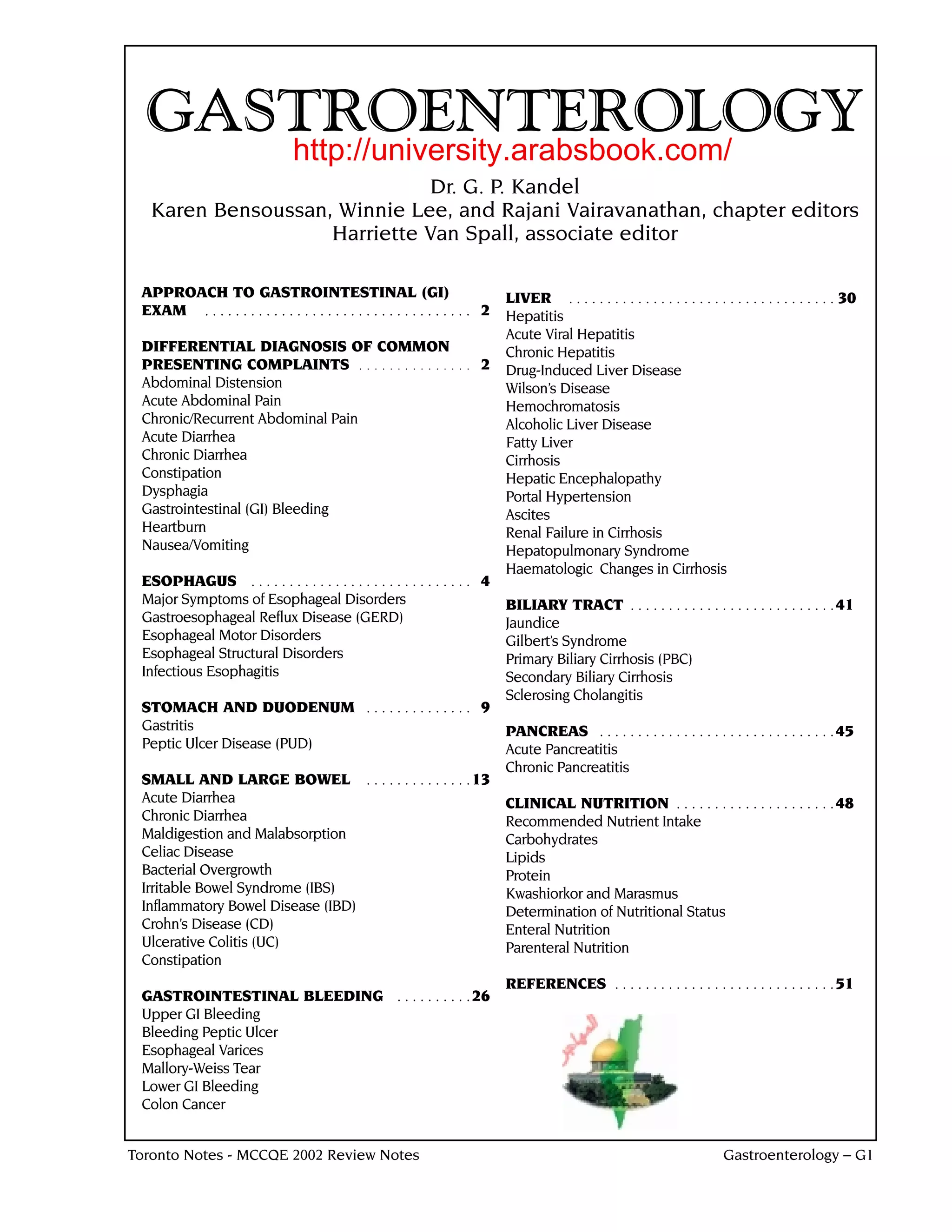
Table 7. Classification of Chronic Diarrhea
Type Characteristics
Inflammatory
Ulcerative colitis (UC) Fever, hematochezia, abdominal pain; usually weight loss with carcinoma
Crohn's disease
Malignancy: lymphoma, adenocarcinoma
Osmotic
Ingestion Stool volume decreases with fasting
Lactose intolerance Increased stool osmotic gap:
Medications, laxatives fecal [Na+] + [K+] < 1/2 serum osmolality – 25 mmol/L
Maldigestion and Malabsorption
Pancreatic insufficiency See Maldigestion and Malabsorption section
Bile salt deficiency Weight loss, fecal fat > 7-10g/24h stool collection
Celiac sprue anemia, hypoalbuminemia
Whipple's disease
Bowel resection
Secretory
Bacterial enterotoxins Large volume (>1L/d); little change with fasting
Secretagogues - VIP, gastrin, carcinoid Normal stool osmotic gap:
secretory: fecal [Na+] + [K+] = 1/2 serum osmolality
Functional
Irritable Bowel Syndrome (IBS) See Irritable Bowel Syndrome section
MALDIGESTION AND MALABSORPTION
Definitions
❏ maldigestion - inability to break down large molecules in the lumen of the intestine into their
component small molecules
❏ malabsorption - inability to transport molecules across the intestinal mucosa to the body fluids
G16 – Gastroenterology Toronto Notes - MCCQE 2002 Review Notes](https://image.slidesharecdn.com/git-111210180758-phpapp02/85/Git-16-320.jpg)
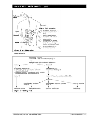

![LIVER . . . CONT.
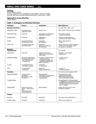
ASCITES
Definition
❏ accumulation of excess free fluid in the peritoneal cavity
Etiology
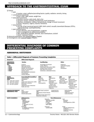
❏ see Table 14
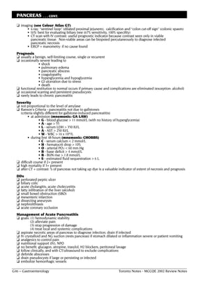
Table 14. Serum-Ascites Albumin Ratio as an Indicator
of the Causes of Ascites
serum [alb] – ascitic [alb] > 11 g/L serum [alb] – ascitic [alb] < 11 g/L
Cirrhosis/severe hepatitis Peritoneal carcinomatosis
Chronic hepatic congestion TB
(right heart failure, Budd-Chiari) Pancreatic disease
Nephrotic syndrome
Massive liver metastases
Myxedema
Pathogenesis of Ascites in Cirrhosis
❏ underfill theory
• portal hypertension and hypoalbuminemia lead to transudation of Na+ and water into peritoneum
• causes decreased intravascular volume and secondary renal Na+ and water retention
❏ overflow theory
• liver disease primarily causes renal retention of Na+ and water which then "overflows"
into peritoneal cavity
❏ combined theory
• liver disease causes vasodilation
• decreased effective intravascular volume (i.e. volume to capacitance ratio low,
but absolute volume is high)
• secondary urinary Na+ and water retention
Diagnosis
❏ ultrasound is gold standard
❏ clinically detectable when > 500 mL (bulging flanks, shifting dullness, fluid wave)
❏ diagnostic paracentesis - send ascitic fluid for:
• cells and differential
• chemistry (albumin, protein, amylase, triglycerides)
• culture and sensitivity and gram stain
• cytology for malignancy
Treatment
❏ paracentesis safe (except large volumes)
❏ medical
• Na+ restriction
• diuretics (spironolactone, furosemide)
• aim for 0.5 kg loss per day (rate of ascitic fluid absorption)
❏ surgical
• peritoneal-systemic (LeVeen) shunts, TIPSS, liver transplantation
• reserved for medically unresponsive cases
Complication - Bacterial Peritonitis
❏ primary/spontaneous bacterial peritonitis (SBP)
• complicates ascites, does not cause it (occurs in 10% of cirrhotic ascites)
• 1/3 of patients are asymptomatic, thus do not hesitate to do a diagnostic paracentesis
• fever, chills, abdominal pain, ileus, hypotension, worsening encephalopathy
• gram negatives compose 70% of bugs - E. coli (most common pathogen), Strep., Klebsiella
❏ secondary bacterial peritonitis
• usually results from perforated viscus or surgical manipulation
❏ diagnosis: absolute neutrophil count in peritoneal fluid > 0.25x10⁹ cells/L or WBC
count > 0.5x10⁹ cells/L + positive culture
❏ gram stain is positive in only 10-50% of patients
❏ treatment
• IV antibiotics (cefotaxime is the treatment of choice until C&S is available) for 10-14 days
• prophylaxis with daily Norfloxacin or TMP-SMX for 5/7 days may decrease the frequency of
recurrent SBP
G40 – Gastroenterology Toronto Notes - MCCQE 2002 Review Notes](https://image.slidesharecdn.com/git-111210180758-phpapp02/85/Git-40-320.jpg)
![LIVER . . . CONT.
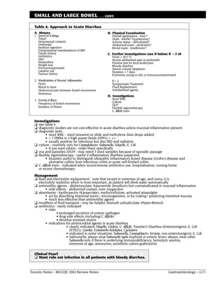
RENAL FAILURE IN CIRRHOSIS
❏ classify as
• pre-renal
• acute tubular necrosis (ATN)
• hepatorenal syndrome
❏ hepatorenal syndrome is secondary to
• overaggressive diuresis or large volume paracentesis
• GI bleeding
• sepsis
❏ differentiate hepatorenal syndrome from pre-renal failure
• clinical (very difficult)
• intravenous fluid challenge (giving volume expanders improves prerenal failure)
• pulmonary capillary wedge measurements (PCWP) (preferable)
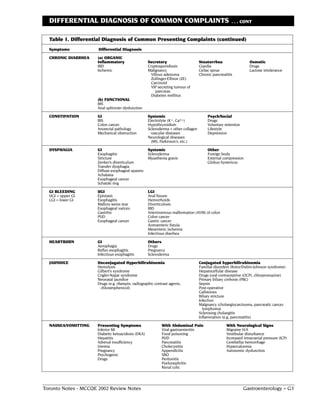
❏ differentiate hepatorenal syndrome from ATN (see Table 15)
❏ treatment for hepatorenal syndrome is generally unsuccessful
• vasopressin, octreotide, or norepinepherine may help (increased renal blood flow by
increased systemic vascular resistance)
• definitive treatment is liver transplant
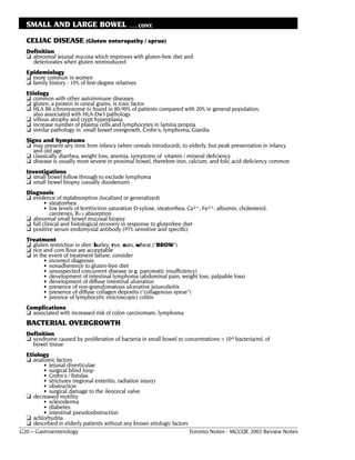
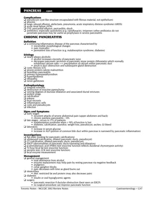
Table 15. Differential Diagnosis of Acute Azotemia in Liver Disease
Laboratory Findings Prerenal Azotemia or Acute Renal Failure
Hepatorenal Syndrome (ATN)
Urine [Na+] (mEq/L) < 10 > 30
Urine:plasma creatinine ratio > 30:1 < 20:1
Urine Osmolality At least 100 mOsm greater Equal to plasma osmolality
than plasma osmolality
Urine Sediment Normal Casts and cellular debris
HEPATOPULMONARY SYNDROME
❏ intrapulmonary vasodilation leading to hypoxia from ventilation/perfusion (V/Q) abnormalities
❏ improves with supplemental oxygen
❏ no proven medical therapy
HAEMATOLOGIC CHANGES IN CIRRHOSIS
❏ pancytopenia from hypersplenism
❏ decreased clotting factors
• fibrin, thrombin, I, II, V, VII, IX, X
BILIARY TRACT
JAUNDICE (see Table 16 and Figure 11)
❏ definition
• yellow pigmentation of skin, sclerae and mucus membranes due to increased serum bilirubin
❏ history
• dark urine, pale stools
• pruritis
• symptoms of biliary colic (obstructive jaundice)
• history of drug and EtOH use, hepatitis
• travel history
• sexual history
• family history
❏ physical exam
• may be unremarkable
❏ investigations
• bilirubin (conjugated and unconjugated)
• AST, ALT, GGT, ALP
• serologic tests for hepatitis
• ultrasound for evidence of obstructive jaundice, CT
• direct duct visualization (ERCP, PTC) (note: PTC only if obstruction is suspected to be periportal
rather than near sphincter or if previous gastric surgery)
• liver biopsy
Toronto Notes - MCCQE 2002 Review Notes Gastroenterology – G41](https://image.slidesharecdn.com/git-111210180758-phpapp02/85/Git-41-320.jpg)

This document provides an overview of gastroenterology. It begins with an approach to the gastrointestinal exam and a differential diagnosis table of common presenting complaints involving various gastrointestinal symptoms. It then covers specific sections on the esophagus, stomach and duodenum, small and large bowel, liver, biliary tract, pancreas, gastrointestinal bleeding, and clinical nutrition. For each section, it lists and briefly describes common disorders. The esophagus section focuses on dysphagia, heartburn, and gastroesophageal reflux disease.


![CASE_PRESENTATION_ON_subdural_hematoma(SDH)[1 FINAL PPT]-1.pptx](https://cdn.slidesharecdn.com/ss_thumbnails/casepresentationonsubduralhematomasdh1finalppt-1-260129172522-d405d375-thumbnail.jpg?width=640&height=640&fit=bounds)


