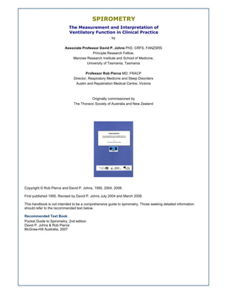
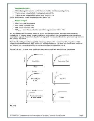
This document provides an overview of spirometry - the measurement and interpretation of lung function. It discusses the different types of spirometry devices, how to properly perform the tests, factors to consider when choosing a device, and guidelines for interpreting test results. The key measurements taken during spirometry include forced vital capacity, forced expiratory volume in one second, and peak expiratory flow. Proper technique and quality control criteria must be followed to obtain accurate and reproducible results. Spirometry is useful for detecting and monitoring respiratory diseases that impact lung function.
Demo Cum Lecture on the topic of Spirometry by Pandian M, Tutor, Dept. of Phy...Pandian M
PY 6.8 Demonstrate the correct technique to perform & interpret Spirometry.
SLOs: After attending lecture & studying the assigned materials, the student will:
1. State the principles of recording of Spirometry.
2. Define & draw different lung volumes & capacities.
3. Explain FEV (TVC) correctly with its clinical significance.
4. Describe the helium dilution method to record the FRC.
5. Demonstrate the correct procedure of recording the spirogram.
6. Interpret the spirogram correctly
Spirometry for Primary Care Physician OfficeRandy Clare
Step by step description of how to collect spirometry tests for Asthma and COPD. Quality control tips supported by literature with links to NIH, NIOSH and the Mayo Clinic. This is a presentation that I use to discuss hand held spirometry products from Carefusion. Micro Loop, Micro Lab, Micro 1 and Pulmolife
What are the pulmonary function tests used?
What are the indications?
What are the contraindications?
How to perform each and prepare patients?
How to interpret and reach a diagnosis?
How to clean and calibrate devices?
A spirometer is an apparatus for measuring the volume of air inspired and expired by the lungs. A spirometer measures ventilation, the movement of air into and out of the lungs. The spirogram will identify two different types of abnormal ventilation patterns, obstructive and restrictive.
A plethysmograph is an instrument for measuring changes in volume within an organ or whole body. The word is derived from the Greek "plethysmos", and "graphos"
Pulmonary function tests: A brief Insight- By RxVichuZ! :)RxVichuZ
This is my 51st powerpoint..deals with PULMONARY FUNCTION TESTS..their uses...details on spirometry, lung volumes and capacities & brief insight into other tests.
Happy reading!!!
This session will provide a quick review of the methodology of early dispatch systems connected to radio, telephone and other resources via circuit switched interfaces such as 4WE&M, 2W analogue etc., and their restricted backhaul capabilities, leading on to the 'stand-alone' RoIP boxes that allowed 4W E&M to be converted to IP and recovered at the other end allowing backhaul via more flexible IP networks.
The next technology is dispatch systems with native IP connectivity allowing the most flexible and functional interfaces between the dispatch system and its connected resources. While some manufacturers equipment uses proprietary IP messaging, most prefer and use open standards such as P25 CSSI (console sub system interface), DFSI (digital fixed system interface) and ISSI (inter sub system interface) or the emerging DMR AIS which ensures that different vendors equipment can interoperate with each other via these interfaces. Open standards provide end users with greatly improved competitive choice and functional capability on these systems.
The session will explore examples of IP interfaces for voice dispatch systems and the functions supported, plus give a background on how these apply to many different technologies and can even be adapted for conventional radio applications:
The workshop will cover on the following issues:
- The difference between RoIP and VoIP - how radio systems differ from phone systems
- Implementing one-to-one connections
- Implementing many-to-many connection
- Risk management: Identifying network issues affecting RoIP/VoIP quality; maintenance; and redundancy
- Design elements :- building blocks; calculating network bandwidth requirements
The implications of RoIP for dispatch consoles will be also be discussed: how dispatch console to radio connections can be implemented with RoIP and how RoIP can be used to provide fault tolerant dispatch architectures.
Finally the workshop will look at the impact of new technologies such as IPv6, Wireless Broadband and the switch to Digital Radio on the RoIP landscape.
Les Scott, Manager, System Sales, Zetron
A Guide to Aerosol Delivery Devices for Respiratory TherapistsUtai Sukviwatsirikul
A Guide to Aerosol Delivery Devices for Respiratory Therapists,
3rd Edition American Association for Respiratory Care
http://www.irccouncil.org/newsite/members/aerosol_guide_rt.pdf
đặC điểm lâm sàng, cận lâm sàng và kết quả điều trị bệnh nhân có hội chứng ch...TÀI LIỆU NGÀNH MAY
Để xem full tài liệu Xin vui long liên hệ page để được hỗ trợ
: https://www.facebook.com/thuvienluanvan01
HOẶC
https://www.facebook.com/garmentspace/
https://www.facebook.com/thuvienluanvan01
tai lieu tong hop, thu vien luan van, luan van tong hop, do an chuyen nganh
Adv. biopharm. APPLICATION OF PHARMACOKINETICS : TARGETED DRUG DELIVERY SYSTEMSAkankshaAshtankar
MIP 201T & MPH 202T
ADVANCED BIOPHARMACEUTICS & PHARMACOKINETICS : UNIT 5
APPLICATION OF PHARMACOKINETICS : TARGETED DRUG DELIVERY SYSTEMS By - AKANKSHA ASHTANKAR
Title: Sense of Taste
Presenter: Dr. Faiza, Assistant Professor of Physiology
Qualifications:
MBBS (Best Graduate, AIMC Lahore)
FCPS Physiology
ICMT, CHPE, DHPE (STMU)
MPH (GC University, Faisalabad)
MBA (Virtual University of Pakistan)
Learning Objectives:
Describe the structure and function of taste buds.
Describe the relationship between the taste threshold and taste index of common substances.
Explain the chemical basis and signal transduction of taste perception for each type of primary taste sensation.
Recognize different abnormalities of taste perception and their causes.
Key Topics:
Significance of Taste Sensation:
Differentiation between pleasant and harmful food
Influence on behavior
Selection of food based on metabolic needs
Receptors of Taste:
Taste buds on the tongue
Influence of sense of smell, texture of food, and pain stimulation (e.g., by pepper)
Primary and Secondary Taste Sensations:
Primary taste sensations: Sweet, Sour, Salty, Bitter, Umami
Chemical basis and signal transduction mechanisms for each taste
Taste Threshold and Index:
Taste threshold values for Sweet (sucrose), Salty (NaCl), Sour (HCl), and Bitter (Quinine)
Taste index relationship: Inversely proportional to taste threshold
Taste Blindness:
Inability to taste certain substances, particularly thiourea compounds
Example: Phenylthiocarbamide
Structure and Function of Taste Buds:
Composition: Epithelial cells, Sustentacular/Supporting cells, Taste cells, Basal cells
Features: Taste pores, Taste hairs/microvilli, and Taste nerve fibers
Location of Taste Buds:
Found in papillae of the tongue (Fungiform, Circumvallate, Foliate)
Also present on the palate, tonsillar pillars, epiglottis, and proximal esophagus
Mechanism of Taste Stimulation:
Interaction of taste substances with receptors on microvilli
Signal transduction pathways for Umami, Sweet, Bitter, Sour, and Salty tastes
Taste Sensitivity and Adaptation:
Decrease in sensitivity with age
Rapid adaptation of taste sensation
Role of Saliva in Taste:
Dissolution of tastants to reach receptors
Washing away the stimulus
Taste Preferences and Aversions:
Mechanisms behind taste preference and aversion
Influence of receptors and neural pathways
Impact of Sensory Nerve Damage:
Degeneration of taste buds if the sensory nerve fiber is cut
Abnormalities of Taste Detection:
Conditions: Ageusia, Hypogeusia, Dysgeusia (parageusia)
Causes: Nerve damage, neurological disorders, infections, poor oral hygiene, adverse drug effects, deficiencies, aging, tobacco use, altered neurotransmitter levels
Neurotransmitters and Taste Threshold:
Effects of serotonin (5-HT) and norepinephrine (NE) on taste sensitivity
Supertasters:
25% of the population with heightened sensitivity to taste, especially bitterness
Increased number of fungiform papillae
Here is the updated list of Top Best Ayurvedic medicine for Gas and Indigestion and those are Gas-O-Go Syp for Dyspepsia | Lavizyme Syrup for Acidity | Yumzyme Hepatoprotective Capsules etc
263778731218 Abortion Clinic /Pills In Harare ,sisternakatoto
263778731218 Abortion Clinic /Pills In Harare ,ABORTION WOMEN’S CLINIC +27730423979 IN women clinic we believe that every woman should be able to make choices in her pregnancy. Our job is to provide compassionate care, safety,affordable and confidential services. That’s why we have won the trust from all generations of women all over the world. we use non surgical method(Abortion pills) to terminate…Dr.LISA +27730423979women Clinic is committed to providing the highest quality of obstetrical and gynecological care to women of all ages. Our dedicated staff aim to treat each patient and her health concerns with compassion and respect.Our dedicated group ABORTION WOMEN’S CLINIC +27730423979 IN women clinic we believe that every woman should be able to make choices in her pregnancy. Our job is to provide compassionate care, safety,affordable and confidential services. That’s why we have won the trust from all generations of women all over the world. we use non surgical method(Abortion pills) to terminate…Dr.LISA +27730423979women Clinic is committed to providing the highest quality of obstetrical and gynecological care to women of all ages. Our dedicated staff aim to treat each patient and her health concerns with compassion and respect.Our dedicated group of receptionists, nurses, and physicians have worked together as a teamof receptionists, nurses, and physicians have worked together as a team wwww.lisywomensclinic.co.za/
Basavarajeeyam is a Sreshta Sangraha grantha (Compiled book ), written by Neelkanta kotturu Basavaraja Virachita. It contains 25 Prakaranas, First 24 Chapters related to Rogas& 25th to Rasadravyas.
Recomendações da OMS sobre cuidados maternos e neonatais para uma experiência pós-natal positiva.
Em consonância com os ODS – Objetivos do Desenvolvimento Sustentável e a Estratégia Global para a Saúde das Mulheres, Crianças e Adolescentes, e aplicando uma abordagem baseada nos direitos humanos, os esforços de cuidados pós-natais devem expandir-se para além da cobertura e da simples sobrevivência, de modo a incluir cuidados de qualidade.
Estas diretrizes visam melhorar a qualidade dos cuidados pós-natais essenciais e de rotina prestados às mulheres e aos recém-nascidos, com o objetivo final de melhorar a saúde e o bem-estar materno e neonatal.
Uma “experiência pós-natal positiva” é um resultado importante para todas as mulheres que dão à luz e para os seus recém-nascidos, estabelecendo as bases para a melhoria da saúde e do bem-estar a curto e longo prazo. Uma experiência pós-natal positiva é definida como aquela em que as mulheres, pessoas que gestam, os recém-nascidos, os casais, os pais, os cuidadores e as famílias recebem informação consistente, garantia e apoio de profissionais de saúde motivados; e onde um sistema de saúde flexível e com recursos reconheça as necessidades das mulheres e dos bebês e respeite o seu contexto cultural.
Estas diretrizes consolidadas apresentam algumas recomendações novas e já bem fundamentadas sobre cuidados pós-natais de rotina para mulheres e neonatos que recebem cuidados no pós-parto em unidades de saúde ou na comunidade, independentemente dos recursos disponíveis.
É fornecido um conjunto abrangente de recomendações para cuidados durante o período puerperal, com ênfase nos cuidados essenciais que todas as mulheres e recém-nascidos devem receber, e com a devida atenção à qualidade dos cuidados; isto é, a entrega e a experiência do cuidado recebido. Estas diretrizes atualizam e ampliam as recomendações da OMS de 2014 sobre cuidados pós-natais da mãe e do recém-nascido e complementam as atuais diretrizes da OMS sobre a gestão de complicações pós-natais.
O estabelecimento da amamentação e o manejo das principais intercorrências é contemplada.
Recomendamos muito.
Vamos discutir essas recomendações no nosso curso de pós-graduação em Aleitamento no Instituto Ciclos.
Esta publicação só está disponível em inglês até o momento.
Prof. Marcus Renato de Carvalho
www.agostodourado.com
Ozempic: Preoperative Management of Patients on GLP-1 Receptor Agonists Saeid Safari
Preoperative Management of Patients on GLP-1 Receptor Agonists like Ozempic and Semiglutide
ASA GUIDELINE
NYSORA Guideline
2 Case Reports of Gastric Ultrasound
Rasamanikya is a excellent preparation in the field of Rasashastra, it is used in various Kushtha Roga, Shwasa, Vicharchika, Bhagandara, Vatarakta, and Phiranga Roga. In this article Preparation& Comparative analytical profile for both Formulationon i.e Rasamanikya prepared by Kushmanda swarasa & Churnodhaka Shodita Haratala. The study aims to provide insights into the comparative efficacy and analytical aspects of these formulations for enhanced therapeutic outcomes.
- Video recording of this lecture in English language: https://youtu.be/kqbnxVAZs-0
- Video recording of this lecture in Arabic language: https://youtu.be/SINlygW1Mpc
- Link to download the book free: https://nephrotube.blogspot.com/p/nephrotube-nephrology-books.html
- Link to NephroTube website: www.NephroTube.com
- Link to NephroTube social media accounts: https://nephrotube.blogspot.com/p/join-nephrotube-on-social-media.html
These simplified slides by Dr. Sidra Arshad present an overview of the non-respiratory functions of the respiratory tract.
Learning objectives:
1. Enlist the non-respiratory functions of the respiratory tract
2. Briefly explain how these functions are carried out
3. Discuss the significance of dead space
4. Differentiate between minute ventilation and alveolar ventilation
5. Describe the cough and sneeze reflexes
Study Resources:
1. Chapter 39, Guyton and Hall Textbook of Medical Physiology, 14th edition
2. Chapter 34, Ganong’s Review of Medical Physiology, 26th edition
3. Chapter 17, Human Physiology by Lauralee Sherwood, 9th edition
4. Non-respiratory functions of the lungs https://academic.oup.com/bjaed/article/13/3/98/278874
These lecture slides, by Dr Sidra Arshad, offer a quick overview of the physiological basis of a normal electrocardiogram.
Learning objectives:
1. Define an electrocardiogram (ECG) and electrocardiography
2. Describe how dipoles generated by the heart produce the waveforms of the ECG
3. Describe the components of a normal electrocardiogram of a typical bipolar lead (limb II)
4. Differentiate between intervals and segments
5. Enlist some common indications for obtaining an ECG
6. Describe the flow of current around the heart during the cardiac cycle
7. Discuss the placement and polarity of the leads of electrocardiograph
8. Describe the normal electrocardiograms recorded from the limb leads and explain the physiological basis of the different records that are obtained
9. Define mean electrical vector (axis) of the heart and give the normal range
10. Define the mean QRS vector
11. Describe the axes of leads (hexagonal reference system)
12. Comprehend the vectorial analysis of the normal ECG
13. Determine the mean electrical axis of the ventricular QRS and appreciate the mean axis deviation
14. Explain the concepts of current of injury, J point, and their significance
Study Resources:
1. Chapter 11, Guyton and Hall Textbook of Medical Physiology, 14th edition
2. Chapter 9, Human Physiology - From Cells to Systems, Lauralee Sherwood, 9th edition
3. Chapter 29, Ganong’s Review of Medical Physiology, 26th edition
4. Electrocardiogram, StatPearls - https://www.ncbi.nlm.nih.gov/books/NBK549803/
5. ECG in Medical Practice by ABM Abdullah, 4th edition
6. Chapter 3, Cardiology Explained, https://www.ncbi.nlm.nih.gov/books/NBK2214/
7. ECG Basics, http://www.nataliescasebook.com/tag/e-c-g-basics
2. SPIROMETRY: The Measurement and Interpretation of Ventilatory Function in Clinical Practice Page 2
Contents
Introduction............................................................................................................................................................... 3
Measurement of Ventilatory Function ....................................................................................................................... 4
Measurement Devices .............................................................................................................................................. 5
Volume-Displacement Spirometers ....................................................................................................................... 5
Flow-Sensing Spirometers .................................................................................................................................... 5
Monitoring Devices................................................................................................................................................ 6
Factors to Consider when Choosing a Spirometer ................................................................................................ 6
The Technique - How to Do it and Common Pitfalls and Problems........................................................................... 7
How to Do It .......................................................................................................................................................... 7
Acceptability Criteria.......................................................................................................................................... 7
Repeatability Criteria ......................................................................................................................................... 8
Results to Report............................................................................................................................................... 8
Patient-Related Problems ..................................................................................................................................... 9
Instrument-Related Problems................................................................................................................................ 9
Predicted Normal Values ........................................................................................................................................ 10
Interpretation of Ventilatory Function Tests............................................................................................................. 10
Classifying Abnormal Ventilatory Function .......................................................................................................... 10
Measuring Reversibility of Airflow Obstruction..................................................................................................... 12
Peak Flow Monitoring.......................................................................................................................................... 13
Choosing an Appropriate Test............................................................................................................................. 13
Infection Control Measures ..................................................................................................................................... 14
Summary ................................................................................................................................................................ 14
Appendix A - Calibration Checks ............................................................................................................................ 15
Appendix B - Predicted Normal Values................................................................................................................... 16
Respiratory function tables.................................................................................................................................. 16
FEV1 (L) Male .................................................................................................................................................. 17
FVC (L) Male ................................................................................................................................................... 17
FEV1/FVC (%) Male......................................................................................................................................... 17
FEV1 (L) Female .............................................................................................................................................. 18
FVC (L) Female ............................................................................................................................................... 18
FEV1/FVC (%) Female..................................................................................................................................... 18
FEV1 (L) Male children (<20 years).................................................................................................................. 19
FVC (L) Male children (<20 years) ................................................................................................................... 19
FEV1/FVC (%) Male Children (<20 years)........................................................................................................ 19
PEF (L/min) Male children (<20 years) ............................................................................................................ 20
FEV1 (L) Female Children (<18 years)............................................................................................................. 21
FVC (L) Female Children (<18 years).............................................................................................................. 21
FEV1/FVC (%) Female Children (<18 years).................................................................................................... 21
PEF (L/min) Female Children (<18 years) ....................................................................................................... 22
Bibliography............................................................................................................................................................ 23
References.......................................................................................................................................................... 23
Further Reading .................................................................................................................................................. 23
Acknowledgements................................................................................................................................................. 23
Copyright & Disclaimer ........................................................................................................................................... 24
3. SPIROMETRY: The Measurement and Interpretation of Ventilatory Function in Clinical Practice Page 3
Introduction
A great deal can be learned about the mechanical properties of the lungs from measurements of forced maximal
expiration and inspiration. Since Hutchinson first developed the spirometer in 1846, measurements of the so-called
dynamic lung volumes and of maximal flow rates have been used in the detection and quantification of diseases
affecting the respiratory system. Over the years it has become obvious that the spirometer and peak flow meter
used to measure ventilatory function are as deserving of a place in the family practitioner's surgery as the
sphygmomanometer. After all, who would dream of managing hypertension without measurement of blood
pressure?
It is important to appreciate that the clinical value of spirometric measurements is critically dependent on the correct
operation and accuracy of the spirometer, performance of the correct breathing manoeuvre and use of relevant
predicted normal values.
Staff performing spirometry should first attend a comprehensive training course. This is important because
inadequate training will result in poor quality spirometry that is of little clinical value.
This handbook was written as a guide for those involved in the performance and interpretation of spirometry in
clinical practice, i.e. medical practitioners and assisting nursing staff, and as an introduction to the topic for
scientists and technicians. It is not intended to be an exhaustive review but rather a guide aiming to help improve
the knowledge and techniques of those already doing and interpreting spirometry, and to introduce spirometry to
those learning how to do it for the first time. The important facts about types of spirometers, how the test is actually
performed and interpreted, and some common pitfalls and problems are covered in the main text.
Those seeking more detailed information, including case histories, are referred to our other publications:
1. Johns DP, Pierce R. Pocket Guide to Spirometry, 2nd edition. Sydney: McGraw-Hill Australia, 2007.
2. Burton D, Johns DP, Swanney M. Spirometer Users’ and Buyers’ Guide. Melbourne: Department of Health and
Ageing, 2005.
3. Johns DP, Pierce R. How to Perform and Interpret Spirometry [CD ROM]. Melbourne: Medi+World
International, 2004.
4. SPIROMETRY: The Measurement and Interpretation of Ventilatory Function in Clinical Practice Page 4
4.
Measurement of Ventilatory Function
Conventionally, a spirometer is a device used to measure timed expired and inspired volumes, and from these we
can calculate how effectively and how quickly the lungs can be emptied and filled.
A spirogram is thus a volume-time curve and Figure 1 shows a typical curve. Alternatively, measures of flow can be
made either absolutely (e.g. peak expiratory flow) or as a function of volume, thus generating a flow-volume curve
(Figure 2), the shape of which is reproducible for any individual but varies considerably between different lung
diseases. A poorly performed manoeuvre is usually characterised by poor reproducibility.
The measurements which are usually made are as follows:
1. VC (vital capacity) is the maximum volume of air which can be exhaled or inspired during either a maximally
forced (FVC) or a slow (VC) manoeuvre. VC is normally equal to FVC unless airflow obstruction is present, in
which case VC is usually higher than FVC.
2. FEV1 (forced expired volume in one second) is the volume expired in the first second of maximal expiration
after a maximal inspiration and is a useful measure of how quickly full lungs can be emptied.
3. FEV1/VC (or FEV1/FVC) is the FEV1 expressed as a percentage of the VC or FVC (whichever volume is larger)
and gives a clinically useful index of airflow limitation.
4. FEF25-75% is the average expired flow over the middle half of the FVC manoeuvre and is regarded as a more
sensitive measure of small airways narrowing than FEV1.
Unfortunately FEF25-75% has a wide range of normality, is less reproducible than FEV1, and is difficult to
interpret if the VC (or FVC) is reduced or increased.
5. PEF (peak expiratory flow) is the maximal expiratory flow rate achieved and this occurs very early in the forced
expiratory manoeuvre.
6. FEF50% and FEF75% (forced expiratory flow at 50% or 75% FVC) is the maximal expiratory flow measured at
the point where 50% of the FVC has been expired (FEF50%) and after 75% has been expired (FEF75%). Both
indices have a wide range of normality but are usually reproducible in a given subject provided the FVC is
reproducible.
7. FVC6 is the forced expiratory volume during the first 6 seconds and is a surrogate of the FVC. The FVC6 (and
FEV1/FVC6) is gaining popularity because stopping the expiratory manoeuvre after 6 seconds is less
demanding and easier to perform for patients with airflow obstruction and the elderly yet is similar to
conventional FVC and FEV1/FVC for diagnosing and grading airflow obstruction.
All indices of ventilatory function should be reported at body temperature and pressure saturated with water vapour
(BTPS). If this is not done the results will be underestimated, because when the patient blows into a ‘cold’
spirometer, the volume recorded by the spirometer is less than that displaced by the lungs.
Figure 1
Normal spirogram showing the measurements of forced vital capacity (FVC), forced expired volume in one second
(FEV1) and forced expiratory flow over the middle half of the FVC (FEF25-75%).
The left panel is a typical recording from a water-sealed (or rolling seal) spirometer with inspired volume upward;
the right panel is a spirogram from a dry wedge-bellows spirometer with expired volume upward.
5. SPIROMETRY: The Measurement and Interpretation of Ventilatory Function in Clinical Practice Page 5
Figure 2
Normal maximal expiratory and inspiratory flow-volume loop.
Measurement Devices
Commonly used devices include volume-displacement and flow-sensing spirometers for use in the office or
laboratory and portable devices suitable for personal use.
Volume-Displacement Spirometers
Conventional spirometers provide a direct measure of respired volume from the:
• displacement of a bell (water sealed);
• piston (rolling seal); or
• bellows (e.g. wedge bellows).
The results are normally presented as a graphic display of expired volume against time (a spirogram). The indices
FEV1, FVC and VC are generally manually calculated (including correction to BTPS) from the spirogram by the
operator and for this reason volume-type spirometers are considered time consuming and less convenient for
routine use in the doctor's surgery.
Generally, volume spirometers are simple to use, accurate, reliable, easy to maintain and provide a clear and
permanent record of the test. They are, however, less portable than flow spirometers, and more difficult to clean
and disinfect.
Flow-Sensing Spirometers
Over recent years advances in electronics and microprocessor technology have led to the development of a new
range of portable spirometers. Flow spirometers generally utilise a sensor that measures flow as the primary signal
and calculate volume by electronic (analog) or numerical (digital) integration of the flow signal. The most commonly
used flow sensors detect and measure flow from:
• the pressure drop across a resistance (e.g. pneumotach or orifice);
• the cooling of a heated wire (anemometer);
• electronically counting the rotation of a turbine blade; or
• the time of flight of an ultrasonic sound pulse directed across the expired gas flow (ultrasonic sensor).
For the general practitioner these devices have largely replaced the volume spirometer because they are usually
portable and they automatically calculate a large range of ventilatory indices, provide immediate feedback on the
quality of each blow, select the best result, store patient results, calculate reference values for the patient being
tested and provide a print-out of the results including the spirogram and flow-volume loop. These features, together
with their portability, ease of use and maintenance (e.g. cleaning and disinfection) have resulted in the increasing
popularity of flow-based spirometers.
Some flow spirometers have single patient use disposable sensors, effectively eliminating the need for cleaning
and disinfection. However, the accuracy of each new sensor may need to be established. Accuracy and
reproducibility depend on the stability and calibration of the electronic circuitry and appropriate correction of flow
and volume to BTPS conditions.
Spirometers need to be calibrated (or their accuracy validated) regularly (see Appendix A).
6. SPIROMETRY: The Measurement and Interpretation of Ventilatory Function in Clinical Practice Page 6
Monitoring Devices
Mechanical devices for personal use by patients, such as the peak flow meter, have been available for several
decades for serial monitoring of lung function and have proven useful in the management of asthma. Most peak
flow meters are robust and provide reproducible results essential for serial monitoring. However, they often have
limited accuracy and, because they provide only a single effort-dependent index of ventilatory function, they have
limited application in the initial assessment of respiratory diseases.
Measurements of PEF are reduced in diseases causing airways obstruction. Peak flow monitoring is particularly
useful for following trends in lung function, quantifying response to treatment and identifying trigger factors in
asthma.
Portable peak flow meters are a reasonably reliable tool for patients to monitor their own airway function.
Recently several small, inexpensive yet accurate battery-powered devices for measuring ventilatory function
(including FEV1) have been developed, some of which can store the test data which can be downloaded onto a
computer for review and statistical analysis.
Factors to Consider when Choosing a Spirometer
• Ease of use
• Provision of easy to read real-time graphic display of the manoeuvre
• Provision of immediate quality feedback concerning the acceptability of blows, including reproducibility
• Provision to interface with clinical software packages
• Provision of customisable final spirometry report
• Provision to print the final report
• Price and running costs
• Reliability and ease of maintenance
• Training, servicing and repair of the spirometer provided by supplier
• Ability to trial the spirometer in your setting before purchase
• Provision of a disposable sensor or a breathing circuit that can be easily cleaned and disinfected
• Provision of appropriate normal reference values with lower limits of normal
• Robustness
• Provision of a comprehensive manual describing the spirometer’s operation, maintenance and calibration
• Calibration requirements
• Conformance with accepted spirometry performance standards
• Compliance with electrical safety standards
A summary of the specifications and features of spirometers currently available in Australia and New Zealand is
provided in the Spirometer Users’ and Buyers’ Guide, which is published on the National Asthma Council Australia
website (http://nationalasthma.org.au).
Faced with such a large variety of spirometers, general practitioners have to choose an instrument suitable for use
in their own surgery. Readers are advised to contact their State Asthma Foundation for further information and
advice on peak flow meters, and local respiratory laboratories regarding spirometers.
7. SPIROMETRY: The Measurement and Interpretation of Ventilatory Function in Clinical Practice Page 7
The Technique - How to Do it and Common Pitfalls and Problems
How to Do It
To ensure an acceptable result, the FVC manoeuvre must be performed with maximum effort immediately following
a maximum inspiration; it should have a rapid start and the spirogram and flow-volume curve should be a smooth
continuous curve.
To achieve good results, carefully explain the procedure to the patient, ensuring that he/she is sitting erect with feet
firmly on the floor (the most comfortable position, though standing gives a similar result in adults, but in children the
vital capacity is often greater in the standing position). Apply a nose clip to the patient's nose (this is recommended
but not essential) and urge the patient to:
• breathe in fully (must be absolutely full)
• seal his/her lips around the mouthpiece
• immediately blast air out as fast and as far as possible until the lungs are completely empty
• breathe in again as forcibly and fully as possible (if inspiratory curve is required and the spirometer is able
to measure inspiration).
If only peak expiratory flow is being measured then the patient need only exhale for a couple of seconds.
Essentials are:
• to breathe in fully (must be absolutely full)
• a good seal on the mouthpiece
• very vigorous effort right from the start of the manoeuvre and continuing until absolutely no more air can be
exhaled
• no leaning forward during the test
• obtain at least 3 acceptable tests that meet repeatability criteria (see below)
Remember, particularly in patients with airflow obstruction, that it may take many seconds to fully exhale. It is also
important to recognise those patients whose efforts are reduced by chest pain or abdominal problems, or by fear of
incontinence, or even just by lack of confidence. There is no substitute for careful explanation and demonstration -
demonstrating the manoeuvre to the patient will overcome 90% of problems encountered and is critical in achieving
satisfactory results. Observation and encouragement of the patient's performance are also crucial.
At least three technically acceptable manoeuvres should be obtained, ideally with less than 0.15 L variability for
FEV1 (and FVC) between the highest and second highest result.
Each individual test is acceptable if it meets the following acceptability and repeatability criteria.
Acceptability Criteria
• The patient followed instructions
• A continuous maximal expiratory manoeuvre throughout the test (i.e. no stops and starts) was achieved
and was initiated from full inspiration
• There was no evidence of hesitation during the test
• The test was performed with a rapid start
• The PEF has a sharp rise (flow-volume)
• No premature termination, i.e. expiration continued until there was no change in volume and the patient
had blown for ≥3 seconds (children aged <10 years) or for ≥6 seconds (patients aged ≥10 years). However,
the patient or practitioner can terminate the blow if the patient cannot or should not continue
• There were no leaks
• No cough (note FEV1 may be valid if cough occurs after the first second)
• No glottis closure (Valsalva)
• No obstruction of the mouthpiece (e.g. by the tongue or teeth)
• No evidence that the patient took an additional breath during the expiratory manoeuvre
8. SPIROMETRY: The Measurement and Interpretation of Ventilatory Function in Clinical Practice Page 8
Repeatability Criteria
• Obtain 3 acceptable tests, i.e. each test should meet the stated acceptability criteria
• The two largest values for FVC should agree to within 0.15L
• The two largest values for FEV1 should agree to within 0.15L
Obtain additional tests if these repeatability criteria are not met.
Results to Report
• FEV1 - report the largest value
• FVC - report the largest value
• PEF - report the largest value
• FEF25-75% - report the value from the test with the highest sum of FEV1 + FVC
It is important that the acceptability criteria be applied and unacceptable tests discarded before assessing
repeatability, as the latter is used to determine whether additional tests from the three acceptable ones already
obtained are required. These criteria (together with a properly maintained and calibrated spirometer) help to ensure
the quality of your results.
Tests that do not fully meet the acceptability criteria may still be useful. For example, FEV1 may still be valid if
cough or premature termination of the blow occurs after the first second. The report should state when the results
are obtained from manoeuvres that do not meet acceptability and repeatability criteria.
Figures 3 (a) and 3 (b) show some problematic examples compared with well-performed manoeuvres.
Fig 3 (a)
9. SPIROMETRY: The Measurement and Interpretation of Ventilatory Function in Clinical Practice Page 9
Fig 3 (b)
Patient-Related Problems
The most common patient-related problems when performing the FVC manoeuvre are:
1. Submaximal effort
2. Leaks between the lips and mouthpiece
3. Incomplete inspiration or expiration (prior to or during the forced manoeuvre)
4. Hesitation at the start of the expiration
5. Cough (particularly within the first second of expiration)
6. Glottic closure
7. Obstruction of the mouthpiece by the tongue
8. Vocalisation during the forced manoeuvre
9. Poor posture.
Once again, demonstration of the procedure will prevent many of these problems, remembering that all effort-
dependent measurements will be variable in patients who are uncooperative or trying to produce low values.
Glottis closure should be suspected if flow ceases abruptly during the test rather than being a continuous smooth
curve. Recordings with cough, particularly if this occurs within the first second, or hesitation at the start should be
rejected. Vocalisation during the test will reduce flows and must be discouraged - performing the manoeuvre with
the neck extended often helps.
The vigorous effort required for spirometry is often facilitated by demonstrating the test yourself.
Instrument-Related Problems
These depend largely on the type of spirometer being used. On volume-displacement spirometers look for leaks in
the hose connections; on flow-sensing spirometers look for rips and tears in the flowhead connector tube; on
electronic spirometers be particularly careful about calibration, accuracy and linearity. Standards recommend
checking the calibration at least daily and a simple self-test of the spirometer is an additional, useful daily check
that the instrument is functioning correctly.
10. SPIROMETRY: The Measurement and Interpretation of Ventilatory Function in Clinical Practice Page 10
Predicted Normal Values
To interpret ventilatory function tests in any individual, compare the results with reference values obtained from a
well-defined population of normal subjects matched for sex, age, height and ethnic origin and using similar test
protocols; and carefully calibrated and validated instruments.1
Normal predicted values for ventilatory function generally vary as follows:
1. Sex: For a given height and age, males have a larger FEV1, FVC, FEF25-75% and PEF, but a
slightly lower FEV1/FVC.
2. Age: FEV1, FVC, FEF25-75% and PEF increase, while FEV1/FVC decreases, with age until about
20 years old in females and 25 years in males.
After this, all indices gradually fall, although the precise rate of decline is probably masked
due to the complex interrelationship between age and height. The fall in FEV1/FVC with
age in adults is due to the greater decline in FEV1 than FVC.
3. Height: All indices other than FEV1/FVC increase with standing height.
4. Ethnic Origin: Caucasians have the largest FEV1 and FVC and, of the various ethnic groups, Polynesians
are among the lowest. The values for black Africans are 10-15% lower than for Caucasians
of similar age, sex and height because for a given standing height their thorax is shorter;
normal values for Indigenous Australians may be even lower. Chinese have been found to
have an FVC about 20% lower and Indians about 10% lower than matched Caucasians.
There is little difference in PEF between ethnic groups.
There is a vast literature of normal population studies, many of which have deficiencies in sample size, definition of
normality, inclusion of smokers and choice of equipment. Appendix B provides tables of mean predicted values
from a well-conducted study on a US Caucasian population2
.
Interpretation of Ventilatory Function Tests
Measurements of ventilatory function may be very useful in a diagnostic sense but they are also useful in following
the natural history of disease over a period of time, assessing preoperative risk and in quantifying the effects of
treatment. The presence of ventilatory abnormality can be inferred if any of FEV1, VC, PEF or FEV1/FVC are
outside the normal range.
Classifying Abnormal Ventilatory Function
The inter-relationships of the various measurements are also important diagnostically (see Table and Figure 4). For
example
1. A reduction of FEV1 in relation to the forced vital capacity will result in a low FEV1/FVC and is typical of
obstructive ventilatory defects (e.g. asthma and emphysema). The lower limit of normal for
FEV1/FVC is around 70-75% but the exact limit is dependent on age. The exact values by age, sex
and height are given in the tables in Appendix B. In obstructive lung disease the FVC may be less than
the slow VC because of earlier airway closure during the forced manoeuvre. This may lead to an
overestimation of the FEV1/FVC. Thus, the FEV1/VC may be a more sensitive index of airflow
obstruction.
2. The FEV1/FVC ratio remains normal or high (typically > 80%) with a reduction in both FEV1 and FVC in
restrictive ventilatory defects (e.g. interstitial lung disease, respiratory muscle weakness, and
thoracic cage deformities such as kypho-scoliosis).
3. A reduced FVC together with a low FEV1/FVC ratio is a feature of a mixed ventilatory defect in which
a combination of both obstruction and restriction appear to be present, or alternatively may occur in
airflow obstruction as a consequence of airway closure resulting in gas trapping, rather than as a result
of small lungs. It is necessary to measure the patient's total lung capacity to distinguish between these
two possibilities.
11. SPIROMETRY: The Measurement and Interpretation of Ventilatory Function in Clinical Practice Page 11
Figure 4
Schematic diagram illustrating idealised shapes of flow-volume curves and spirograms for obstructing, restrictive
and mixed ventilatory defects.
Classification Of Ventilatory Abnormalities by Spirometry
OBSTRUCTIVE RESTRICTIVE MIXED
FEV1
or Normal
FVC
or Normal
FEV1/FVC
Normal or
The shape of the expiratory flow-volume curve varies between obstructive ventilatory defects where maximal
flow rates are diminished and the expiratory curve is scooped out or concave to the x-axis, and restrictive
diseases where flows may be increased in relation to lung volume (convex).
A "tail" on the expiratory curve as residual volume is approached is suggestive of obstruction in the small peripheral
airways. Examination of the shape of the flow-volume curve can help to distinguish different disease states, but
note that the inspiratory curve is effort-dependent.
For example, a plateau of inspiratory flow may result from a floppy extra-thoracic airway, whereas both inspiratory
and expiratory flow are truncated for fixed lesions.
Expiratory flows alone are reduced for intra-thoracic obstruction (Figure 5).
12. SPIROMETRY: The Measurement and Interpretation of Ventilatory Function in Clinical Practice Page 12
Figure 5
Maximum expiratory and inspiratory flow volume curves with examples of how respiratory disease can alter its
shape
Measuring Reversibility of Airflow Obstruction
To measure the degree of reversibility (typically increased in asthma) of airflow obstruction, perform spirometry
before and 10 to 15 minutes after administering a bronchodilator by metered dose inhaler or jet nebuliser. Beta2
agonists (e.g. salbutamol, terbutaline, etc.) are generally considered the benchmark bronchodilator.
To express the degree of improvement,
• calculate the absolute change in FEV1 (i.e. post-bronchodilator FEV1 minus baseline FEV1) and
• calculate the percentage improvement from the baseline FEV1.
FEV1 (post-bronchodilator) - FEV1 (baseline)
% Improvement =100 X
FEV1 (baseline)
There is presently no universal agreement on the definition of significant bronchodilator reversibility. According to
the ATS/ERS the criteria for a significant response in adults is:
>12% improvement in FEV1 (or FVC) and an absolute improvement of >0.2 L
Normal subjects generally exhibit a smaller degree of reversibility (up to 8% in most studies). The absence of
reversibility does not exclude asthma because an asthmatic person’s response can vary from time to time and at
times airway calibre in asthmatic subjects is clearly normal and incapable of dramatic improvement.
13. SPIROMETRY: The Measurement and Interpretation of Ventilatory Function in Clinical Practice Page 13
Peak Flow Monitoring
When peak expiratory flow is measured repeatedly over a period and plotted against time (e.g. by patients with
asthma), the pattern of the graph can be helpful in identifying particular aspects of the patient's disease. Typical
patterns are
• the fall in PEF during the week with improvement on weekends and holidays which occurs in occupational
asthma; and
• the ‘morning dipper’ pattern of some patients with asthma due to a fall in PEF in the early morning hours.
Isolated falls in PEF in relation to specific allergens or trigger factors can help to identify and quantify these for the
doctor and patient. A downward trend in PEF and an increase in its variability can identify worsening asthma and
can be used by the doctor or patient to modify therapy. PEF monitoring is particularly useful for people with poor
perception of their own airway calibre. Response to asthma treatment is usually accompanied by an increase in
PEF and a decrease in its variability.
Further practical information about measuring peak flow is given in the National Asthma Council’s Asthma
Management Handbook.
PEF self-monitoring can be useful in asthma management, particularly in those with poor perception of
their own airway calibre.
Choosing an Appropriate Test
It is worth trying to recognise clinical situations and choosing the appropriate test for each. For example,
• If upper airway obstruction is suspected, flow-volume curve with particular emphasis on inspiration is the
best test.
• For the diagnosis of asthma, spirometry before and after the administration of a bronchodilator, looking for
an obstructive pattern with significant improvement, would apply. It is usually necessary to repeat
spirometric assessment of airway function at follow-up visits in asthma and other lung conditions where
change can occur over short periods of time.
• In patients suspected of having asthma but in whom baseline spirometry is normal, it may be appropriate to
try bronchial challenge testing with measurement of spirometry before and after provocation by exercise or
by inhalation of histamine, methacholine or hypertonic saline.
To identify asthma triggers or treatment responses over long periods of time, regular PEF monitoring by the patient
can be helpful.
Spirometry is most useful for:
• Detection of disease and its severity
• Identification of asthma triggers
• Progress/natural history monitoring
• Treatment response assessment
• Preoperative assessment
14. SPIROMETRY: The Measurement and Interpretation of Ventilatory Function in Clinical Practice Page 14
Infection Control Measures
In patients with a known infectious disease, many laboratories prefer to measure ventilatory function using a
pneumotachograph or other electronic sensor, as these can be more easily cleaned and sterilised than
conventional bellows or water-sealed spirometers.
Although the transmission of respiratory pathogens (e.g. Mycobacterium avium, M. tuberculosis and aspergillus
species) via spirometers has not been fully established, the potential risks are difficult to disprove. During
spirometry patients can generate flows up to 14 L/sec (840 L/min) which can easily mobilise saliva and create
dense macro- and micro-aerosols by entrainment of the fluid lining the mucous membranes. These can then be
deposited in the equipment. Unless such deposition is prevented or the equipment is rigorously cleaned and
decontaminated, the chance of cross-infection exists.
Mouthpieces must be disposed of or cleaned and disinfected between patients because the greatest
danger of cross-infection is via direct contact with bodily fluids.
Since it is usually impractical to effectively decontaminate the interior surfaces of a spirometer between patients,
most lung function laboratories clean and disinfect their equipment periodically (weekly or monthly) or use a
disposable, low-resistance micro-aerosol filter inserted between the subject and spirometer to prevent
contamination. Filters also have the advantage of protecting sensors and the internal surfaces of the spirometer
from damage and reduce the corroding effects of cleaning agents and disinfectants. The extent to which the use of
filters can effectively obviate the need for cleaning and disinfection is unclear. The cost of filters may be offset by
reduced cleaning and disinfection costs. Other laboratories use disposable mouthpieces containing a one-way
valve to prevent inspiration from equipment, but this is only possible when performing solely expiratory spirometry.
However, their effectiveness at reducing the risk of cross-infection does not appear to have been studied.
If disassembling the spirometer for cleaning, it is essential to:
• thoroughly dry the components before reassembling
• check the spirometer for correct operation
• adjust the calibration, if necessary.
Summary
Measurements of ventilatory function should be part of the routine assessment of patients with respiratory disease.
Spirometry measurements can detect respiratory abnormalities and help to differentiate the various disease
processes which result in ventilatory impairment.
They also have an important role in following the natural history of respiratory disease and its treatment.
15. SPIROMETRY: The Measurement and Interpretation of Ventilatory Function in Clinical Practice Page 15
Appendix A - Calibration Checks
From a practical point of view it is necessary to perform calibration checks on spirometers: a calibration syringe is
generally needed. The frequency of performing checks will vary with the clinical setting and the type of instrument
being used, and the need to adjust the calibration will depend on whether it is outside control limits. Flow-type
spirometers generally require daily calibration checks. An important determinant is the stability of the calibration
over time and this can only be established with hindsight, having performed many calibration checks on the
instrument. All spirometers must be recalibrated after cleaning or disinfection, or if an unusual or unexpected result
indicates a problem.
Typically, spirometers should be accurate (volume to within ± 0.05 L or ±3%, whichever is greater; flow to within
±0.2 L/sec or ±5%, whichever is greater) and calibrated periodically with an accurate (certified) 3 L syringe. When a
spirometer is moved into a cooler or hotter environment, it is important to allow time for it to reach the new
temperature and to measure it, otherwise the BTPS correction factor will be incorrect. Similarly, the calibration
syringe needs to be at the same temperature as the spirometer and for this reason it is usually stored near the
spirometer. In order to detect changes in overall spirometer performance, the ventilatory function of one or more
subjects with stable respiratory function should be measured and recorded regularly as part of an ongoing quality
control programme.
Records of calibration checks, quality control and service history should be kept with the equipment. In the
surgery, testing yourself (if you have stable function) on your spirometer every week or two is a practical way of
ensuring quality control. A variation of >5% in FEV1 or FVC should alert you to a problem and the need to have
your instrument properly checked and serviced
Flow measurement devices (e.g. pneumotachographs, turbinometers) should be checked regularly for linearity
over the physiological range of flows (0-14 L per second). A good test of linearity is to deliver a given volume (e.g.
with a 3 L syringe) at a wide variety of flows, ensuring that the volume recorded by the instrument is close to 3.00 L
over the whole range of flows. When 3 L is passed into the spirometer it should record a volume to within ±3.5%;
that is, a spirometer is accurate if the recorded volume is between 2.895 L and 3.105 L.
Peak flow meters can generally be expected to wear out after about 12 to 24 months of heavy use, although there
is little published data to support this, whereas a volume-displacement spirometer will usually last years if properly
maintained and serviced.
16. SPIROMETRY: The Measurement and Interpretation of Ventilatory Function in Clinical Practice Page 16
Appendix B - Predicted Normal Values
There is a vast literature of normal population studies, many of which have deficiencies in sample size, ethnicity,
definition of normality, inclusion of smokers and choice of equipment. In the absence of population-specific
reference values, measurements for patients of mainly European origin are better predicted by European values
while those for patients of mainly British and Irish origin are better predicted by North American values.
The following tables provide mean predicted values for Caucasians of both sexes for FEV1, FVC, FEV1/FVC,
FEF25–75% and PEF. The tables are based on a large and well-conducted study in asymptomatic, lifelong non-
smokers aged 8 to 80 years who participated in the National Health and Nutrition Examination Survey (NHANES
III).2,3
It is not recommended to extrapolate beyond the age and height range of the population used to obtain the
reference equations.
Age is shown in years, height is shown in centimetres.
Respiratory function tables
From the National Asthma Council Australia. Asthma Management Handbook 2006. Melbourne: National Asthma
Council Australia, 2006. Used with permission.
Table Page
FEV1 (L) Male 19
FVC (L) Male 19
FEV1/FVC (%) Male 19
FEV1 (L) Female 20
FVC (L) Female 20
FEV1/FVC (%) Female 20
FEV1 (L) Male children (<20 years) 21
FVC (L) Male children (<20 years) 21
FEV1/FVC (%) Male Children (<20 years) 21
PEF (L/min) Male children (<20 years) 22
FEV1 (L) Female Children (<18 years) 23
FVC (L) Female Children (<18 years) 23
FEV1/FVC (%) Female Children (<18 years) 23
PEF (L/min) Female Children (<18 years) 24
19. SPIROMETRY: The Measurement and Interpretation of Ventilatory Function in Clinical Practice Page 19
FEV1 (L) Male children (<20 years)
Age 8 10 12 14 16 18 20
125 cm 1.42 1.49 1.61 1.76 1.95 2.17 2.43
130 cm 1.60 1.67 1.79 1.94 2.13 2.35 2.61
135 cm 1.78 1.86 1.98 2.13 2.31 2.54 2.79
140 cm 1.98 2.06 2.17 2.32 2.51 2.73 2.99
145 cm 2.18 2.26 2.37 2.52 2.71 2.93 3.19
150 cm 2.38 2.46 2.58 2.73 2.92 3.14 3.40
155 cm 2.60 2.68 2.79 2.94 3.13 3.35 3.61
160 cm 2.82 2.90 3.02 3.17 3.35 3.58 3.83
165 cm 3.05 3.13 3.24 3.40 3.58 3.80 4.06
170 cm 3.29 3.37 3.48 3.63 3.82 4.04 4.30
175 cm 3.53 3.61 3.72 3.87 4.06 4.28 4.54
180 cm 3.78 3.86 3.97 4.13 4.31 4.53 4.79
185 cm 4.04 4.12 4.23 4.38 4.57 4.79 5.05
FVC (L) Male children (<20 years)
Age 8 10 12 14 16 18 20
125 cm 1.67 1.63 1.66 1.78 1.98 2.26 2.62
130 cm 1.91 1.86 1.90 2.02 2.22 2.50 2.86
135 cm 2.15 2.11 2.15 2.27 2.47 2.75 3.11
140 cm 2.41 2.37 2.40 2.52 2.72 3.00 3.37
145 cm 2.68 2.63 2.67 2.79 2.99 3.27 3.63
150 cm 2.95 2.91 2.95 3.06 3.26 3.54 3.91
155 cm 3.24 3.19 3.23 3.35 3.55 3.83 4.19
160 cm 3.53 3.49 3.52 3.64 3.84 4.12 4.48
165 cm 3.83 3.79 3.83 3.94 4.14 4.43 4.79
170 cm 4.14 4.10 4.14 4.26 4.46 4.74 5.10
175 cm 4.47 4.42 4.46 4.58 4.78 5.06 5.42
180 cm 4.80 4.75 4.79 4.91 5.11 5.39 5.75
185 cm 5.14 5.09 5.13 5.25 5.45 5.73 6.09
FEV1/FVC (%) Male Children (<20 years)
Age 8 10 12 14 16 18 20
All Heights 86.4 86.0 85.6 85.2 84.8 84.3 83.9
20. SPIROMETRY: The Measurement and Interpretation of Ventilatory Function in Clinical Practice Page 20
PEF (L/min) Male children (<20 years)
Age 8 10 12 14 16 18 20
125 cm 189 203 223 249 281 320 365
130 cm 208 222 242 268 300 339 384
135 cm 228 242 262 288 320 359 404
140 cm 249 262 282 308 341 380 425
145 cm 270 284 304 330 362 401 446
150 cm 292 306 326 352 384 423 468
155 cm 315 329 349 375 407 446 491
160 cm 339 352 372 398 431 470 515
165 cm 363 377 396 423 455 494 539
170 cm 388 402 422 448 480 519 564
175 cm 414 428 447 474 506 545 590
180 cm 441 454 474 500 533 571 616
185 cm 468 481 501 527 560 599 644
21. SPIROMETRY: The Measurement and Interpretation of Ventilatory Function in Clinical Practice Page 21
FEV1 (L) Female Children (<18 years)
Age 8 10 12 14 16 18
125 cm 1.45 1.58 1.71 1.84 1.97 2.10
130 cm 1.59 1.73 1.86 1.99 2.12 2.25
135 cm 1.75 1.88 2.01 2.14 2.27 2.40
140 cm 1.91 2.04 2.17 2.30 2.43 2.56
145 cm 2.07 2.20 2.33 2.46 2.59 2.72
150 cm 2.24 2.37 2.50 2.63 2.76 2.89
155 cm 2.41 2.54 2.68 2.81 2.94 3.07
160 cm 2.59 2.73 2.86 2.99 3.12 3.25
165 cm 2.78 2.91 3.04 3.17 3.30 3.44
170 cm 2.97 3.11 3.24 3.37 3.50 3.63
175 cm 3.17 3.30 3.43 3.56 3.70 3.83
180 cm 3.38 3.51 3.64 3.77 3.90 4.03
185 cm 3.59 3.72 3.85 3.98 4.11 4.24
FVC (L) Female Children (<18 years)
Age 8 10 12 14 16 18
125 cm 1.58 1.70 1.82 1.93 2.05 2.17
130 cm 1.77 1.89 2.01 2.12 2.24 2.36
135 cm 1.97 2.08 2.20 2.32 2.44 2.56
140 cm 2.17 2.29 2.41 2.52 2.64 2.76
145 cm 2.38 2.50 2.62 2.73 2.85 2.97
150 cm 2.60 2.72 2.84 2.95 3.07 3.19
155 cm 2.82 2.94 3.06 3.18 3.30 3.42
160 cm 3.06 3.18 3.29 3.41 3.53 3.65
165 cm 3.30 3.42 3.54 3.65 3.77 3.89
170 cm 3.55 3.66 3.78 3.90 4.02 4.14
175 cm 3.80 3.92 4.04 4.16 4.28 4.39
180 cm 4.07 4.18 4.30 4.42 4.54 4.66
185 cm 4.34 4.45 4.57 4.69 4.81 4.93
FEV1/FVC (%) Female Children (<18 years)
Age 8 10 12 14 16 18
All Heights 89.1 88.7 88.3 87.8 87.4 87.0
22. SPIROMETRY: The Measurement and Interpretation of Ventilatory Function in Clinical Practice Page 22
PEF (L/min) Female Children (<18 years)
Age 8 10 12 14 16 18
125 cm 184 220 249 269 281 285
130 cm 198 235 263 283 295 299
135 cm 213 249 278 298 310 314
140 cm 228 265 293 313 325 329
145 cm 244 281 309 329 341 345
150 cm 261 297 325 346 358 362
155 cm 278 314 342 363 375 379
160 cm 295 332 360 380 392 396
165 cm 314 350 378 398 411 415
170 cm 332 369 397 417 429 433
175 cm 352 388 416 436 449 453
180 cm 371 408 436 456 468 472
185 cm 392 428 456 477 489 493
23. SPIROMETRY: The Measurement and Interpretation of Ventilatory Function in Clinical Practice Page 23
Bibliography
References
1. Miller MR, Hankinson JL, Brusasco V et al. Standardisation of spirometry. Eur Respir J 2005; 26: 319–38
2. Hankinson JL, Odencrantz JR, Fedan KB. Spirometric reference values from a sample of the general US
population. Am J Respir Crit Care Med 1999; 159: 179–87
3. Hankinson JL, Crapo RO, Jensen RL. Spirometric reference values for the 6-s FVC manoeuvre. Chest 2003;
124: 1805–11.
Further Reading
a. Burton D, Johns DP, Swanney M. Spirometer Users’ and Buyers’ Guide. Melbourne: Department of Health and
Ageing, 2005. (http://www.nationalasthma.org.au/html/management/spiro_guide/index.asp)
b. Johns DP, Pierce R. Pocket Guide to Spirometry, 2nd edition. Sydney: McGraw-Hill Australia, 2007
c. Johns DP, Pierce R. How to Perform and Interpret Spirometry [CD ROM]. Melbourne: Medi+World
International, 2004.
d. Kendrick AH, Johns DP, Leeming JP. Infection control of lung function equipment: a practical approach. Respir
Med 2003; 97: 1163–79.
e. McKenzie, DK, Abramson M, Crockett AJ et al. The COPD-X Plan: Australian and New Zealand Guidelines for
the Management of Chronic Obstructive Pulmonary Disease. Brisbane: Australian Lung Foundation, 2007.
f. National Asthma Council Australia. Asthma Management Handbook 2006. Melbourne: National Asthma
Council Australia, 2006. (http://www.nationalasthma.org.au/cms/index.php)
Acknowledgements
This handbook was commissioned by The Thoracic Society of Australia and New Zealand. It carries the
endorsement of:
• The Thoracic Society of Australia and New Zealand
• The Australian and New Zealand Society of Respiratory Science
• The National Asthma Council Australia
• The Australian Lung Foundation
Comments to:
Associate Professor David P. Johns
Menzies Research Institute and School of Medicine
University of Tasmania,
Hobart, Australia
Email: david.johns@utas.edu.au