Download to read offline

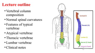
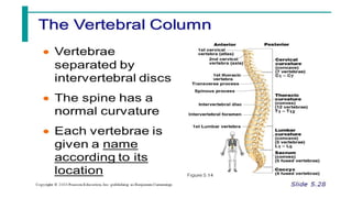
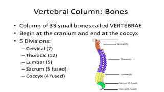
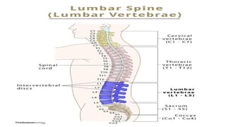
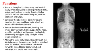


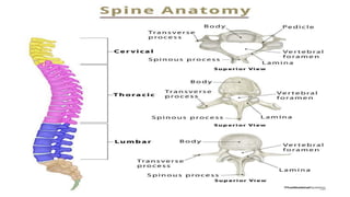
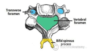
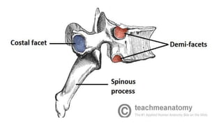
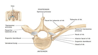
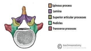
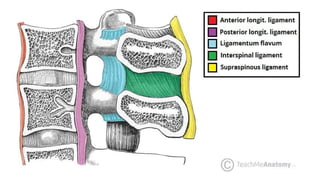
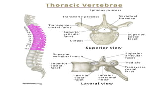
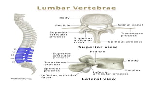
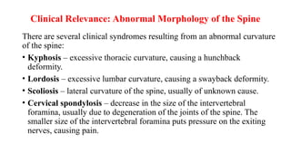
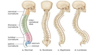
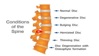
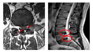

The document outlines the anatomy of the vertebral column, focusing on the thoracic and lumbar sections, detailing the composition, normal spinal curvatures, and the features of typical and atypical vertebrae. It discusses the functions of the vertebral column, including protection of the spinal cord, weight bearing, and facilitating body movement, alongside clinical relevance related to spinal deformities like kyphosis, lordosis, and scoliosis. Additionally, it covers the intervertebral discs and the implications of herniation on spinal health.


