SlideShare a Scribd company logo
SURGICAL ONCOLOGY AND RECONSTRUCTION
Two-Step Reconstruction of Non-Marginal
Auricular Defects
Maria Barbara Heinz, MD, DMD,* Hosein Ghanepur, DMD,y
and Alireza Ghassemi, MD, DMD, PhDz
Purpose: Auricular reconstruction is an extensively discussed topic in facial reconstructive surgery and
poses an immense challenge to the reconstructive surgeon. This report describes a 2-stage technique to
repair non-marginal full-thickness defects of the auricle.
Materials and Methods: Thirteen patients underwent surgery after partial to complete loss of the
auricular concha using an improved and refined method. Tissue from the pre- and retroauricular regions
was used to replace nonhelical auricular tissue loss in 2 surgical steps. All procedures were performed in
an ambulatory setting using local anesthesia.
Results: All 13 patients (age range, 37 to 82 yr; mean age, 68 yr; 4 women and 9 men) had excellent
esthetic outcomes with low surgical morbidity and were satisfied with the achieved results. No flap necro-
sis was observed. Auricular vertical and horizontal dimensions changed minimally (0 to 4 mm). A tension-
free closure of the donor-site defects could be achieved primarily.
Conclusion: The present method uses 2 separate donor sites to reconstruct centrally located
full-thickness defects of the auricle. It is straightforward to perform, minimizes the surgical steps required,
shows excellent outcomes, and allows easier closure of the donor site because of the distribution of the
harvested tissue.
Ó 2016 American Association of Oral and Maxillofacial Surgeons
J Oral Maxillofac Surg -:1-5, 2016
Defects of the auricle are often caused by trauma or
partial amputation after excision of cancerous
lesions.1
Of all carcinomas involving the ear, 45 to
55% are located on the helical rim and only few lesions
are located exclusively in the central part of
the auricle.2
As a protruded part of the facial soft tissue envelope,
the ear plays a crucial role in the overall esthetics of
the face. Even minor asymmetry in the size, shape,
color, surface, and outer contours can considerably
affect the overall appearance and bring about unpleas-
ant psychological distress in patients.3
Therefore,
ear reconstruction has always been considered a
challenging intervention, which requires knowledge
of facial esthetics and adequate surgical skills.
The method selected should be tailored to the general
medical condition of the patient and the patient’s
esthetic goal. Moreover, the proposed operative steps
should be thoroughly planned with utmost regard to
the local and regional anatomy and accounting for
any possible complications.
Previously published reconstruction methods have
included primary closure of auricular defects and
different reconstructive procedures ranging from local
flaps in single or multiple stages, use of grafts, implant-
able materials, and an auricular prosthesis.4
In accor-
dance to the clinical abundance and distribution
of lesions across the ear, only a few reconstructive
*Resident, Department of Oral and Maxillofacial Surgery,
Bethesda Hospital M€onchengladbach, Academic Hospital of RWTH
Aachen, M€onchengladbach, Germany.
yResident, Department of Oral and Maxillofacial Surgery, Babol
University of Medical Sciences, Babol, Iran.
zConsultant, Department of Oral and Maxillofacial Surgery,
Hospital Detmold, Academic Hospital of the University of
Hannover, Detmold; Medical Faculty University RWTH Aachen,
Aachen, Germany.
Address correspondence and reprint requests to Dr Heinz:
Department of Oral and Maxillofacial Surgery, Bethesda Hospital
M€onchengladbach, Academic Hospital of RWTH Aachen, Ludwig-
Weber-Straße 15, 41061 M€onchengladbach, Germany; e-mail:
mbheinz@gmx.net
Received December 15 2015
Accepted January 15 2016
Ó 2016 American Association of Oral and Maxillofacial Surgeons
0278-2391/16/00117-8
http://dx.doi.org/10.1016/j.joms.2016.01.025
1
methods have been described for the surgical recon-
struction of centrally located non-marginal defects of
the auricle.5,6
Even fewer reports have evaluated the
treatment of full-thickness defects of this region. In
consequence, reconstruction of these infrequent
defects is very important.
The anterior pedicled retroauricular flap (APRF)
was first described in 2012.7,8
In 2015, the authors
introduced the APRF for the repair of centrally
located perforating defects. The described method
needed 3 surgical stages.9
Since then, the method
has been refined for centrally located perforating
defects and the APRF was combined with a flap from
the preauricular region, the cranial pedicled preauric-
ular flap (CPPF). The present report describes the
technique of 2-step reconstruction and the complica-
tions, changes in auricular size, esthetic outcome,
and patient satisfaction.
Materials and Methods
Thirteenpatientswithcentrallylocatedfull-thickness
auricle defects were treated. Defect size ranged from 10
to 40 mm in diameter. Vertical and transversal dimen-
sions were measured pre- and postoperatively. The
reconstruction procedures were performed in 2 steps
in ambulatory settings using local anesthesia. A CPPF
was used to cover the anterior surface and an APRF
was used for the posterior defect site.
Surgical interventions were carried out in compli-
ance with the World Medical Association Declaration
of Helsinki on medical research protocols and ethics.
SURGICAL PROCEDURE
A precisely defined donor-site area, mirroring the
size of the existing defect, is outlined on the preauric-
ular and retroauricular skin (Figs 1A, 2A). First, the
FIGURE 1. Illustrations of the reconstruction procedure after full-thickness excision of the central part of the auricle. A, Designing the flaps in
the pre- and retroauricular regions. B, Harvesting and transposition of the anterior pedicled retroauricular flap into the defect area to reconstruct
the dorsal surface of the auricle. C, Incision and transposition of the cranial pedicled preauricular flap to reconstruct the anterior surface of the
auricle. The donor sites are closed primarily. D, Separation of the 2 pedicles and adaptation of the flaps in the second step.
Heinz, Ghanepur, and Ghassemi. Reconstruction of Auricular Defects. J Oral Maxillofac Surg 2016.
2 RECONSTRUCTION OF AURICULAR DEFECTS
dorsal incision is performed to elevate an APRF from
the retroauricular region. Then, the posterior tip of
the flap is folded into the anterior edge of the dorsal
defect to replace the dorsal surface (Fig 1B). As a
result, the subcutaneous part of the APRF is seen ante-
riorly. Second, the skin of the preauricular region is
incised with the pedicle based cranially. The skin is
used to reconstruct the anterior surface of the ear
(Fig 1C). The pre- and retroauricular donor sites are
closed primarily without tension (Fig 2B). After
2 weeks, the pedicles of the 2 flaps can be separated
surgically and adjusted accordingly (Fig 1D).
Results
Tissue from the pre- and retroauricular regions was
used to reconstruct full-thickness conchal defects in
13 patients (age range, 37 to 82 yr; mean age, 68 yr;
4 women and 9men). Postoperatively, vertical and hor-
izontal decreases in auricle size were 0 to 4 mm
(Table 1). Flap necrosis or flap loss, clinically notice-
able hematoma, or wound infection were not
observed. Scars were placed in hidden regions and
were barely visible. Skin color, texture, and thickness
of the reconstructed part matched well with the sur-
rounding tissue (Figs 2C, D). Tension-free wound
closure of the donor sites was achieved by the distribu-
tion of the needed skin into 2 separate regions. All 13
reconstructive treatments could be performed in 2 sur-
gical steps. Esthetic results were excellent and all pa-
tients were pleased with the outcome.
Discussion
The authors have presented a method to recon-
struct auricular conchal defects using the APRF
combined with a CPPF. Their aim was to maintain
the original size of the ear, achieve an optimal esthetic
outcome, distribute the donor-site defect wound, and
shorten the treatment time needed for reconstruction
FIGURE 2. Photographs of an 82-year-old patient after excision of a basal cell carcinoma of the central part of the left ear. A, Full-thickness
defect of the antihelix (diameter, 3.5 Â 3.0 cm) (posterior view). B, The anterior pedicled retroauricular and cranial pedicled preauricular flaps
are adapted. The 2 donor sites are closed primarily. (Fig 2 continued on next page.)
Heinz, Ghanepur, and Ghassemi. Reconstruction of Auricular Defects. J Oral Maxillofac Surg 2016.
HEINZ, GHANEPUR, AND GHASSEMI 3
(from 3 to 2 surgical steps). The CPPF was used to
cover the anterior surface of the defect. Because of
the similarity in texture, this tissue replaced the ante-
rior surface more adequately. The posterior surface
was reconstructed by the APRF, which consists of
thicker skin from the retroauricular region and offers
greater stability. In contrast to the previous technique,
there was no need for dividing any transitional part of
the APRF after passing the edge of the cartilaginous
framework, which would require another step.9
Because the tissue needed originated from 2 separate
regions, the size of the resulting defect of each donor
site was decreased and could be closed primarily and
without tension. The resultant scars were located in
invisible areas.
The goal of reconstructive surgery of the auricle is
to achieve morphologically acceptable auricular size
and shape and proportionally integrated anatomic
landmarks. Moreover, the proposed reconstructive
procedure should consider the patient’s existing
medical condition and should aim for a positive image
by achieving an adequate esthetic outcome. The first
reported method used a composite graft from the
contralateral ear.10
Since then, different methods
have been developed. Many reported technical
methods involve reconstruction of the helical rim,11-
17
because it is the prominent part of the ear and
most neoplastic lesions are found in this region.
Although some investigators have suggested
reconstructive techniques of central nonperforating
defects of the ear,4,18-21
only a few methods have
described full-thickness reconstruction of this region.
Nevertheless, most of these described methods result
in visible deformities and a smaller ear overall.22
In summary, there is a lack in the literature of tech-
nical reconstruction methods to close full-thickness
nonhelical defects of the ear in a convenient way
without changing the size. In their previous report,
the authors introduced a reconstruction technique
using the APRF, which required 3 steps. To decrease
the stages, the authors refined their technique and
used tissue from 2 separate pre- and retroauricular
donor sites for reconstruction. Because of the distribu-
tion of the donor-site defect, the present technique
allowed tension-free wound closure of the 2 donor
regions. In addition, sufficient tissue was available to
FIGURE 2 (cont’d). C, Frontal view of the patient 7 months postoperatively. D, Posterior aspect of the patient during follow-up period. Overall
size and shape of the reconstructed ear matches the contralateral side.
Heinz, Ghanepur, and Ghassemi. Reconstruction of Auricular Defects. J Oral Maxillofac Surg 2016.
4 RECONSTRUCTION OF AURICULAR DEFECTS
reconstruct the complete non-marginal part of the
auricle easily without obvious decrease of auricular
size. Although the method required 2 surgical steps,
it was reliable for flap survival. Esthetic outcome
was excellent because of the similarity of tissue
texture between the preauricular region and the
anterior surface of the auricle. Moreover, the thick
skin from the retroauricular region offered sufficient
stability and there was no need for cartilage grafting.
The new method was associated with improved
patient satisfaction because of fewer surgical steps
and shorter reconstruction time.
Combining flaps from pre- and retroauricular
regions simplified the full-thickness reconstruction
of non-marginal auricular defects, resulting in minimal
horizontal and vertical size changes. Because of the
distribution of required tissue into 2 anatomically
separate regions, primary wound closure was easier
to achieve. However, 2 surgical steps were required.
Acknowledgment
The authors thank Mr Wolfgang Graulich, from the Institute of
Anatomy, RWTH Aachen, for his contribution in creating and permit-
ting the use of his illustrations.
References
1. Antia NH, Busch V: Chondrocutaneous advancement flap for
the marginal defect of the ear. Plast Reconstr Surg 39:472,
1967
2. Leferink VJ, Nicolai JP: Malignant tumors of the external ear. Ann
Plast Surg 21:550, 1988
3. Hyckel P, Robotta C, Schumann D: Partial loss of the auricle:
Multiphase reconstruction and complete preservation of the
helix. Mund Kiefer Gesichtschir 3:131, 1999
4. Mellette JR: Ear reconstruction with local flaps. J Dermatol Surg
Oncol 17:176, 1991
5. Elsahy NI: Ear reconstruction with rotation-advancement com-
posite flaps. Plast Reconstr Surg 75:567, 1985
6. Elsahy NI: Reconstruction of the ear after skin and perichon-
drium loss. Clin Plast Surg 29:187, 2002
7. Stiller MB, Gerressen M, Modabber A, et al: Anteriorly pedicled
retroauricular flap for repair of auricular defects. Aesthetic Plast
Surg 36:623, 2012
8. Ghassemi A, Modabber A, Talebzadeh M, et al: Surgical
management of auricular defect depending on the size,
location, and tissue involved. J Oral Maxillofac Surg 71:
232, 2013
9. Heinz MB, H€olzle F, Ghassemi A: Repairing a non-marginal full-
thickness auricular defect using a reversed flap from the postaur-
icular area. J Oral Maxillofac Surg 73:764, 2015
10. Pegram M, Peterson R: Repair of partial defects of the ear. Plast
Reconstr Surg 18:305, 1956
11. Butler C: Extended retroauricular advancement flap reconstruc-
tion of a full-thickness auricular defect including posteromedial
and retroauricular skin. Ann Plast Surg 49:317, 2002
12. Goldberg LH, Maulsin DV, Humphreys TR: The postauricular
cutaneous advancement flap for repairing ear rim defects. Der-
matol Surg 22:28, 1995
13. Fata JJ: Composite chondrocutaneous advancement flap: A tech-
nique for reconstruction of marginal defects of the ear. Plast Re-
constr Surg 99:1172, 1997
14. Johnson TM, Fader DJ, Arbor A: The staged retroauricular to
auricular direct pedicle (interpolation) flap for helical ear recon-
struction. J Am Acad Dermatol 37:975, 1997
15. Kaminsky A: Repair of the partial loss of the helix. Aesthetic
Plast Surg 21:427, 1997
16. Lewin ML: Reconstruction of the helix. Arch Otolaryngol 47:
802, 1948
17. Stefanoff DN: Retroauricular tubed skin flap to the helical rim, in
Strauch B, Vasconez LO, Hall-Findlay EJ (eds): Grabb’s Encyclo-
pedia of Flaps. Volume 1, Head and Neck. Boston, MA, Little
Brown & Co, 1990, pp 295–297
18. Chen C, Chen ZJ: Reconstruction of the concha of the ear
using a postauricular island flap. Plast Reconstr Surg 86:569,
1990
19. Cordova A, D’Arpa S, Pirrello R, et al: Retroauricular skin: A flaps
bank for ear reconstruction. J Plast Reconstr Aesthet Surg 61:44,
2008
20. Ohsumi N, Iida N: Ear reconstruction with chondrocutane-
ous postauricular island flap. Plast Reconstr Surg 96:718,
1995
21. Renard A: Postauricular flap based on a dermal pedicle for ear
reconstruction. Plast Reconstr Surg 68:159, 1981
22. Ramirez OM, Heckler FR: Reconstruction of nonmarginal ear de-
fects with chondrocutaneous advancement flaps. Plast Reconstr
Surg 84:32, 1989
Table 1. AURICULAR DEFECT SIZE COMPARED WITH
POSTOPERATIVE DECREASE OF VERTICAL AND
HORIZONTAL AURICULAR DIMENSIONS
Patient
Number
Defect
Size (mm)
Postoperative Decrease (mm)
Vertical Horizontal
1 18 3 2
2 25 2 2
3 40 4 3
4 32 3 3
5 23 3 3
6 35 4 3
7 10 1 0
8 28 2 1
9 36 4 1
10 20 2 1
11 30 3 4
12 24 4 3
13 26 2 2
Heinz, Ghanepur, and Ghassemi. Reconstruction of Auricular De-
fects. J Oral Maxillofac Surg 2016.
HEINZ, GHANEPUR, AND GHASSEMI 5

More Related Content

What's hot

Microsurgery
MicrosurgeryMicrosurgery
Microsurgery
OpenWideGroup
 
DO for osa
DO for osaDO for osa
DO for osa
Dr. SHEETAL KAPSE
 
Dr Rahul VC Tiwari - Novel Transoral Approach to the Posterolateral Maxilla a...
Dr Rahul VC Tiwari - Novel Transoral Approach to the Posterolateral Maxilla a...Dr Rahul VC Tiwari - Novel Transoral Approach to the Posterolateral Maxilla a...
Dr Rahul VC Tiwari - Novel Transoral Approach to the Posterolateral Maxilla a...
CLOVE Dental OMNI Hospitals Andhra Hospital
 
Zygomaticomaxillary buttress as a donor site
Zygomaticomaxillary buttress as a donor siteZygomaticomaxillary buttress as a donor site
Zygomaticomaxillary buttress as a donor siteDr. SHEETAL KAPSE
 
Modified preauricular approach for treating intracapsular condylar fractures ...
Modified preauricular approach for treating intracapsular condylar fractures ...Modified preauricular approach for treating intracapsular condylar fractures ...
Modified preauricular approach for treating intracapsular condylar fractures ...
Dr. SHEETAL KAPSE
 
How do bisphosphonated affect # healing
How do bisphosphonated affect # healingHow do bisphosphonated affect # healing
How do bisphosphonated affect # healing
Dr. SHEETAL KAPSE
 
Journal Club On Pre-extractive Interradicular Implant Bed Preparation: Case P...
Journal Club On Pre-extractive Interradicular Implant Bed Preparation: Case P...Journal Club On Pre-extractive Interradicular Implant Bed Preparation: Case P...
Journal Club On Pre-extractive Interradicular Implant Bed Preparation: Case P...
Shilpa Shiv
 
Creating papilla implant (dentalxp) by Stuart
Creating papilla implant (dentalxp) by StuartCreating papilla implant (dentalxp) by Stuart
Creating papilla implant (dentalxp) by Stuart
Muaiyed Mahmoud Buzayan
 
Effect of Calcium Hydroxide on Deep Caries Dentin: A Clinical Study
Effect of Calcium Hydroxide on Deep Caries Dentin: A Clinical StudyEffect of Calcium Hydroxide on Deep Caries Dentin: A Clinical Study
Effect of Calcium Hydroxide on Deep Caries Dentin: A Clinical Study
asclepiuspdfs
 
Dr. Rahul VC Tiwari - Fellowship In Orthognathic Surgery - Jubilee Mission Me...
Dr. Rahul VC Tiwari - Fellowship In Orthognathic Surgery - Jubilee Mission Me...Dr. Rahul VC Tiwari - Fellowship In Orthognathic Surgery - Jubilee Mission Me...
Dr. Rahul VC Tiwari - Fellowship In Orthognathic Surgery - Jubilee Mission Me...
CLOVE Dental OMNI Hospitals Andhra Hospital
 
Surgical Endodontics
Surgical Endodontics Surgical Endodontics
Surgical Endodontics
DrOsamaMushtaq
 
Journal Club on Clinical comparison of ultrasonic surgery and conventional su...
Journal Club on Clinical comparison of ultrasonic surgery and conventional su...Journal Club on Clinical comparison of ultrasonic surgery and conventional su...
Journal Club on Clinical comparison of ultrasonic surgery and conventional su...
Dr Bhavik Miyani
 
Analysis of buccolingual dimensional changes of the extraction socket using t...
Analysis of buccolingual dimensional changes of the extraction socket using t...Analysis of buccolingual dimensional changes of the extraction socket using t...
Analysis of buccolingual dimensional changes of the extraction socket using t...
MD Abdul Haleem
 
Assessment of lingual nerve injury using different surgical variables for man...
Assessment of lingual nerve injury using different surgical variables for man...Assessment of lingual nerve injury using different surgical variables for man...
Assessment of lingual nerve injury using different surgical variables for man...
DrKamini Dadsena
 
Akhil jc expandable
Akhil jc expandableAkhil jc expandable
Akhil jc expandable
Akhil Sankar
 
CARNOY’S SOLUTION AS A SURGICAL MEDICAMENT IN THE TREATMENT OF KERATOCYSTIC O...
CARNOY’S SOLUTION AS A SURGICAL MEDICAMENT IN THETREATMENT OF KERATOCYSTIC O...CARNOY’S SOLUTION AS A SURGICAL MEDICAMENT IN THETREATMENT OF KERATOCYSTIC O...
CARNOY’S SOLUTION AS A SURGICAL MEDICAMENT IN THE TREATMENT OF KERATOCYSTIC O...
DrKamini Dadsena
 
Prosthodontics Journal Club: Altered cast impression techniques
Prosthodontics Journal Club: Altered cast impression techniquesProsthodontics Journal Club: Altered cast impression techniques
Prosthodontics Journal Club: Altered cast impression techniques
NeerajaMenon4
 
Journal Club On Combination Flap Surgery with Resin-Modified Glass Ionomer fo...
Journal Club On Combination Flap Surgery with Resin-Modified Glass Ionomer fo...Journal Club On Combination Flap Surgery with Resin-Modified Glass Ionomer fo...
Journal Club On Combination Flap Surgery with Resin-Modified Glass Ionomer fo...
Shilpa Shiv
 
Entire papilla preservation technique in the regenerative treatment of deep i...
Entire papilla preservation technique in the regenerative treatment of deep i...Entire papilla preservation technique in the regenerative treatment of deep i...
Entire papilla preservation technique in the regenerative treatment of deep i...
MD Abdul Haleem
 
Case of bilateral tmj dislocation
Case of bilateral tmj dislocationCase of bilateral tmj dislocation
Case of bilateral tmj dislocation
Dr Bhavik Miyani
 

What's hot (20)

Microsurgery
MicrosurgeryMicrosurgery
Microsurgery
 
DO for osa
DO for osaDO for osa
DO for osa
 
Dr Rahul VC Tiwari - Novel Transoral Approach to the Posterolateral Maxilla a...
Dr Rahul VC Tiwari - Novel Transoral Approach to the Posterolateral Maxilla a...Dr Rahul VC Tiwari - Novel Transoral Approach to the Posterolateral Maxilla a...
Dr Rahul VC Tiwari - Novel Transoral Approach to the Posterolateral Maxilla a...
 
Zygomaticomaxillary buttress as a donor site
Zygomaticomaxillary buttress as a donor siteZygomaticomaxillary buttress as a donor site
Zygomaticomaxillary buttress as a donor site
 
Modified preauricular approach for treating intracapsular condylar fractures ...
Modified preauricular approach for treating intracapsular condylar fractures ...Modified preauricular approach for treating intracapsular condylar fractures ...
Modified preauricular approach for treating intracapsular condylar fractures ...
 
How do bisphosphonated affect # healing
How do bisphosphonated affect # healingHow do bisphosphonated affect # healing
How do bisphosphonated affect # healing
 
Journal Club On Pre-extractive Interradicular Implant Bed Preparation: Case P...
Journal Club On Pre-extractive Interradicular Implant Bed Preparation: Case P...Journal Club On Pre-extractive Interradicular Implant Bed Preparation: Case P...
Journal Club On Pre-extractive Interradicular Implant Bed Preparation: Case P...
 
Creating papilla implant (dentalxp) by Stuart
Creating papilla implant (dentalxp) by StuartCreating papilla implant (dentalxp) by Stuart
Creating papilla implant (dentalxp) by Stuart
 
Effect of Calcium Hydroxide on Deep Caries Dentin: A Clinical Study
Effect of Calcium Hydroxide on Deep Caries Dentin: A Clinical StudyEffect of Calcium Hydroxide on Deep Caries Dentin: A Clinical Study
Effect of Calcium Hydroxide on Deep Caries Dentin: A Clinical Study
 
Dr. Rahul VC Tiwari - Fellowship In Orthognathic Surgery - Jubilee Mission Me...
Dr. Rahul VC Tiwari - Fellowship In Orthognathic Surgery - Jubilee Mission Me...Dr. Rahul VC Tiwari - Fellowship In Orthognathic Surgery - Jubilee Mission Me...
Dr. Rahul VC Tiwari - Fellowship In Orthognathic Surgery - Jubilee Mission Me...
 
Surgical Endodontics
Surgical Endodontics Surgical Endodontics
Surgical Endodontics
 
Journal Club on Clinical comparison of ultrasonic surgery and conventional su...
Journal Club on Clinical comparison of ultrasonic surgery and conventional su...Journal Club on Clinical comparison of ultrasonic surgery and conventional su...
Journal Club on Clinical comparison of ultrasonic surgery and conventional su...
 
Analysis of buccolingual dimensional changes of the extraction socket using t...
Analysis of buccolingual dimensional changes of the extraction socket using t...Analysis of buccolingual dimensional changes of the extraction socket using t...
Analysis of buccolingual dimensional changes of the extraction socket using t...
 
Assessment of lingual nerve injury using different surgical variables for man...
Assessment of lingual nerve injury using different surgical variables for man...Assessment of lingual nerve injury using different surgical variables for man...
Assessment of lingual nerve injury using different surgical variables for man...
 
Akhil jc expandable
Akhil jc expandableAkhil jc expandable
Akhil jc expandable
 
CARNOY’S SOLUTION AS A SURGICAL MEDICAMENT IN THE TREATMENT OF KERATOCYSTIC O...
CARNOY’S SOLUTION AS A SURGICAL MEDICAMENT IN THETREATMENT OF KERATOCYSTIC O...CARNOY’S SOLUTION AS A SURGICAL MEDICAMENT IN THETREATMENT OF KERATOCYSTIC O...
CARNOY’S SOLUTION AS A SURGICAL MEDICAMENT IN THE TREATMENT OF KERATOCYSTIC O...
 
Prosthodontics Journal Club: Altered cast impression techniques
Prosthodontics Journal Club: Altered cast impression techniquesProsthodontics Journal Club: Altered cast impression techniques
Prosthodontics Journal Club: Altered cast impression techniques
 
Journal Club On Combination Flap Surgery with Resin-Modified Glass Ionomer fo...
Journal Club On Combination Flap Surgery with Resin-Modified Glass Ionomer fo...Journal Club On Combination Flap Surgery with Resin-Modified Glass Ionomer fo...
Journal Club On Combination Flap Surgery with Resin-Modified Glass Ionomer fo...
 
Entire papilla preservation technique in the regenerative treatment of deep i...
Entire papilla preservation technique in the regenerative treatment of deep i...Entire papilla preservation technique in the regenerative treatment of deep i...
Entire papilla preservation technique in the regenerative treatment of deep i...
 
Case of bilateral tmj dislocation
Case of bilateral tmj dislocationCase of bilateral tmj dislocation
Case of bilateral tmj dislocation
 

Similar to 2016 heinz-two-step reconstruction of non-marginal auricular defects

2013 gerressen-micro sug
2013 gerressen-micro sug2013 gerressen-micro sug
2013 gerressen-micro sug
Klinikum Lippe GmbH
 
Anaplastology dr jehad al sukhun
Anaplastology  dr jehad al sukhunAnaplastology  dr jehad al sukhun
Anaplastology dr jehad al sukhun
jehadsukhun
 
Preventing elevated radix deformity in asian
Preventing elevated radix deformity in asianPreventing elevated radix deformity in asian
Preventing elevated radix deformity in asian
Nhat Nguyen
 
538-Article Text-7432-1-10-202000805.pdf
538-Article Text-7432-1-10-202000805.pdf538-Article Text-7432-1-10-202000805.pdf
538-Article Text-7432-1-10-202000805.pdf
Ankita Gurav
 
Salivary glands anatomy applied aspects 140608050047-phpapp01
Salivary glands anatomy applied aspects 140608050047-phpapp01Salivary glands anatomy applied aspects 140608050047-phpapp01
Salivary glands anatomy applied aspects 140608050047-phpapp01
Abhishek PT
 
2016 movahedian- acellular-dermal-graft
2016 movahedian- acellular-dermal-graft2016 movahedian- acellular-dermal-graft
2016 movahedian- acellular-dermal-graft
Klinikum Lippe GmbH
 
Connective tissue grafts
Connective tissue grafts Connective tissue grafts
Connective tissue grafts
Raveena Bhanushali
 
estlander.flap.ppt
estlander.flap.pptestlander.flap.ppt
estlander.flap.ppt
ContactNovaderm
 
Acs0205 Oral Cavity Procedures
Acs0205 Oral Cavity ProceduresAcs0205 Oral Cavity Procedures
Acs0205 Oral Cavity Proceduresmedbookonline
 
2014 ghassemi-u-considering the f
2014 ghassemi-u-considering the f2014 ghassemi-u-considering the f
2014 ghassemi-u-considering the f
Klinikum Lippe GmbH
 
48th Publication- JDHR-3rd Name.pdf
48th Publication- JDHR-3rd Name.pdf48th Publication- JDHR-3rd Name.pdf
48th Publication- JDHR-3rd Name.pdf
CLOVE Dental OMNI Hospitals Andhra Hospital
 
Cancer of Oral Cavity Abutting the Mandible; Predictors of Loco-regional Fail...
Cancer of Oral Cavity Abutting the Mandible; Predictors of Loco-regional Fail...Cancer of Oral Cavity Abutting the Mandible; Predictors of Loco-regional Fail...
Cancer of Oral Cavity Abutting the Mandible; Predictors of Loco-regional Fail...
Dr./ Ihab Samy
 
Short nose correction_suh_mk
Short nose correction_suh_mkShort nose correction_suh_mk
Short nose correction_suh_mk
Man Koon SUH
 
Short nose correction_by Man Koon Suh
Short nose correction_by Man Koon SuhShort nose correction_by Man Koon Suh
Short nose correction_by Man Koon Suh
韩国JW整形医院
 
2016 bartella-face-lift-gesichtsrekonstruktion-2
2016 bartella-face-lift-gesichtsrekonstruktion-22016 bartella-face-lift-gesichtsrekonstruktion-2
2016 bartella-face-lift-gesichtsrekonstruktion-2
Klinikum Lippe GmbH
 
74th Publication- JPBS- 7th Name.pdf
74th Publication- JPBS- 7th Name.pdf74th Publication- JPBS- 7th Name.pdf
74th Publication- JPBS- 7th Name.pdf
CLOVE Dental OMNI Hospitals Andhra Hospital
 
connectivetissuegraftswithnonincisedpapillaesurgicalapproachforperiodontalrec...
connectivetissuegraftswithnonincisedpapillaesurgicalapproachforperiodontalrec...connectivetissuegraftswithnonincisedpapillaesurgicalapproachforperiodontalrec...
connectivetissuegraftswithnonincisedpapillaesurgicalapproachforperiodontalrec...
MohammadEissaAhmadi
 
Further experience with the double onlay preputial flap
Further experience with the double onlay preputial flapFurther experience with the double onlay preputial flap
Further experience with the double onlay preputial flap
asopahospital
 
model planing mock up for orthognathic surgery
 model planing mock up for orthognathic surgery  model planing mock up for orthognathic surgery
model planing mock up for orthognathic surgery
bilal falahi
 
Forehead Reconstruction Using an A to T
Forehead Reconstruction Using an A to TForehead Reconstruction Using an A to T
Forehead Reconstruction Using an A to T
W. Thomas McClellan, MD FACS
 

Similar to 2016 heinz-two-step reconstruction of non-marginal auricular defects (20)

2013 gerressen-micro sug
2013 gerressen-micro sug2013 gerressen-micro sug
2013 gerressen-micro sug
 
Anaplastology dr jehad al sukhun
Anaplastology  dr jehad al sukhunAnaplastology  dr jehad al sukhun
Anaplastology dr jehad al sukhun
 
Preventing elevated radix deformity in asian
Preventing elevated radix deformity in asianPreventing elevated radix deformity in asian
Preventing elevated radix deformity in asian
 
538-Article Text-7432-1-10-202000805.pdf
538-Article Text-7432-1-10-202000805.pdf538-Article Text-7432-1-10-202000805.pdf
538-Article Text-7432-1-10-202000805.pdf
 
Salivary glands anatomy applied aspects 140608050047-phpapp01
Salivary glands anatomy applied aspects 140608050047-phpapp01Salivary glands anatomy applied aspects 140608050047-phpapp01
Salivary glands anatomy applied aspects 140608050047-phpapp01
 
2016 movahedian- acellular-dermal-graft
2016 movahedian- acellular-dermal-graft2016 movahedian- acellular-dermal-graft
2016 movahedian- acellular-dermal-graft
 
Connective tissue grafts
Connective tissue grafts Connective tissue grafts
Connective tissue grafts
 
estlander.flap.ppt
estlander.flap.pptestlander.flap.ppt
estlander.flap.ppt
 
Acs0205 Oral Cavity Procedures
Acs0205 Oral Cavity ProceduresAcs0205 Oral Cavity Procedures
Acs0205 Oral Cavity Procedures
 
2014 ghassemi-u-considering the f
2014 ghassemi-u-considering the f2014 ghassemi-u-considering the f
2014 ghassemi-u-considering the f
 
48th Publication- JDHR-3rd Name.pdf
48th Publication- JDHR-3rd Name.pdf48th Publication- JDHR-3rd Name.pdf
48th Publication- JDHR-3rd Name.pdf
 
Cancer of Oral Cavity Abutting the Mandible; Predictors of Loco-regional Fail...
Cancer of Oral Cavity Abutting the Mandible; Predictors of Loco-regional Fail...Cancer of Oral Cavity Abutting the Mandible; Predictors of Loco-regional Fail...
Cancer of Oral Cavity Abutting the Mandible; Predictors of Loco-regional Fail...
 
Short nose correction_suh_mk
Short nose correction_suh_mkShort nose correction_suh_mk
Short nose correction_suh_mk
 
Short nose correction_by Man Koon Suh
Short nose correction_by Man Koon SuhShort nose correction_by Man Koon Suh
Short nose correction_by Man Koon Suh
 
2016 bartella-face-lift-gesichtsrekonstruktion-2
2016 bartella-face-lift-gesichtsrekonstruktion-22016 bartella-face-lift-gesichtsrekonstruktion-2
2016 bartella-face-lift-gesichtsrekonstruktion-2
 
74th Publication- JPBS- 7th Name.pdf
74th Publication- JPBS- 7th Name.pdf74th Publication- JPBS- 7th Name.pdf
74th Publication- JPBS- 7th Name.pdf
 
connectivetissuegraftswithnonincisedpapillaesurgicalapproachforperiodontalrec...
connectivetissuegraftswithnonincisedpapillaesurgicalapproachforperiodontalrec...connectivetissuegraftswithnonincisedpapillaesurgicalapproachforperiodontalrec...
connectivetissuegraftswithnonincisedpapillaesurgicalapproachforperiodontalrec...
 
Further experience with the double onlay preputial flap
Further experience with the double onlay preputial flapFurther experience with the double onlay preputial flap
Further experience with the double onlay preputial flap
 
model planing mock up for orthognathic surgery
 model planing mock up for orthognathic surgery  model planing mock up for orthognathic surgery
model planing mock up for orthognathic surgery
 
Forehead Reconstruction Using an A to T
Forehead Reconstruction Using an A to TForehead Reconstruction Using an A to T
Forehead Reconstruction Using an A to T
 

More from Klinikum Lippe GmbH

Kongressplakat pathologie lunge recurrent pleural effusions_prof. hansen
Kongressplakat pathologie lunge recurrent pleural effusions_prof. hansenKongressplakat pathologie lunge recurrent pleural effusions_prof. hansen
Kongressplakat pathologie lunge recurrent pleural effusions_prof. hansen
Klinikum Lippe GmbH
 
Gunnemann harnleiterstenose v2
Gunnemann harnleiterstenose v2Gunnemann harnleiterstenose v2
Gunnemann harnleiterstenose v2
Klinikum Lippe GmbH
 
Kongressplakat durasinusmalformation kinderklinik zurebiani
Kongressplakat durasinusmalformation kinderklinik zurebianiKongressplakat durasinusmalformation kinderklinik zurebiani
Kongressplakat durasinusmalformation kinderklinik zurebiani
Klinikum Lippe GmbH
 
Kongressplakat innere amputation dermatologie quellmalz_din a0
Kongressplakat innere amputation dermatologie quellmalz_din a0Kongressplakat innere amputation dermatologie quellmalz_din a0
Kongressplakat innere amputation dermatologie quellmalz_din a0
Klinikum Lippe GmbH
 
Kongressplakat pathologie dworak grading system prof. hansen_din a0
Kongressplakat pathologie dworak grading system prof. hansen_din a0Kongressplakat pathologie dworak grading system prof. hansen_din a0
Kongressplakat pathologie dworak grading system prof. hansen_din a0
Klinikum Lippe GmbH
 
2018 kleinfeld-speech-paper-1
2018 kleinfeld-speech-paper-12018 kleinfeld-speech-paper-1
2018 kleinfeld-speech-paper-1
Klinikum Lippe GmbH
 
2018 ghassemi-parotis-bjoms
2018 ghassemi-parotis-bjoms2018 ghassemi-parotis-bjoms
2018 ghassemi-parotis-bjoms
Klinikum Lippe GmbH
 
2018 ghaneh-compsarsion
2018 ghaneh-compsarsion2018 ghaneh-compsarsion
2018 ghaneh-compsarsion
Klinikum Lippe GmbH
 
2018 behrens-patient-spezcific
2018 behrens-patient-spezcific2018 behrens-patient-spezcific
2018 behrens-patient-spezcific
Klinikum Lippe GmbH
 
2017 modabber-medial-approach-dcia-cad-cam
2017 modabber-medial-approach-dcia-cad-cam2017 modabber-medial-approach-dcia-cad-cam
2017 modabber-medial-approach-dcia-cad-cam
Klinikum Lippe GmbH
 
2017 modabber-ear-aps
2017 modabber-ear-aps2017 modabber-ear-aps
2017 modabber-ear-aps
Klinikum Lippe GmbH
 
2017 jamilian-family history-cleft
2017 jamilian-family history-cleft2017 jamilian-family history-cleft
2017 jamilian-family history-cleft
Klinikum Lippe GmbH
 
2016 wahl-immunotherapy with imiquimod and interferon alfa for metastasized m...
2016 wahl-immunotherapy with imiquimod and interferon alfa for metastasized m...2016 wahl-immunotherapy with imiquimod and interferon alfa for metastasized m...
2016 wahl-immunotherapy with imiquimod and interferon alfa for metastasized m...
Klinikum Lippe GmbH
 
2016 ghassemi-maxillary advancement versus man
2016 ghassemi-maxillary advancement versus man2016 ghassemi-maxillary advancement versus man
2016 ghassemi-maxillary advancement versus man
Klinikum Lippe GmbH
 
2016 ghassemi-clinically-usable-fib-ilium
2016 ghassemi-clinically-usable-fib-ilium2016 ghassemi-clinically-usable-fib-ilium
2016 ghassemi-clinically-usable-fib-ilium
Klinikum Lippe GmbH
 
2015 ghassemi-maxillary advancment2015
2015 ghassemi-maxillary advancment20152015 ghassemi-maxillary advancment2015
2015 ghassemi-maxillary advancment2015
Klinikum Lippe GmbH
 
2015 ghassemi-histologie-vessel-pedicled
2015 ghassemi-histologie-vessel-pedicled2015 ghassemi-histologie-vessel-pedicled
2015 ghassemi-histologie-vessel-pedicled
Klinikum Lippe GmbH
 
2014 ghassemi-factor-influencing
2014 ghassemi-factor-influencing2014 ghassemi-factor-influencing
2014 ghassemi-factor-influencing
Klinikum Lippe GmbH
 
2013 teilseite 11za14- (1)
2013 teilseite 11za14- (1)2013 teilseite 11za14- (1)
2013 teilseite 11za14- (1)
Klinikum Lippe GmbH
 
2013 modabber-zygoma-reconstruction
2013 modabber-zygoma-reconstruction2013 modabber-zygoma-reconstruction
2013 modabber-zygoma-reconstruction
Klinikum Lippe GmbH
 

More from Klinikum Lippe GmbH (20)

Kongressplakat pathologie lunge recurrent pleural effusions_prof. hansen
Kongressplakat pathologie lunge recurrent pleural effusions_prof. hansenKongressplakat pathologie lunge recurrent pleural effusions_prof. hansen
Kongressplakat pathologie lunge recurrent pleural effusions_prof. hansen
 
Gunnemann harnleiterstenose v2
Gunnemann harnleiterstenose v2Gunnemann harnleiterstenose v2
Gunnemann harnleiterstenose v2
 
Kongressplakat durasinusmalformation kinderklinik zurebiani
Kongressplakat durasinusmalformation kinderklinik zurebianiKongressplakat durasinusmalformation kinderklinik zurebiani
Kongressplakat durasinusmalformation kinderklinik zurebiani
 
Kongressplakat innere amputation dermatologie quellmalz_din a0
Kongressplakat innere amputation dermatologie quellmalz_din a0Kongressplakat innere amputation dermatologie quellmalz_din a0
Kongressplakat innere amputation dermatologie quellmalz_din a0
 
Kongressplakat pathologie dworak grading system prof. hansen_din a0
Kongressplakat pathologie dworak grading system prof. hansen_din a0Kongressplakat pathologie dworak grading system prof. hansen_din a0
Kongressplakat pathologie dworak grading system prof. hansen_din a0
 
2018 kleinfeld-speech-paper-1
2018 kleinfeld-speech-paper-12018 kleinfeld-speech-paper-1
2018 kleinfeld-speech-paper-1
 
2018 ghassemi-parotis-bjoms
2018 ghassemi-parotis-bjoms2018 ghassemi-parotis-bjoms
2018 ghassemi-parotis-bjoms
 
2018 ghaneh-compsarsion
2018 ghaneh-compsarsion2018 ghaneh-compsarsion
2018 ghaneh-compsarsion
 
2018 behrens-patient-spezcific
2018 behrens-patient-spezcific2018 behrens-patient-spezcific
2018 behrens-patient-spezcific
 
2017 modabber-medial-approach-dcia-cad-cam
2017 modabber-medial-approach-dcia-cad-cam2017 modabber-medial-approach-dcia-cad-cam
2017 modabber-medial-approach-dcia-cad-cam
 
2017 modabber-ear-aps
2017 modabber-ear-aps2017 modabber-ear-aps
2017 modabber-ear-aps
 
2017 jamilian-family history-cleft
2017 jamilian-family history-cleft2017 jamilian-family history-cleft
2017 jamilian-family history-cleft
 
2016 wahl-immunotherapy with imiquimod and interferon alfa for metastasized m...
2016 wahl-immunotherapy with imiquimod and interferon alfa for metastasized m...2016 wahl-immunotherapy with imiquimod and interferon alfa for metastasized m...
2016 wahl-immunotherapy with imiquimod and interferon alfa for metastasized m...
 
2016 ghassemi-maxillary advancement versus man
2016 ghassemi-maxillary advancement versus man2016 ghassemi-maxillary advancement versus man
2016 ghassemi-maxillary advancement versus man
 
2016 ghassemi-clinically-usable-fib-ilium
2016 ghassemi-clinically-usable-fib-ilium2016 ghassemi-clinically-usable-fib-ilium
2016 ghassemi-clinically-usable-fib-ilium
 
2015 ghassemi-maxillary advancment2015
2015 ghassemi-maxillary advancment20152015 ghassemi-maxillary advancment2015
2015 ghassemi-maxillary advancment2015
 
2015 ghassemi-histologie-vessel-pedicled
2015 ghassemi-histologie-vessel-pedicled2015 ghassemi-histologie-vessel-pedicled
2015 ghassemi-histologie-vessel-pedicled
 
2014 ghassemi-factor-influencing
2014 ghassemi-factor-influencing2014 ghassemi-factor-influencing
2014 ghassemi-factor-influencing
 
2013 teilseite 11za14- (1)
2013 teilseite 11za14- (1)2013 teilseite 11za14- (1)
2013 teilseite 11za14- (1)
 
2013 modabber-zygoma-reconstruction
2013 modabber-zygoma-reconstruction2013 modabber-zygoma-reconstruction
2013 modabber-zygoma-reconstruction
 

Recently uploaded

Surat @ℂall @Girls ꧁❤8527049040❤꧂@ℂall @Girls Service Vip Top Model Safe
Surat @ℂall @Girls ꧁❤8527049040❤꧂@ℂall @Girls Service Vip Top Model SafeSurat @ℂall @Girls ꧁❤8527049040❤꧂@ℂall @Girls Service Vip Top Model Safe
Surat @ℂall @Girls ꧁❤8527049040❤꧂@ℂall @Girls Service Vip Top Model Safe
Savita Shen $i11
 
Are There Any Natural Remedies To Treat Syphilis.pdf
Are There Any Natural Remedies To Treat Syphilis.pdfAre There Any Natural Remedies To Treat Syphilis.pdf
Are There Any Natural Remedies To Treat Syphilis.pdf
Little Cross Family Clinic
 
Prix Galien International 2024 Forum Program
Prix Galien International 2024 Forum ProgramPrix Galien International 2024 Forum Program
Prix Galien International 2024 Forum Program
Levi Shapiro
 
Flu Vaccine Alert in Bangalore Karnataka
Flu Vaccine Alert in Bangalore KarnatakaFlu Vaccine Alert in Bangalore Karnataka
Flu Vaccine Alert in Bangalore Karnataka
addon Scans
 
Hemodialysis: Chapter 3, Dialysis Water Unit - Dr.Gawad
Hemodialysis: Chapter 3, Dialysis Water Unit - Dr.GawadHemodialysis: Chapter 3, Dialysis Water Unit - Dr.Gawad
Hemodialysis: Chapter 3, Dialysis Water Unit - Dr.Gawad
NephroTube - Dr.Gawad
 
Knee anatomy and clinical tests 2024.pdf
Knee anatomy and clinical tests 2024.pdfKnee anatomy and clinical tests 2024.pdf
Knee anatomy and clinical tests 2024.pdf
vimalpl1234
 
TEST BANK for Operations Management, 14th Edition by William J. Stevenson, Ve...
TEST BANK for Operations Management, 14th Edition by William J. Stevenson, Ve...TEST BANK for Operations Management, 14th Edition by William J. Stevenson, Ve...
TEST BANK for Operations Management, 14th Edition by William J. Stevenson, Ve...
kevinkariuki227
 
Pulmonary Thromboembolism - etilogy, types, medical- Surgical and nursing man...
Pulmonary Thromboembolism - etilogy, types, medical- Surgical and nursing man...Pulmonary Thromboembolism - etilogy, types, medical- Surgical and nursing man...
Pulmonary Thromboembolism - etilogy, types, medical- Surgical and nursing man...
VarunMahajani
 
New Drug Discovery and Development .....
New Drug Discovery and Development .....New Drug Discovery and Development .....
New Drug Discovery and Development .....
NEHA GUPTA
 
How to Give Better Lectures: Some Tips for Doctors
How to Give Better Lectures: Some Tips for DoctorsHow to Give Better Lectures: Some Tips for Doctors
How to Give Better Lectures: Some Tips for Doctors
LanceCatedral
 
Superficial & Deep Fascia of the NECK.pptx
Superficial & Deep Fascia of the NECK.pptxSuperficial & Deep Fascia of the NECK.pptx
Superficial & Deep Fascia of the NECK.pptx
Dr. Rabia Inam Gandapore
 
263778731218 Abortion Clinic /Pills In Harare ,
263778731218 Abortion Clinic /Pills In Harare ,263778731218 Abortion Clinic /Pills In Harare ,
263778731218 Abortion Clinic /Pills In Harare ,
sisternakatoto
 
Non-respiratory Functions of the Lungs.pdf
Non-respiratory Functions of the Lungs.pdfNon-respiratory Functions of the Lungs.pdf
Non-respiratory Functions of the Lungs.pdf
MedicoseAcademics
 
Alcohol_Dr. Jeenal Mistry MD Pharmacology.pdf
Alcohol_Dr. Jeenal Mistry MD Pharmacology.pdfAlcohol_Dr. Jeenal Mistry MD Pharmacology.pdf
Alcohol_Dr. Jeenal Mistry MD Pharmacology.pdf
Dr Jeenal Mistry
 
ACUTE SCROTUM.....pdf. ACUTE SCROTAL CONDITIOND
ACUTE SCROTUM.....pdf. ACUTE SCROTAL CONDITIONDACUTE SCROTUM.....pdf. ACUTE SCROTAL CONDITIOND
ACUTE SCROTUM.....pdf. ACUTE SCROTAL CONDITIOND
DR SETH JOTHAM
 
Ozempic: Preoperative Management of Patients on GLP-1 Receptor Agonists
Ozempic: Preoperative Management of Patients on GLP-1 Receptor Agonists  Ozempic: Preoperative Management of Patients on GLP-1 Receptor Agonists
Ozempic: Preoperative Management of Patients on GLP-1 Receptor Agonists
Saeid Safari
 
Pharynx and Clinical Correlations BY Dr.Rabia Inam Gandapore.pptx
Pharynx and Clinical Correlations BY Dr.Rabia Inam Gandapore.pptxPharynx and Clinical Correlations BY Dr.Rabia Inam Gandapore.pptx
Pharynx and Clinical Correlations BY Dr.Rabia Inam Gandapore.pptx
Dr. Rabia Inam Gandapore
 
New Directions in Targeted Therapeutic Approaches for Older Adults With Mantl...
New Directions in Targeted Therapeutic Approaches for Older Adults With Mantl...New Directions in Targeted Therapeutic Approaches for Older Adults With Mantl...
New Directions in Targeted Therapeutic Approaches for Older Adults With Mantl...
i3 Health
 
ANATOMY AND PHYSIOLOGY OF URINARY SYSTEM.pptx
ANATOMY AND PHYSIOLOGY OF URINARY SYSTEM.pptxANATOMY AND PHYSIOLOGY OF URINARY SYSTEM.pptx
ANATOMY AND PHYSIOLOGY OF URINARY SYSTEM.pptx
Swetaba Besh
 
Cervical & Brachial Plexus By Dr. RIG.pptx
Cervical & Brachial Plexus By Dr. RIG.pptxCervical & Brachial Plexus By Dr. RIG.pptx
Cervical & Brachial Plexus By Dr. RIG.pptx
Dr. Rabia Inam Gandapore
 

Recently uploaded (20)

Surat @ℂall @Girls ꧁❤8527049040❤꧂@ℂall @Girls Service Vip Top Model Safe
Surat @ℂall @Girls ꧁❤8527049040❤꧂@ℂall @Girls Service Vip Top Model SafeSurat @ℂall @Girls ꧁❤8527049040❤꧂@ℂall @Girls Service Vip Top Model Safe
Surat @ℂall @Girls ꧁❤8527049040❤꧂@ℂall @Girls Service Vip Top Model Safe
 
Are There Any Natural Remedies To Treat Syphilis.pdf
Are There Any Natural Remedies To Treat Syphilis.pdfAre There Any Natural Remedies To Treat Syphilis.pdf
Are There Any Natural Remedies To Treat Syphilis.pdf
 
Prix Galien International 2024 Forum Program
Prix Galien International 2024 Forum ProgramPrix Galien International 2024 Forum Program
Prix Galien International 2024 Forum Program
 
Flu Vaccine Alert in Bangalore Karnataka
Flu Vaccine Alert in Bangalore KarnatakaFlu Vaccine Alert in Bangalore Karnataka
Flu Vaccine Alert in Bangalore Karnataka
 
Hemodialysis: Chapter 3, Dialysis Water Unit - Dr.Gawad
Hemodialysis: Chapter 3, Dialysis Water Unit - Dr.GawadHemodialysis: Chapter 3, Dialysis Water Unit - Dr.Gawad
Hemodialysis: Chapter 3, Dialysis Water Unit - Dr.Gawad
 
Knee anatomy and clinical tests 2024.pdf
Knee anatomy and clinical tests 2024.pdfKnee anatomy and clinical tests 2024.pdf
Knee anatomy and clinical tests 2024.pdf
 
TEST BANK for Operations Management, 14th Edition by William J. Stevenson, Ve...
TEST BANK for Operations Management, 14th Edition by William J. Stevenson, Ve...TEST BANK for Operations Management, 14th Edition by William J. Stevenson, Ve...
TEST BANK for Operations Management, 14th Edition by William J. Stevenson, Ve...
 
Pulmonary Thromboembolism - etilogy, types, medical- Surgical and nursing man...
Pulmonary Thromboembolism - etilogy, types, medical- Surgical and nursing man...Pulmonary Thromboembolism - etilogy, types, medical- Surgical and nursing man...
Pulmonary Thromboembolism - etilogy, types, medical- Surgical and nursing man...
 
New Drug Discovery and Development .....
New Drug Discovery and Development .....New Drug Discovery and Development .....
New Drug Discovery and Development .....
 
How to Give Better Lectures: Some Tips for Doctors
How to Give Better Lectures: Some Tips for DoctorsHow to Give Better Lectures: Some Tips for Doctors
How to Give Better Lectures: Some Tips for Doctors
 
Superficial & Deep Fascia of the NECK.pptx
Superficial & Deep Fascia of the NECK.pptxSuperficial & Deep Fascia of the NECK.pptx
Superficial & Deep Fascia of the NECK.pptx
 
263778731218 Abortion Clinic /Pills In Harare ,
263778731218 Abortion Clinic /Pills In Harare ,263778731218 Abortion Clinic /Pills In Harare ,
263778731218 Abortion Clinic /Pills In Harare ,
 
Non-respiratory Functions of the Lungs.pdf
Non-respiratory Functions of the Lungs.pdfNon-respiratory Functions of the Lungs.pdf
Non-respiratory Functions of the Lungs.pdf
 
Alcohol_Dr. Jeenal Mistry MD Pharmacology.pdf
Alcohol_Dr. Jeenal Mistry MD Pharmacology.pdfAlcohol_Dr. Jeenal Mistry MD Pharmacology.pdf
Alcohol_Dr. Jeenal Mistry MD Pharmacology.pdf
 
ACUTE SCROTUM.....pdf. ACUTE SCROTAL CONDITIOND
ACUTE SCROTUM.....pdf. ACUTE SCROTAL CONDITIONDACUTE SCROTUM.....pdf. ACUTE SCROTAL CONDITIOND
ACUTE SCROTUM.....pdf. ACUTE SCROTAL CONDITIOND
 
Ozempic: Preoperative Management of Patients on GLP-1 Receptor Agonists
Ozempic: Preoperative Management of Patients on GLP-1 Receptor Agonists  Ozempic: Preoperative Management of Patients on GLP-1 Receptor Agonists
Ozempic: Preoperative Management of Patients on GLP-1 Receptor Agonists
 
Pharynx and Clinical Correlations BY Dr.Rabia Inam Gandapore.pptx
Pharynx and Clinical Correlations BY Dr.Rabia Inam Gandapore.pptxPharynx and Clinical Correlations BY Dr.Rabia Inam Gandapore.pptx
Pharynx and Clinical Correlations BY Dr.Rabia Inam Gandapore.pptx
 
New Directions in Targeted Therapeutic Approaches for Older Adults With Mantl...
New Directions in Targeted Therapeutic Approaches for Older Adults With Mantl...New Directions in Targeted Therapeutic Approaches for Older Adults With Mantl...
New Directions in Targeted Therapeutic Approaches for Older Adults With Mantl...
 
ANATOMY AND PHYSIOLOGY OF URINARY SYSTEM.pptx
ANATOMY AND PHYSIOLOGY OF URINARY SYSTEM.pptxANATOMY AND PHYSIOLOGY OF URINARY SYSTEM.pptx
ANATOMY AND PHYSIOLOGY OF URINARY SYSTEM.pptx
 
Cervical & Brachial Plexus By Dr. RIG.pptx
Cervical & Brachial Plexus By Dr. RIG.pptxCervical & Brachial Plexus By Dr. RIG.pptx
Cervical & Brachial Plexus By Dr. RIG.pptx
 

2016 heinz-two-step reconstruction of non-marginal auricular defects

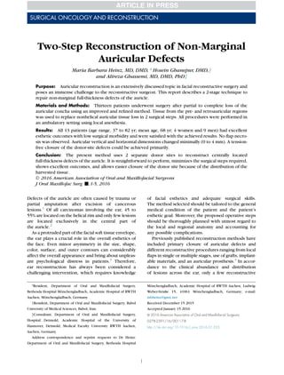
  • 1. SURGICAL ONCOLOGY AND RECONSTRUCTION Two-Step Reconstruction of Non-Marginal Auricular Defects Maria Barbara Heinz, MD, DMD,* Hosein Ghanepur, DMD,y and Alireza Ghassemi, MD, DMD, PhDz Purpose: Auricular reconstruction is an extensively discussed topic in facial reconstructive surgery and poses an immense challenge to the reconstructive surgeon. This report describes a 2-stage technique to repair non-marginal full-thickness defects of the auricle. Materials and Methods: Thirteen patients underwent surgery after partial to complete loss of the auricular concha using an improved and refined method. Tissue from the pre- and retroauricular regions was used to replace nonhelical auricular tissue loss in 2 surgical steps. All procedures were performed in an ambulatory setting using local anesthesia. Results: All 13 patients (age range, 37 to 82 yr; mean age, 68 yr; 4 women and 9 men) had excellent esthetic outcomes with low surgical morbidity and were satisfied with the achieved results. No flap necro- sis was observed. Auricular vertical and horizontal dimensions changed minimally (0 to 4 mm). A tension- free closure of the donor-site defects could be achieved primarily. Conclusion: The present method uses 2 separate donor sites to reconstruct centrally located full-thickness defects of the auricle. It is straightforward to perform, minimizes the surgical steps required, shows excellent outcomes, and allows easier closure of the donor site because of the distribution of the harvested tissue. Ó 2016 American Association of Oral and Maxillofacial Surgeons J Oral Maxillofac Surg -:1-5, 2016 Defects of the auricle are often caused by trauma or partial amputation after excision of cancerous lesions.1 Of all carcinomas involving the ear, 45 to 55% are located on the helical rim and only few lesions are located exclusively in the central part of the auricle.2 As a protruded part of the facial soft tissue envelope, the ear plays a crucial role in the overall esthetics of the face. Even minor asymmetry in the size, shape, color, surface, and outer contours can considerably affect the overall appearance and bring about unpleas- ant psychological distress in patients.3 Therefore, ear reconstruction has always been considered a challenging intervention, which requires knowledge of facial esthetics and adequate surgical skills. The method selected should be tailored to the general medical condition of the patient and the patient’s esthetic goal. Moreover, the proposed operative steps should be thoroughly planned with utmost regard to the local and regional anatomy and accounting for any possible complications. Previously published reconstruction methods have included primary closure of auricular defects and different reconstructive procedures ranging from local flaps in single or multiple stages, use of grafts, implant- able materials, and an auricular prosthesis.4 In accor- dance to the clinical abundance and distribution of lesions across the ear, only a few reconstructive *Resident, Department of Oral and Maxillofacial Surgery, Bethesda Hospital M€onchengladbach, Academic Hospital of RWTH Aachen, M€onchengladbach, Germany. yResident, Department of Oral and Maxillofacial Surgery, Babol University of Medical Sciences, Babol, Iran. zConsultant, Department of Oral and Maxillofacial Surgery, Hospital Detmold, Academic Hospital of the University of Hannover, Detmold; Medical Faculty University RWTH Aachen, Aachen, Germany. Address correspondence and reprint requests to Dr Heinz: Department of Oral and Maxillofacial Surgery, Bethesda Hospital M€onchengladbach, Academic Hospital of RWTH Aachen, Ludwig- Weber-Straße 15, 41061 M€onchengladbach, Germany; e-mail: mbheinz@gmx.net Received December 15 2015 Accepted January 15 2016 Ó 2016 American Association of Oral and Maxillofacial Surgeons 0278-2391/16/00117-8 http://dx.doi.org/10.1016/j.joms.2016.01.025 1
  • 2. methods have been described for the surgical recon- struction of centrally located non-marginal defects of the auricle.5,6 Even fewer reports have evaluated the treatment of full-thickness defects of this region. In consequence, reconstruction of these infrequent defects is very important. The anterior pedicled retroauricular flap (APRF) was first described in 2012.7,8 In 2015, the authors introduced the APRF for the repair of centrally located perforating defects. The described method needed 3 surgical stages.9 Since then, the method has been refined for centrally located perforating defects and the APRF was combined with a flap from the preauricular region, the cranial pedicled preauric- ular flap (CPPF). The present report describes the technique of 2-step reconstruction and the complica- tions, changes in auricular size, esthetic outcome, and patient satisfaction. Materials and Methods Thirteenpatientswithcentrallylocatedfull-thickness auricle defects were treated. Defect size ranged from 10 to 40 mm in diameter. Vertical and transversal dimen- sions were measured pre- and postoperatively. The reconstruction procedures were performed in 2 steps in ambulatory settings using local anesthesia. A CPPF was used to cover the anterior surface and an APRF was used for the posterior defect site. Surgical interventions were carried out in compli- ance with the World Medical Association Declaration of Helsinki on medical research protocols and ethics. SURGICAL PROCEDURE A precisely defined donor-site area, mirroring the size of the existing defect, is outlined on the preauric- ular and retroauricular skin (Figs 1A, 2A). First, the FIGURE 1. Illustrations of the reconstruction procedure after full-thickness excision of the central part of the auricle. A, Designing the flaps in the pre- and retroauricular regions. B, Harvesting and transposition of the anterior pedicled retroauricular flap into the defect area to reconstruct the dorsal surface of the auricle. C, Incision and transposition of the cranial pedicled preauricular flap to reconstruct the anterior surface of the auricle. The donor sites are closed primarily. D, Separation of the 2 pedicles and adaptation of the flaps in the second step. Heinz, Ghanepur, and Ghassemi. Reconstruction of Auricular Defects. J Oral Maxillofac Surg 2016. 2 RECONSTRUCTION OF AURICULAR DEFECTS
  • 3. dorsal incision is performed to elevate an APRF from the retroauricular region. Then, the posterior tip of the flap is folded into the anterior edge of the dorsal defect to replace the dorsal surface (Fig 1B). As a result, the subcutaneous part of the APRF is seen ante- riorly. Second, the skin of the preauricular region is incised with the pedicle based cranially. The skin is used to reconstruct the anterior surface of the ear (Fig 1C). The pre- and retroauricular donor sites are closed primarily without tension (Fig 2B). After 2 weeks, the pedicles of the 2 flaps can be separated surgically and adjusted accordingly (Fig 1D). Results Tissue from the pre- and retroauricular regions was used to reconstruct full-thickness conchal defects in 13 patients (age range, 37 to 82 yr; mean age, 68 yr; 4 women and 9men). Postoperatively, vertical and hor- izontal decreases in auricle size were 0 to 4 mm (Table 1). Flap necrosis or flap loss, clinically notice- able hematoma, or wound infection were not observed. Scars were placed in hidden regions and were barely visible. Skin color, texture, and thickness of the reconstructed part matched well with the sur- rounding tissue (Figs 2C, D). Tension-free wound closure of the donor sites was achieved by the distribu- tion of the needed skin into 2 separate regions. All 13 reconstructive treatments could be performed in 2 sur- gical steps. Esthetic results were excellent and all pa- tients were pleased with the outcome. Discussion The authors have presented a method to recon- struct auricular conchal defects using the APRF combined with a CPPF. Their aim was to maintain the original size of the ear, achieve an optimal esthetic outcome, distribute the donor-site defect wound, and shorten the treatment time needed for reconstruction FIGURE 2. Photographs of an 82-year-old patient after excision of a basal cell carcinoma of the central part of the left ear. A, Full-thickness defect of the antihelix (diameter, 3.5 Â 3.0 cm) (posterior view). B, The anterior pedicled retroauricular and cranial pedicled preauricular flaps are adapted. The 2 donor sites are closed primarily. (Fig 2 continued on next page.) Heinz, Ghanepur, and Ghassemi. Reconstruction of Auricular Defects. J Oral Maxillofac Surg 2016. HEINZ, GHANEPUR, AND GHASSEMI 3
  • 4. (from 3 to 2 surgical steps). The CPPF was used to cover the anterior surface of the defect. Because of the similarity in texture, this tissue replaced the ante- rior surface more adequately. The posterior surface was reconstructed by the APRF, which consists of thicker skin from the retroauricular region and offers greater stability. In contrast to the previous technique, there was no need for dividing any transitional part of the APRF after passing the edge of the cartilaginous framework, which would require another step.9 Because the tissue needed originated from 2 separate regions, the size of the resulting defect of each donor site was decreased and could be closed primarily and without tension. The resultant scars were located in invisible areas. The goal of reconstructive surgery of the auricle is to achieve morphologically acceptable auricular size and shape and proportionally integrated anatomic landmarks. Moreover, the proposed reconstructive procedure should consider the patient’s existing medical condition and should aim for a positive image by achieving an adequate esthetic outcome. The first reported method used a composite graft from the contralateral ear.10 Since then, different methods have been developed. Many reported technical methods involve reconstruction of the helical rim,11- 17 because it is the prominent part of the ear and most neoplastic lesions are found in this region. Although some investigators have suggested reconstructive techniques of central nonperforating defects of the ear,4,18-21 only a few methods have described full-thickness reconstruction of this region. Nevertheless, most of these described methods result in visible deformities and a smaller ear overall.22 In summary, there is a lack in the literature of tech- nical reconstruction methods to close full-thickness nonhelical defects of the ear in a convenient way without changing the size. In their previous report, the authors introduced a reconstruction technique using the APRF, which required 3 steps. To decrease the stages, the authors refined their technique and used tissue from 2 separate pre- and retroauricular donor sites for reconstruction. Because of the distribu- tion of the donor-site defect, the present technique allowed tension-free wound closure of the 2 donor regions. In addition, sufficient tissue was available to FIGURE 2 (cont’d). C, Frontal view of the patient 7 months postoperatively. D, Posterior aspect of the patient during follow-up period. Overall size and shape of the reconstructed ear matches the contralateral side. Heinz, Ghanepur, and Ghassemi. Reconstruction of Auricular Defects. J Oral Maxillofac Surg 2016. 4 RECONSTRUCTION OF AURICULAR DEFECTS
  • 5. reconstruct the complete non-marginal part of the auricle easily without obvious decrease of auricular size. Although the method required 2 surgical steps, it was reliable for flap survival. Esthetic outcome was excellent because of the similarity of tissue texture between the preauricular region and the anterior surface of the auricle. Moreover, the thick skin from the retroauricular region offered sufficient stability and there was no need for cartilage grafting. The new method was associated with improved patient satisfaction because of fewer surgical steps and shorter reconstruction time. Combining flaps from pre- and retroauricular regions simplified the full-thickness reconstruction of non-marginal auricular defects, resulting in minimal horizontal and vertical size changes. Because of the distribution of required tissue into 2 anatomically separate regions, primary wound closure was easier to achieve. However, 2 surgical steps were required. Acknowledgment The authors thank Mr Wolfgang Graulich, from the Institute of Anatomy, RWTH Aachen, for his contribution in creating and permit- ting the use of his illustrations. References 1. Antia NH, Busch V: Chondrocutaneous advancement flap for the marginal defect of the ear. Plast Reconstr Surg 39:472, 1967 2. Leferink VJ, Nicolai JP: Malignant tumors of the external ear. Ann Plast Surg 21:550, 1988 3. Hyckel P, Robotta C, Schumann D: Partial loss of the auricle: Multiphase reconstruction and complete preservation of the helix. Mund Kiefer Gesichtschir 3:131, 1999 4. Mellette JR: Ear reconstruction with local flaps. J Dermatol Surg Oncol 17:176, 1991 5. Elsahy NI: Ear reconstruction with rotation-advancement com- posite flaps. Plast Reconstr Surg 75:567, 1985 6. Elsahy NI: Reconstruction of the ear after skin and perichon- drium loss. Clin Plast Surg 29:187, 2002 7. Stiller MB, Gerressen M, Modabber A, et al: Anteriorly pedicled retroauricular flap for repair of auricular defects. Aesthetic Plast Surg 36:623, 2012 8. Ghassemi A, Modabber A, Talebzadeh M, et al: Surgical management of auricular defect depending on the size, location, and tissue involved. J Oral Maxillofac Surg 71: 232, 2013 9. Heinz MB, H€olzle F, Ghassemi A: Repairing a non-marginal full- thickness auricular defect using a reversed flap from the postaur- icular area. J Oral Maxillofac Surg 73:764, 2015 10. Pegram M, Peterson R: Repair of partial defects of the ear. Plast Reconstr Surg 18:305, 1956 11. Butler C: Extended retroauricular advancement flap reconstruc- tion of a full-thickness auricular defect including posteromedial and retroauricular skin. Ann Plast Surg 49:317, 2002 12. Goldberg LH, Maulsin DV, Humphreys TR: The postauricular cutaneous advancement flap for repairing ear rim defects. Der- matol Surg 22:28, 1995 13. Fata JJ: Composite chondrocutaneous advancement flap: A tech- nique for reconstruction of marginal defects of the ear. Plast Re- constr Surg 99:1172, 1997 14. Johnson TM, Fader DJ, Arbor A: The staged retroauricular to auricular direct pedicle (interpolation) flap for helical ear recon- struction. J Am Acad Dermatol 37:975, 1997 15. Kaminsky A: Repair of the partial loss of the helix. Aesthetic Plast Surg 21:427, 1997 16. Lewin ML: Reconstruction of the helix. Arch Otolaryngol 47: 802, 1948 17. Stefanoff DN: Retroauricular tubed skin flap to the helical rim, in Strauch B, Vasconez LO, Hall-Findlay EJ (eds): Grabb’s Encyclo- pedia of Flaps. Volume 1, Head and Neck. Boston, MA, Little Brown & Co, 1990, pp 295–297 18. Chen C, Chen ZJ: Reconstruction of the concha of the ear using a postauricular island flap. Plast Reconstr Surg 86:569, 1990 19. Cordova A, D’Arpa S, Pirrello R, et al: Retroauricular skin: A flaps bank for ear reconstruction. J Plast Reconstr Aesthet Surg 61:44, 2008 20. Ohsumi N, Iida N: Ear reconstruction with chondrocutane- ous postauricular island flap. Plast Reconstr Surg 96:718, 1995 21. Renard A: Postauricular flap based on a dermal pedicle for ear reconstruction. Plast Reconstr Surg 68:159, 1981 22. Ramirez OM, Heckler FR: Reconstruction of nonmarginal ear de- fects with chondrocutaneous advancement flaps. Plast Reconstr Surg 84:32, 1989 Table 1. AURICULAR DEFECT SIZE COMPARED WITH POSTOPERATIVE DECREASE OF VERTICAL AND HORIZONTAL AURICULAR DIMENSIONS Patient Number Defect Size (mm) Postoperative Decrease (mm) Vertical Horizontal 1 18 3 2 2 25 2 2 3 40 4 3 4 32 3 3 5 23 3 3 6 35 4 3 7 10 1 0 8 28 2 1 9 36 4 1 10 20 2 1 11 30 3 4 12 24 4 3 13 26 2 2 Heinz, Ghanepur, and Ghassemi. Reconstruction of Auricular De- fects. J Oral Maxillofac Surg 2016. HEINZ, GHANEPUR, AND GHASSEMI 5