Download to read offline
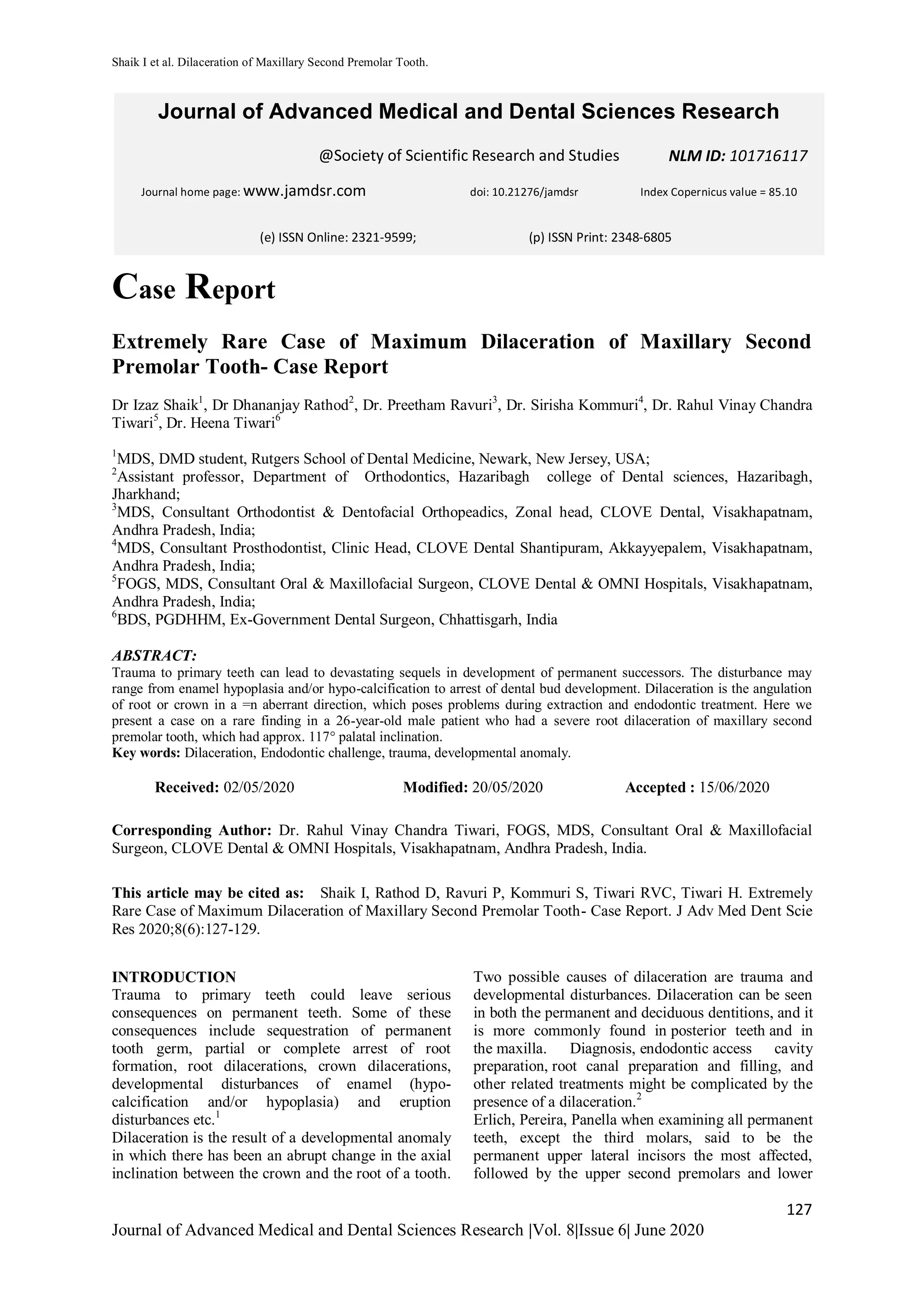

This case report describes an extremely rare case of severe dilaceration (117° palatal inclination) of the root of a maxillary second premolar tooth. Trauma to primary teeth can result in developmental disturbances to permanent successor teeth, including crown and root dilaceration. Dilacerated teeth pose challenges for diagnosis, treatment planning, endodontic access, and extraction. In this case, the maxillary second premolar tooth was severely dilacerated and had to be extracted. Dilaceration is an abnormality that requires a multidisciplinary approach and modified treatment procedures.


![CTEV [ clubfoot] DR ARUN LAL ,DR MOHAMED ASHRAF travancore medical college k...](https://cdn.slidesharecdn.com/ss_thumbnails/ctevclubfootdrarunlaldrmohamedashraftravancoremedicalcollegekollamkeralaindia-260208063247-18fc466c-thumbnail.jpg?width=640&height=640&fit=bounds)

