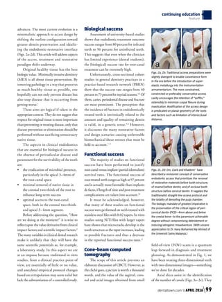
Course description
This article is an overview of the essen-
tials that accept that the endorestorative
continuum is not a concept. Rather, it is a
de facto treatment approach. “Clean, shape
and pack,” with hindsight, should have
been “shape, clean and pack”—but even
that was a somewhat simplistic approach.
Debridement, disinfection and microstruc-
tural replication due to the introduction of
adaptive bioceramic technologies for root
filling are increasingly being recognized as
the standard for optimal endodontic therapy.
Objectives
1. 	 To have a better understanding of
the dynamic changes in mainstream
endodontic instrumentation.
2. 	 To have a better understanding of
latest and most advanced obturation
techniques.
3.	 To understand the synergism between
the endodontic and restorative
continuum of tooth rehabilitation.
4.	 To dispel the myths of past
“concepts” and evaluate the shifts
in treatment protocols with better
evidentiary understanding.
History
The recent history of endodontics has
beenanevolutionthroughseveraltransitional
stages: nickel-titanium (NiTi) instruments
for sculpting and debridement; microscopes
for illumination and magnification—indis-
pensable for detection and exploration of the
inner space of teeth (Figs. 1a–1d); and, of
late, the inclusion of cone-beam computed
tomography for diagnosis and treatment
planning.
The clinician can thus anticipate, visu-
alize and treat with unprecedented levels of
accuracy and safety. However, the failure
to overcome all potential mechanical and
pathologic vectors that negate treatment
success continues in spite of these technologic
continuing education
feature
Dr. Martin Trope
served as the chair
of endodontology at
Temple University,
School of Dentistry,
and is currently
clinical professor in the department
of endodontics at the School of Dental
Medicine at the University of Pennsylvania.
In 1993, he accepted the J.B. Freedland
professorship in the department of
endodontics at the University of North
Carolina at Chapel Hill, School of Dentistry.
Trope has been director of the American
Board of Endodontics; editor-in-chief of
both Dental Traumatology and Endodontic
Topics; a member of the editorial board of
Oral Surgery, Oral Medicine, Oral Pathology;
a member of the advisory board of Esthetic
Dentistry; and has received The Louis
I. Grossman Award for publication of
significant research.
by Dr. Martin Trope
Dr. Kenneth Serota
received his certificate
in endodontics and
master of medical
sciences degree in
nuclear medicine from
the Harvard-Forsyth Dental Center in
Boston. In 1981, he was the recipient of
the American Association of Endodontics
Memorial Research Award for his work in
nuclear-medicine screening procedures
related to dental pathology. Active in online
education since 1998, he is the founder
of the online endodontic forum ROOTS,
the Facebook forums NEXUS and Dental
Divas Global. He is currently global clinical
director for Dentaltown.
and Dr. Kenneth Serota
Approved PACE Program Provider FAGD/
MAGD Credit
Approval does not imply acceptance by a
state or provincial board of dentistry or
AGD endorsement.
1/1/2016 to 4/30/2016
Provider ID#304396
This print or PDF course is a written self-instructional
article with adjunct images and is designated for 1.5
hours of CE credit by Farran Media. Participants will
receive verification shortly after Farran Media receives
the completed post-test. See instructions on page 102.
AGD
Code:
070
Bio-Minimalism: Trends and
Transitions in Endodontics
Four-rooted
maxillary molar
Maxillary molar
fused roots
Mandibular molar BMandibular molar A
Fig. 1a Fig. 1b Fig. 1c Fig. 1d
The root-canal system is an arborizational, anastomotic, byzantine, labyrinthine complexity,
morphologically comparable to the passages of a maze. While primary canals exist, the tributaries,
accessory branches and luminae of the dentinal tubuli harbor extensive tissue and microflora, which if
left untreated remain vectors for persistent and refractory pathology.
Figs.1a-1dprovidedbyDr.MarcoVersiani,
JesusD.PécoraandManoelD.SousaNeto
98 APRIL 2016 // dentaltown.com
advances. The most current evolution is a
minimalistic approach to access design by
shifting the outline configuration toward
greater dentin preservation and idealiz-
ing the endodontic-restorative interface
(Figs. 2a–2d).Thisarticlewilladdressaspects
of the access, treatment and restorative
paradigm shifts underway.
Original healthy tissue has the best
biologic value. Minimally invasive dentistry
(MID) is all about tissue preservation. By
removing pathology in a way that preserves
as much healthy tissue as possible, one
hopefully can not only prevent disease but
also stop disease that is occurring from
getting worse.1
These aims are logical if taken in the
appropriate context. They do not suggest that
respect for original tissue is more important
than preventing or treating disease, only that
disease prevention or elimination should be
performed without sacrificing unnecessary
native tissue.
The aspects in clinical endodontics
that are essential for biological success in
the absence of periradicular disease and
paramount for the survivability of the tooth
are:
•	 the eradication of microbial presence,
particularly in the apical 3–4mm of
the root-canal space
•	 minimal removal of native tissue in
the coronal two-thirds of the root to
enhance long-term success
•	 optimal access to the root-canal
space, both in the coronal two-thirds
and apical 3–4mm segment.
Before addressing the question, “How
are we doing at the moment?” it is wise to
reflect upon the value derivative from clinical
impact factors and scientific impact factors.2
The many variables in clinical dental research
make it unlikely that they will have the
same scientific potentials as, for example,
a laboratory study. In this aspect we are
at an impasse because traditional in vitro
studies, from a clinical practice point of
view, are essentially of little or no value,
and anecdotal empirical protocol changes
based on extrapolation may seem valid but
lack the substantiation of a controlled study.
Biological success
Assessment of university-based studies
shows that endodontic treatment outcome
success ranges from 80 percent for infected
teeth to 96 percent for uninfected teeth.
This suggests that even when the clinician
has limited experience (dental students),
the (biological) success rate for root-canal
treatment can be extremely high.
Unfortunately, cross-sectional cohort
studies in general dentistry practices in a
practice-based research network (PBRN)
show that the success rate ranges from 40
percentto73percentformyriadreasons.3,4
Of
them, caries, periodontal disease and fracture
are most prominent. The perception that
the incidence of fracture in endodontically
treated teeth is intrinsically related to the
amount and quality of remaining dentin
is valid, in a generic sense.5,6
However,
it discounts the many restorative factors
and design scenarios causing unfavorable
biomechanical shear stresses that must be
held to account.7–9
Functional success
The majority of studies on functional
success have been performed to justify
root-canalversusimplant(partialedentulism)
survival rates. The functional success of
root-treatedteethrangesashighas97percent
and is actually more favorable than implants
de facto, if length of time and post-treatment
complications are taken into account.10
It must be acknowledged, however,
that many of these studies on functional
success were performed on teeth treated with
stainless-steel files with 0.02 tapers. In vitro
studies using NiTi files with larger tapers
have shown that microcracks develop in the
tooth structure as the taper increases, leading
to possible fractures and thus a decrease
in the reported functional success rates.11
Cone-beam computed
tomography
The scope of this article prevents an
elaborate discussion of CBCT. However, as
the cliché goes, a picture is worth a thousand
words, and the value of the sagittal, coro-
nal and axial images obtained from small
field-of-view (FOV) scans is a quantum
leap forward in diagnosis and treatment
planning. As demonstrated in Fig. 1, we
have been treating three-dimensional teeth
with two-dimensional images—something
we’ve done for decades.
Axial slices assist in the identification
of the number of canals (Figs. 3a–3c). They
continuing education
feature
Figs. 2a, 2b: Traditional access preparations were
slightly divergent to enable convenience form
in the era before the introduction of super-
elastic metallurgy into the instrumentation
armamentarium. The more constrained,
constricted or preferably conservative access
cavity encourages the retention of “soffits,”
ostensibly to minimize cuspal flexure during
mastication. Modification of the access design
is predicated on planar geometry of the roots
and factors such as limitation of interocclusal
distance.
Figs. 2c, 2d: Drs. Clark and Khademi  1
have
described a revisionist concept of conservative
endodontic access that prioritizes the removal
of restorative materials before tooth structure;
of enamel before dentin; and of occlusal tooth
structure before cervical dentin. It negates the
traditionalist straight-line access protocol and
the totality of deroofing the pulp chamber.
The biologic mandate of greatest imperative is
the preservation of the critical region of peri-
cervical dentin (PCD)—4mm above and below
the crestal bone—to the paramount achievable
degree without compromising debridement or
inducing iatrogenic misadventure. (With sincere
appreciation to Dr. Hany Mohamed Aly Ahmed of
the Universiti Sains Malaysia.)
Fig. 2a
Fig. 2c
Fig. 2b
Fig. 2d
dentaltown.com  APRIL 2016 99
continuing education
feature
are an aid in determining the location of
external root resorptions, bifidities, diametral
width and curvature at various levels along
the length of the root.
Coronal slices offer an evaluation of
the many configurations characteristic of
premolars and molars, predominantly in the
mandible.Theyassistinguidingtheclinician
to avoid removing hard tissue in exploring
for canal angulation and orientation (Fig. 4).
Sagittal slices assist in eliminating the
anatomical noise of overlapping structures,
particularly in the maxilla, and enable
a clarity of a Y-axis perspective in both
orthograde and retrograde treatment that
has been previously unobtainable (Fig. 5).
The challenges and changes
Bio-minimalism recognizes that the
pericervical dentinal (PCD) zone is crucial
for buttressing the residual coronal tooth
structure during functional loading stress,
and ostensibly acts to minimize cuspal
flexure during mastication. The challenge
to the clinician is to negotiate all canals,
debride residual pulp tissue from all loci
and avoid iatrogenic complications while
working through a restricted access without
“convenience form.”12
This begs the question:
“How can we remove as many microbes as
possible while still maintaining the strength
of the tooth?”
Fortunately, there are technological
advances in the instrumentation armamen-
tarium that bring us closer to the aims of
minimally invasive endodontics. We are in a
newerainendodontics,withtheintroduction
of disinfection systems that do not require
changing the nonround canal to a round
shape. This previous formulistic alteration
meant that sculpting the inner space was
overly minimized, or that too much native
tissue was removed.
We now have files and finishers that
adjust to the original shape of the canal,
scrape biofilm in a manner similar to peri-
odontal scalers, and facilitate irrigants to act
upon exposed microbes. These systems will
continue to iterate, enabling the clinician
to retain more native root structure while
more effectively removing inflammatory
vectors.
From the standpoint of survivability,
the future looks increasingly bright, not
only due to bio-minimalistic technical
changes, but also to the introduction of
new obturation materials. Until recently,
the preparation of the coronal two-thirds
of the canal was designed to facilitate
plugger placement to within 4mm of the
apical terminus to generate the hydraulic
forces necessary to deform thermolabile
gutta-percha in the apical capture zone
Fig. 4
The coronal view can assist in determining branching otherwise undetectable and assist in access
orientation to ensure optimal preservation of pericervical dentin.
Fig. 3a
Fig. 3b
Fig. 3c
The traditional flat film used to preoperatively
assess the number of roots, the degree of
calcification and the planar geometries of the
roots along their length has now been augmented
by axial slices which assist in identification of
the number of canals and various anatomical
configurations and aberrations.
Three canals untreated in MB root of tooth #2.
Canals obturated.
Fig3providedbyDr.WilliamNuderaFig3providedbyDr.WilliamNuderaFig3providedbyDr.WilliamNudera
Fig.4providedDr.CharlesBoveda
100 APRIL 2016 // dentaltown.com
with optimal gravitometric density. This
necessitated large tapers destructive to the
native root structure with minimal benefit
to microbial removal.
The introduction of bioceramics brings
a material with antimicrobial potential
(high-alkaline pH), biocompatibility, bio-
activity and no evidence of shrinkage upon
setting. The myriad applications—from
orthograde to retrograde to resorptive treat-
ment—facilitate enhanced sealing without
the need for removal of excess inner or outer
root structure.13
Unfortunately, minimally invasive access
design has dominated the discussion of
bio-minimalism in endodontics. Traditional
endodontic access design adhered to the
restorative axiom “extension for prevention,”
which manifested as straight-line access,
including deroofing the pulp chamber and
preflaring the canal orifice to facilitate the
subsequent shaping of the entire root-canal
system to negotiate the apical terminus.
However, optimal diametral retention of
coronal/radicular dentin and maintenance
of the planar geometry of the root-canal
anatomy essential for the structural integrity
of endodontically restored teeth was unduly
compromised.14
Preparing the cavity
There are many limitations to what is
preached as the perfect minimally invasive
cavity design (conservative access prepara-
tion). In teeth requiring root-canal treatment,
the shape and size of the access opening is
invariably dictated by caries, or failing or
lost restorations. It is imprudent and unlikely
that a clinician would leave diseased tooth
structureintacttoconfigureatextbook-access
cavity. Removal of unnecessary dentin
during access preparation was never the
biologic mandate of access design. Even in
the Schilderian era, the goal was to retain
as much of the cervical dentin as possible,
while still achieving straight-line access to
the apical termini. (The greater the curvature
in the canal, the greater the relevance for a
glide path to achieve a straighter-line access.)
Perhaps the aim of conservative cav-
ity preparation should be reframed from
“removal of as little tooth structure as pos-
sible” to “removal of as little as necessary.”
As discussed previously, clinicians now have
a multitude of technologies at their disposal
to assist in the execution of this redefined
objective. Restoratively in the adhesion era,
we are now blessed with an abundance of
riches to move away from the post/core and
full-coverage paradigm of the past, which
only further compromised the retention
of foundational tooth structure, to a truly
bio-minimalistic restorative template.
One can barely skim the surface in a
publication of this brevity; however, the
purpose is to create an energy dynamic anal-
ogous to the ripples produced by dropping
a stone in a pool of water. With minimal
resistance, they can expand concentrically
in greater and greater diameters. Or one
could be a curious octopus and go off in
all directions. That will be forthcoming in
the online curriculum under development
for Dentaltown.com.
Perhaps the answer to all of the issues
that remain to be corrected in endodontics
was articulated eloquently more than 100
years ago by the man who predicted waves in
the gravitation fields of the universe, Albert
Einstein. “We cannot solve our problems
with the same thinking we used when we
created them,” he said. ■
References
1.	 Ericson D. What is minimally invasive dentistry? Oral
Health Prev Dent. 2004;2(sup1):287-92
2.	 Mjor IA. Scientific Impact and Clinical Impact. J Adhesion
Dent. 2009;11(3)Letter to the editor
3. 	 Gilbert GH et al. Outcomes of root canal treatment in Den-
tal Practice-Based Research Network practices. Gen Dent;
January/February 2010 www.agd.org 28-36
4.	 Kabak Y, Abbott PV. Prevalence of apical periodontitis and
the quality of endodontic treatment in an adult Belarusian
population. In Endo J April 2005;38(4):238-245
5.	 Kishen A. Mechanisms and risk factors for fracture predi-
lection in endodontically treated teeth. Endodontic Topics
2006;13:57-83
6.	 Trope M, Maltz DO, Tronstad L. Resistance to fracture of
restored endodontically treated teeth. Endod Dent Traumatol
1985 June;1(3):108-11
7.	 Schmitter M, Sterzenbach et al. A flood tide of systematic
reviews on endodontic posts: methodological assessment using
of R-AMSTAR. Clin Oral Investig 2013;17:1287-1294
8.	 Naumann M, Blankenstein et al. Risk factors for failure of
glass fiber-reinforced composite post restorations: a prospective
observational clinical study. Eur J Oral Sci 2005;113:519-
524
9.	 Torbjorner A. Fransson B. Biomechanical aspects of prosthetic
treatment of structurally compromised teeth. Int J Prostho-
dont 2004;17:135-141
10.	 Salehrabi R, Rotstein I. Endodontic Treatment Outcomes in
a Large Patient Population in the USA: An Epidemiological
Study. J Endo 2004;30(12): 846-850
11.	 Pop I et al. Synchroton light-based microCT to analyse the
presence of dentinal microcracks post-rotary and reciprocating
instrumentation. Clin Oral Invest 2014 Feb (published
online)
12.	 Christie WH, Thompson GK. The importance of endodontic
access in locating maxillary and mandibular molar canals.
JCDA 1994;60(6):527-536
13.	 Haapasalo M. Clinical use of bioceramic materials. Endo
Topics 2015;32:97-117
14.	 Castellucci A. Endodontics; Access Cavity and Endodontic
Anatomy. Editions Martina, Bologna IT;244-329
Fig. 5
Sagittal views provide the clinician with the
z-axis perspective to identify the distance
between splits in roots and the orientation of the
roots about themselves. This is of paramount
importance particularly in microsurgical
treatment.
The clinician can thus anticipate, visualize and treat with
unprecedented levels of accuracy and safety. However, the
failure to overcome all potential mechanical and pathologic
vectors that negate treatment success continues in spite of
these technologic advances.
Fig.5providedbyDr.WiliamNudera
dentaltown.com  APRIL 2016 101
continuing education
feature
Legal Disclaimer: The CE provider uses reasonable care in selecting and providing content that is accurate. The CE provider, however, does not independently verify the content or materials. The
CE provider does not represent that the instructional materials are error-free or that the content or materials are comprehensive. Any opinions expressed in the materials are those of the author
of the materials and not the CE provider. Completing one or more continuing education courses does not provide sufficient information to qualify participant as an expert in the field related to the
course topic or in any specific technique or procedure. The instructional materials are intended to supplement, but are not a substitute for, the knowledge, expertise, skill and judgment of a trained
health-care professional. You may be contacted by the sponsor of this course.
Licensure: Continuing education credits issued for completion of online CE courses may not apply toward license renewal in all licensing jurisdictions. It is the responsibility of each registrant to
verify the CE requirements of his/her licensing or regulatory agency.
Claim Your CE Credits
P O S T-T E S T
Answer the test in the Continuing Education Answer Sheet and submit it by mail or fax with a
processing fee of $36. You can also answer the post-test questions online at dentaltown.com/
onlinece. We invite you to view all of our CE courses online by going to dentaltown.com/onlinece and
clicking the View All Courses button. Note: If you are not already registered on Dentaltown, you will
be prompted to do so. Registration is fast, easy and, of course, free.
continuing education
feature
1.	 Which of the below statements are true?
A)		G.V Black’s principles are an intriguing premise for addressing the restor-
ative needs of the tooth. However, in endodontics, there is no rationale for
removal of the restoration, eradication of decay and assessment of struc-
tural fractures, until the root-canal treatment is completed.
B)		Ninja access, mousehole access, soffits and trusses have long-term stud-
ies that verify their validity in the transition of access-preparation design
shapes and configuration.
C)		Residue in the pulp chamber is entombed within the final core restoration
and of no infective concern.
D)		None of the above.
2.	 Which of the below is true?
A)		Calcification occurs apical to coronal in the root-canal space.
B)		Calcification occurs coronal to apical in the root-canal space.
C) 	As long as you can access the canal orifice, residual pulp stones are not an
impediment to root-canal success.
3.	 Which of the below is true?
A) 	Preservation of pericervical dentin or “girth” is not relevant to ensuring the
structural integrity of residual tooth structure.
B) 	Post placement reinforces the structure and ensures elimination of the
potential for stress fractures.
C)		The use of Gates Glidden drills or Peezo reamers will not compromise den-
tinal girth.
D)		All of the above.
E)		None of the above.
4.	 Which of the below is true?
A)		Gates Glidden drills should be used to the interface of the coronal and mid-
dle thirds of the root-canal space to enable straight-line access.
B)		Peezo reamers are a better alternative than Gates Glidden drills in anterior
teeth.
C) 	Any nickel-titanium instrument can go around any curve, because the met-
al is super-elastic.
D)		None of the above.
5.	 True or False?
Irrigants present little or no concern in regard to the biochemical composition of
residual structural dentin after root canal therapy.
A) 	 True
B) 	 False
6.	 True or False?
A)		To establish conservative access, it is reasonable to allow loci of infective
organic material to remain in the pulp chamber and the isthmus areas of
the root-canal space.
B)		Conservative access implies that we no longer need worry about glide path
before the use of nickel-titanium instruments.
C)		Residue in seams between orifices and canal isthmi can be completed
cleared with irrigants and passive ultrasonic irrigation or sonic activators.
D)		 All of the above are true.
E)		All of the above are false.
7.	 Which of the below is true?
A) 	The ideal obturation material adapts to, but does not adhere to, the
root-canal walls. Sealer ensures that no leakage can occur once it has set.
B) 	 Gutta-percha is the ideal root filling material.
C) 	 Gutta-percha will not shrink on cooling.
D) 	 Gutta-percha’s gravitometric density is enhanced on cooling.
E) 	Bioceramic sealers are hydrophilic and will penetrate dentinal tubuli.
8.	 Which of the below is true?
A)		Full coverage of the endodontically treated teeth is more likely to result in
long-term success rather than the use of inlay/onlay adhesive restorations.
B) 	Occlusal coverage of the endodontically treated tooth is not required.
C) 	In the absence of a suitable ferrule of more than 1.5mm in anterior teeth, a
feather-edged preparation is acceptable.
D) 	 All the above statements are false.
9.	Preservation of the tooth structure 4mm above and 4mm below the CEJ is not
desirable nor essential to a positive treatment outcome.
A) 	 The above statement is true.
B) 	 The above statement is false.
10.	 Which of the below is true?
A) 	The greater the taper in a funneling canal, the better the apical seal.
B) 	Regardless of the root curvature, the condenser must be accommodated to
within 4mm of the apical terminus.
C) 	 The above statements are true.
D) 	 The above statements are false.
102 APRIL 2016 // dentaltown.com
CONTINUING
EDUCATION
ANSWER
SHEET
1.	a	 b	 c	 d
2.	a	 b	 c	 d
3.	a	 b	 c	 d
4.	a	 b	 c	 d
5.	a	 b	 c	 d
6.	a	 b	 c	 d
7.	a	 b	c	 d
8.	a	 b	 c	 d
9.	a	 b	 c	 d
10.	a	 b	 c	 d
Please circle your answers.
CE Post-TestBio-Minimalism
by Dr. Martin Trope and Dr. Kenneth Serota
License Number ______ ______ ______ ______ ______ ______ ______ ______ ______ ______
AGD# _____________________________________________________________________________________________________
Name ______________________________________________________________________________________________________
Address ___________________________________________________________________________________________________
City ____________________________________________________ State ___________ ZIP ________________________
Daytime phone _____________________________________________________________________________________________
E-mail (required for certificate) _______________________________________________________________________________
o Check (payable to Dentaltown.com, Inc.)
o Credit Card (please complete the information below and sign; we accept Visa, MasterCard and American Express.)
Card Number ______ ______ ______ ______ ______ ______ ______ ______ ______ ______ ______ ______ ______ ______ ______ ______
Expiration Date – Month / Year ______ ______ / ______ ______ ______ ______
Signature ___________________________________________________________________________	 Date __________________________________________________	
Program Evaluation (required)
Please evaluate this program by circling the corresponding numbers: (5 = Strongly Agree to 1 = Strongly Disagree)
1.	 Course administration was efficient and friendly					 5	 4	 3	 2	 1
2.	 Course objectives were consistent with the course as advertised			 5	 4	 3	 2	 1
3.	 COURSE OBJECTIVE #1 was adequately addressed and achieved			 5	 4	 3	 2	 1
4.	 COURSE OBJECTIVE #2 was adequately addressed and achieved			 5	 4	 3	 2	 1
5.	 COURSE OBJECTIVE #3 was adequately addressed and achieved			 5	 4	 3	 2	 1
6.	 COURSE OBJECTIVE #4 was adequately addressed and achieved			 5	 4	 3	 2	 1
7.	 Course material was up-to-date, well-organized and presented in sufficient depth		 5	 4	 3	 2	 1
8.	 Instructor demonstrated a comprehensive knowledge of the subject			 5	 4	 3	 2	 1
9.	 Instructor appeared to be interested and enthusiastic about the subject			 5	 4	 3	 2	 1
10. 	 Audio-visual materials used were relevant and of high quality				 5	 4	 3	 2	 1
11. 	 Handout materials enhanced course content					 5	 4	 3	 2	 1
12. 	 Overall, I would rate this course (5 = Excellent to 1 = Poor):				 5	 4	 3	 2	 1
13. 	 Overall, I would rate this instructor (5 = Excellent to 1 = Poor):				 5	 4	 3	 2	 1
14. 	 Overall, this course met my expectations					 5	 4	 3	 2	 1
Comments (positive or negative): _________________________________________________________________________________________________________________
____________________________________________________________________________________________________________________________________________________
For questions, contact Director of Continuing Education Howard Goldstein at hogo@dentaltown.com.
Instructions: To receive credit, complete the answer sheet and mail it, along with a check or credit card pay-
ment of $36 to: Dentaltown.com, Inc., 9633 S. 48th St., Suite 200, Phoenix, AZ 85044. You may also fax this
form to (480) 598-3450 or answer the post-test questions online at dentaltown.com/onlinece. This written
self-instructional program is designated for 1.5 hours of CE credit by Farran Media. You will need a minimum
score of 70 percent to receive your credits. Participants pay only if they wish to receive CE credits, thus
no refunds are available. Please print clearly. This course is available to be taken for credit April 1, 2016,
through its expiration on April 1, 2019. Your certificate will be emailed to you within 3–4 weeks.
continuing education
feature
dentaltown.com  APRIL 2016 103

Bio minimalism

  • 1.
    Course description This articleis an overview of the essen- tials that accept that the endorestorative continuum is not a concept. Rather, it is a de facto treatment approach. “Clean, shape and pack,” with hindsight, should have been “shape, clean and pack”—but even that was a somewhat simplistic approach. Debridement, disinfection and microstruc- tural replication due to the introduction of adaptive bioceramic technologies for root filling are increasingly being recognized as the standard for optimal endodontic therapy. Objectives 1. To have a better understanding of the dynamic changes in mainstream endodontic instrumentation. 2. To have a better understanding of latest and most advanced obturation techniques. 3. To understand the synergism between the endodontic and restorative continuum of tooth rehabilitation. 4. To dispel the myths of past “concepts” and evaluate the shifts in treatment protocols with better evidentiary understanding. History The recent history of endodontics has beenanevolutionthroughseveraltransitional stages: nickel-titanium (NiTi) instruments for sculpting and debridement; microscopes for illumination and magnification—indis- pensable for detection and exploration of the inner space of teeth (Figs. 1a–1d); and, of late, the inclusion of cone-beam computed tomography for diagnosis and treatment planning. The clinician can thus anticipate, visu- alize and treat with unprecedented levels of accuracy and safety. However, the failure to overcome all potential mechanical and pathologic vectors that negate treatment success continues in spite of these technologic continuing education feature Dr. Martin Trope served as the chair of endodontology at Temple University, School of Dentistry, and is currently clinical professor in the department of endodontics at the School of Dental Medicine at the University of Pennsylvania. In 1993, he accepted the J.B. Freedland professorship in the department of endodontics at the University of North Carolina at Chapel Hill, School of Dentistry. Trope has been director of the American Board of Endodontics; editor-in-chief of both Dental Traumatology and Endodontic Topics; a member of the editorial board of Oral Surgery, Oral Medicine, Oral Pathology; a member of the advisory board of Esthetic Dentistry; and has received The Louis I. Grossman Award for publication of significant research. by Dr. Martin Trope Dr. Kenneth Serota received his certificate in endodontics and master of medical sciences degree in nuclear medicine from the Harvard-Forsyth Dental Center in Boston. In 1981, he was the recipient of the American Association of Endodontics Memorial Research Award for his work in nuclear-medicine screening procedures related to dental pathology. Active in online education since 1998, he is the founder of the online endodontic forum ROOTS, the Facebook forums NEXUS and Dental Divas Global. He is currently global clinical director for Dentaltown. and Dr. Kenneth Serota Approved PACE Program Provider FAGD/ MAGD Credit Approval does not imply acceptance by a state or provincial board of dentistry or AGD endorsement. 1/1/2016 to 4/30/2016 Provider ID#304396 This print or PDF course is a written self-instructional article with adjunct images and is designated for 1.5 hours of CE credit by Farran Media. Participants will receive verification shortly after Farran Media receives the completed post-test. See instructions on page 102. AGD Code: 070 Bio-Minimalism: Trends and Transitions in Endodontics Four-rooted maxillary molar Maxillary molar fused roots Mandibular molar BMandibular molar A Fig. 1a Fig. 1b Fig. 1c Fig. 1d The root-canal system is an arborizational, anastomotic, byzantine, labyrinthine complexity, morphologically comparable to the passages of a maze. While primary canals exist, the tributaries, accessory branches and luminae of the dentinal tubuli harbor extensive tissue and microflora, which if left untreated remain vectors for persistent and refractory pathology. Figs.1a-1dprovidedbyDr.MarcoVersiani, JesusD.PécoraandManoelD.SousaNeto 98 APRIL 2016 // dentaltown.com
  • 2.
    advances. The mostcurrent evolution is a minimalistic approach to access design by shifting the outline configuration toward greater dentin preservation and idealiz- ing the endodontic-restorative interface (Figs. 2a–2d).Thisarticlewilladdressaspects of the access, treatment and restorative paradigm shifts underway. Original healthy tissue has the best biologic value. Minimally invasive dentistry (MID) is all about tissue preservation. By removing pathology in a way that preserves as much healthy tissue as possible, one hopefully can not only prevent disease but also stop disease that is occurring from getting worse.1 These aims are logical if taken in the appropriate context. They do not suggest that respect for original tissue is more important than preventing or treating disease, only that disease prevention or elimination should be performed without sacrificing unnecessary native tissue. The aspects in clinical endodontics that are essential for biological success in the absence of periradicular disease and paramount for the survivability of the tooth are: • the eradication of microbial presence, particularly in the apical 3–4mm of the root-canal space • minimal removal of native tissue in the coronal two-thirds of the root to enhance long-term success • optimal access to the root-canal space, both in the coronal two-thirds and apical 3–4mm segment. Before addressing the question, “How are we doing at the moment?” it is wise to reflect upon the value derivative from clinical impact factors and scientific impact factors.2 The many variables in clinical dental research make it unlikely that they will have the same scientific potentials as, for example, a laboratory study. In this aspect we are at an impasse because traditional in vitro studies, from a clinical practice point of view, are essentially of little or no value, and anecdotal empirical protocol changes based on extrapolation may seem valid but lack the substantiation of a controlled study. Biological success Assessment of university-based studies shows that endodontic treatment outcome success ranges from 80 percent for infected teeth to 96 percent for uninfected teeth. This suggests that even when the clinician has limited experience (dental students), the (biological) success rate for root-canal treatment can be extremely high. Unfortunately, cross-sectional cohort studies in general dentistry practices in a practice-based research network (PBRN) show that the success rate ranges from 40 percentto73percentformyriadreasons.3,4 Of them, caries, periodontal disease and fracture are most prominent. The perception that the incidence of fracture in endodontically treated teeth is intrinsically related to the amount and quality of remaining dentin is valid, in a generic sense.5,6 However, it discounts the many restorative factors and design scenarios causing unfavorable biomechanical shear stresses that must be held to account.7–9 Functional success The majority of studies on functional success have been performed to justify root-canalversusimplant(partialedentulism) survival rates. The functional success of root-treatedteethrangesashighas97percent and is actually more favorable than implants de facto, if length of time and post-treatment complications are taken into account.10 It must be acknowledged, however, that many of these studies on functional success were performed on teeth treated with stainless-steel files with 0.02 tapers. In vitro studies using NiTi files with larger tapers have shown that microcracks develop in the tooth structure as the taper increases, leading to possible fractures and thus a decrease in the reported functional success rates.11 Cone-beam computed tomography The scope of this article prevents an elaborate discussion of CBCT. However, as the cliché goes, a picture is worth a thousand words, and the value of the sagittal, coro- nal and axial images obtained from small field-of-view (FOV) scans is a quantum leap forward in diagnosis and treatment planning. As demonstrated in Fig. 1, we have been treating three-dimensional teeth with two-dimensional images—something we’ve done for decades. Axial slices assist in the identification of the number of canals (Figs. 3a–3c). They continuing education feature Figs. 2a, 2b: Traditional access preparations were slightly divergent to enable convenience form in the era before the introduction of super- elastic metallurgy into the instrumentation armamentarium. The more constrained, constricted or preferably conservative access cavity encourages the retention of “soffits,” ostensibly to minimize cuspal flexure during mastication. Modification of the access design is predicated on planar geometry of the roots and factors such as limitation of interocclusal distance. Figs. 2c, 2d: Drs. Clark and Khademi  1 have described a revisionist concept of conservative endodontic access that prioritizes the removal of restorative materials before tooth structure; of enamel before dentin; and of occlusal tooth structure before cervical dentin. It negates the traditionalist straight-line access protocol and the totality of deroofing the pulp chamber. The biologic mandate of greatest imperative is the preservation of the critical region of peri- cervical dentin (PCD)—4mm above and below the crestal bone—to the paramount achievable degree without compromising debridement or inducing iatrogenic misadventure. (With sincere appreciation to Dr. Hany Mohamed Aly Ahmed of the Universiti Sains Malaysia.) Fig. 2a Fig. 2c Fig. 2b Fig. 2d dentaltown.com APRIL 2016 99
  • 3.
    continuing education feature are anaid in determining the location of external root resorptions, bifidities, diametral width and curvature at various levels along the length of the root. Coronal slices offer an evaluation of the many configurations characteristic of premolars and molars, predominantly in the mandible.Theyassistinguidingtheclinician to avoid removing hard tissue in exploring for canal angulation and orientation (Fig. 4). Sagittal slices assist in eliminating the anatomical noise of overlapping structures, particularly in the maxilla, and enable a clarity of a Y-axis perspective in both orthograde and retrograde treatment that has been previously unobtainable (Fig. 5). The challenges and changes Bio-minimalism recognizes that the pericervical dentinal (PCD) zone is crucial for buttressing the residual coronal tooth structure during functional loading stress, and ostensibly acts to minimize cuspal flexure during mastication. The challenge to the clinician is to negotiate all canals, debride residual pulp tissue from all loci and avoid iatrogenic complications while working through a restricted access without “convenience form.”12 This begs the question: “How can we remove as many microbes as possible while still maintaining the strength of the tooth?” Fortunately, there are technological advances in the instrumentation armamen- tarium that bring us closer to the aims of minimally invasive endodontics. We are in a newerainendodontics,withtheintroduction of disinfection systems that do not require changing the nonround canal to a round shape. This previous formulistic alteration meant that sculpting the inner space was overly minimized, or that too much native tissue was removed. We now have files and finishers that adjust to the original shape of the canal, scrape biofilm in a manner similar to peri- odontal scalers, and facilitate irrigants to act upon exposed microbes. These systems will continue to iterate, enabling the clinician to retain more native root structure while more effectively removing inflammatory vectors. From the standpoint of survivability, the future looks increasingly bright, not only due to bio-minimalistic technical changes, but also to the introduction of new obturation materials. Until recently, the preparation of the coronal two-thirds of the canal was designed to facilitate plugger placement to within 4mm of the apical terminus to generate the hydraulic forces necessary to deform thermolabile gutta-percha in the apical capture zone Fig. 4 The coronal view can assist in determining branching otherwise undetectable and assist in access orientation to ensure optimal preservation of pericervical dentin. Fig. 3a Fig. 3b Fig. 3c The traditional flat film used to preoperatively assess the number of roots, the degree of calcification and the planar geometries of the roots along their length has now been augmented by axial slices which assist in identification of the number of canals and various anatomical configurations and aberrations. Three canals untreated in MB root of tooth #2. Canals obturated. Fig3providedbyDr.WilliamNuderaFig3providedbyDr.WilliamNuderaFig3providedbyDr.WilliamNudera Fig.4providedDr.CharlesBoveda 100 APRIL 2016 // dentaltown.com
  • 4.
    with optimal gravitometricdensity. This necessitated large tapers destructive to the native root structure with minimal benefit to microbial removal. The introduction of bioceramics brings a material with antimicrobial potential (high-alkaline pH), biocompatibility, bio- activity and no evidence of shrinkage upon setting. The myriad applications—from orthograde to retrograde to resorptive treat- ment—facilitate enhanced sealing without the need for removal of excess inner or outer root structure.13 Unfortunately, minimally invasive access design has dominated the discussion of bio-minimalism in endodontics. Traditional endodontic access design adhered to the restorative axiom “extension for prevention,” which manifested as straight-line access, including deroofing the pulp chamber and preflaring the canal orifice to facilitate the subsequent shaping of the entire root-canal system to negotiate the apical terminus. However, optimal diametral retention of coronal/radicular dentin and maintenance of the planar geometry of the root-canal anatomy essential for the structural integrity of endodontically restored teeth was unduly compromised.14 Preparing the cavity There are many limitations to what is preached as the perfect minimally invasive cavity design (conservative access prepara- tion). In teeth requiring root-canal treatment, the shape and size of the access opening is invariably dictated by caries, or failing or lost restorations. It is imprudent and unlikely that a clinician would leave diseased tooth structureintacttoconfigureatextbook-access cavity. Removal of unnecessary dentin during access preparation was never the biologic mandate of access design. Even in the Schilderian era, the goal was to retain as much of the cervical dentin as possible, while still achieving straight-line access to the apical termini. (The greater the curvature in the canal, the greater the relevance for a glide path to achieve a straighter-line access.) Perhaps the aim of conservative cav- ity preparation should be reframed from “removal of as little tooth structure as pos- sible” to “removal of as little as necessary.” As discussed previously, clinicians now have a multitude of technologies at their disposal to assist in the execution of this redefined objective. Restoratively in the adhesion era, we are now blessed with an abundance of riches to move away from the post/core and full-coverage paradigm of the past, which only further compromised the retention of foundational tooth structure, to a truly bio-minimalistic restorative template. One can barely skim the surface in a publication of this brevity; however, the purpose is to create an energy dynamic anal- ogous to the ripples produced by dropping a stone in a pool of water. With minimal resistance, they can expand concentrically in greater and greater diameters. Or one could be a curious octopus and go off in all directions. That will be forthcoming in the online curriculum under development for Dentaltown.com. Perhaps the answer to all of the issues that remain to be corrected in endodontics was articulated eloquently more than 100 years ago by the man who predicted waves in the gravitation fields of the universe, Albert Einstein. “We cannot solve our problems with the same thinking we used when we created them,” he said. ■ References 1. Ericson D. What is minimally invasive dentistry? Oral Health Prev Dent. 2004;2(sup1):287-92 2. Mjor IA. Scientific Impact and Clinical Impact. J Adhesion Dent. 2009;11(3)Letter to the editor 3. Gilbert GH et al. Outcomes of root canal treatment in Den- tal Practice-Based Research Network practices. Gen Dent; January/February 2010 www.agd.org 28-36 4. Kabak Y, Abbott PV. Prevalence of apical periodontitis and the quality of endodontic treatment in an adult Belarusian population. In Endo J April 2005;38(4):238-245 5. Kishen A. Mechanisms and risk factors for fracture predi- lection in endodontically treated teeth. Endodontic Topics 2006;13:57-83 6. Trope M, Maltz DO, Tronstad L. Resistance to fracture of restored endodontically treated teeth. Endod Dent Traumatol 1985 June;1(3):108-11 7. Schmitter M, Sterzenbach et al. A flood tide of systematic reviews on endodontic posts: methodological assessment using of R-AMSTAR. Clin Oral Investig 2013;17:1287-1294 8. Naumann M, Blankenstein et al. Risk factors for failure of glass fiber-reinforced composite post restorations: a prospective observational clinical study. Eur J Oral Sci 2005;113:519- 524 9. Torbjorner A. Fransson B. Biomechanical aspects of prosthetic treatment of structurally compromised teeth. Int J Prostho- dont 2004;17:135-141 10. Salehrabi R, Rotstein I. Endodontic Treatment Outcomes in a Large Patient Population in the USA: An Epidemiological Study. J Endo 2004;30(12): 846-850 11. Pop I et al. Synchroton light-based microCT to analyse the presence of dentinal microcracks post-rotary and reciprocating instrumentation. Clin Oral Invest 2014 Feb (published online) 12. Christie WH, Thompson GK. The importance of endodontic access in locating maxillary and mandibular molar canals. JCDA 1994;60(6):527-536 13. Haapasalo M. Clinical use of bioceramic materials. Endo Topics 2015;32:97-117 14. Castellucci A. Endodontics; Access Cavity and Endodontic Anatomy. Editions Martina, Bologna IT;244-329 Fig. 5 Sagittal views provide the clinician with the z-axis perspective to identify the distance between splits in roots and the orientation of the roots about themselves. This is of paramount importance particularly in microsurgical treatment. The clinician can thus anticipate, visualize and treat with unprecedented levels of accuracy and safety. However, the failure to overcome all potential mechanical and pathologic vectors that negate treatment success continues in spite of these technologic advances. Fig.5providedbyDr.WiliamNudera dentaltown.com APRIL 2016 101 continuing education feature
  • 5.
    Legal Disclaimer: TheCE provider uses reasonable care in selecting and providing content that is accurate. The CE provider, however, does not independently verify the content or materials. The CE provider does not represent that the instructional materials are error-free or that the content or materials are comprehensive. Any opinions expressed in the materials are those of the author of the materials and not the CE provider. Completing one or more continuing education courses does not provide sufficient information to qualify participant as an expert in the field related to the course topic or in any specific technique or procedure. The instructional materials are intended to supplement, but are not a substitute for, the knowledge, expertise, skill and judgment of a trained health-care professional. You may be contacted by the sponsor of this course. Licensure: Continuing education credits issued for completion of online CE courses may not apply toward license renewal in all licensing jurisdictions. It is the responsibility of each registrant to verify the CE requirements of his/her licensing or regulatory agency. Claim Your CE Credits P O S T-T E S T Answer the test in the Continuing Education Answer Sheet and submit it by mail or fax with a processing fee of $36. You can also answer the post-test questions online at dentaltown.com/ onlinece. We invite you to view all of our CE courses online by going to dentaltown.com/onlinece and clicking the View All Courses button. Note: If you are not already registered on Dentaltown, you will be prompted to do so. Registration is fast, easy and, of course, free. continuing education feature 1. Which of the below statements are true? A) G.V Black’s principles are an intriguing premise for addressing the restor- ative needs of the tooth. However, in endodontics, there is no rationale for removal of the restoration, eradication of decay and assessment of struc- tural fractures, until the root-canal treatment is completed. B) Ninja access, mousehole access, soffits and trusses have long-term stud- ies that verify their validity in the transition of access-preparation design shapes and configuration. C) Residue in the pulp chamber is entombed within the final core restoration and of no infective concern. D) None of the above. 2. Which of the below is true? A) Calcification occurs apical to coronal in the root-canal space. B) Calcification occurs coronal to apical in the root-canal space. C) As long as you can access the canal orifice, residual pulp stones are not an impediment to root-canal success. 3. Which of the below is true? A) Preservation of pericervical dentin or “girth” is not relevant to ensuring the structural integrity of residual tooth structure. B) Post placement reinforces the structure and ensures elimination of the potential for stress fractures. C) The use of Gates Glidden drills or Peezo reamers will not compromise den- tinal girth. D) All of the above. E) None of the above. 4. Which of the below is true? A) Gates Glidden drills should be used to the interface of the coronal and mid- dle thirds of the root-canal space to enable straight-line access. B) Peezo reamers are a better alternative than Gates Glidden drills in anterior teeth. C) Any nickel-titanium instrument can go around any curve, because the met- al is super-elastic. D) None of the above. 5. True or False? Irrigants present little or no concern in regard to the biochemical composition of residual structural dentin after root canal therapy. A) True B) False 6. True or False? A) To establish conservative access, it is reasonable to allow loci of infective organic material to remain in the pulp chamber and the isthmus areas of the root-canal space. B) Conservative access implies that we no longer need worry about glide path before the use of nickel-titanium instruments. C) Residue in seams between orifices and canal isthmi can be completed cleared with irrigants and passive ultrasonic irrigation or sonic activators. D) All of the above are true. E) All of the above are false. 7. Which of the below is true? A) The ideal obturation material adapts to, but does not adhere to, the root-canal walls. Sealer ensures that no leakage can occur once it has set. B) Gutta-percha is the ideal root filling material. C) Gutta-percha will not shrink on cooling. D) Gutta-percha’s gravitometric density is enhanced on cooling. E) Bioceramic sealers are hydrophilic and will penetrate dentinal tubuli. 8. Which of the below is true? A) Full coverage of the endodontically treated teeth is more likely to result in long-term success rather than the use of inlay/onlay adhesive restorations. B) Occlusal coverage of the endodontically treated tooth is not required. C) In the absence of a suitable ferrule of more than 1.5mm in anterior teeth, a feather-edged preparation is acceptable. D) All the above statements are false. 9. Preservation of the tooth structure 4mm above and 4mm below the CEJ is not desirable nor essential to a positive treatment outcome. A) The above statement is true. B) The above statement is false. 10. Which of the below is true? A) The greater the taper in a funneling canal, the better the apical seal. B) Regardless of the root curvature, the condenser must be accommodated to within 4mm of the apical terminus. C) The above statements are true. D) The above statements are false. 102 APRIL 2016 // dentaltown.com
  • 6.
    CONTINUING EDUCATION ANSWER SHEET 1. a b c d 2. a b c d 3. a b c d 4. a b c d 5. a b c d 6. a b c d 7. a b c d 8. a b c d 9. a b c d 10. a b c d Please circle your answers. CE Post-TestBio-Minimalism by Dr. Martin Trope and Dr. Kenneth Serota License Number ______ ______ ______ ______ ______ ______ ______ ______ ______ ______ AGD# _____________________________________________________________________________________________________ Name ______________________________________________________________________________________________________ Address ___________________________________________________________________________________________________ City ____________________________________________________ State ___________ ZIP ________________________ Daytime phone _____________________________________________________________________________________________ E-mail (required for certificate) _______________________________________________________________________________ o Check (payable to Dentaltown.com, Inc.) o Credit Card (please complete the information below and sign; we accept Visa, MasterCard and American Express.) Card Number ______ ______ ______ ______ ______ ______ ______ ______ ______ ______ ______ ______ ______ ______ ______ ______ Expiration Date – Month / Year ______ ______ / ______ ______ ______ ______ Signature ___________________________________________________________________________ Date __________________________________________________ Program Evaluation (required) Please evaluate this program by circling the corresponding numbers: (5 = Strongly Agree to 1 = Strongly Disagree) 1. Course administration was efficient and friendly 5 4 3 2 1 2. Course objectives were consistent with the course as advertised 5 4 3 2 1 3. COURSE OBJECTIVE #1 was adequately addressed and achieved 5 4 3 2 1 4. COURSE OBJECTIVE #2 was adequately addressed and achieved 5 4 3 2 1 5. COURSE OBJECTIVE #3 was adequately addressed and achieved 5 4 3 2 1 6. COURSE OBJECTIVE #4 was adequately addressed and achieved 5 4 3 2 1 7. Course material was up-to-date, well-organized and presented in sufficient depth 5 4 3 2 1 8. Instructor demonstrated a comprehensive knowledge of the subject 5 4 3 2 1 9. Instructor appeared to be interested and enthusiastic about the subject 5 4 3 2 1 10. Audio-visual materials used were relevant and of high quality 5 4 3 2 1 11. Handout materials enhanced course content 5 4 3 2 1 12. Overall, I would rate this course (5 = Excellent to 1 = Poor): 5 4 3 2 1 13. Overall, I would rate this instructor (5 = Excellent to 1 = Poor): 5 4 3 2 1 14. Overall, this course met my expectations 5 4 3 2 1 Comments (positive or negative): _________________________________________________________________________________________________________________ ____________________________________________________________________________________________________________________________________________________ For questions, contact Director of Continuing Education Howard Goldstein at hogo@dentaltown.com. Instructions: To receive credit, complete the answer sheet and mail it, along with a check or credit card pay- ment of $36 to: Dentaltown.com, Inc., 9633 S. 48th St., Suite 200, Phoenix, AZ 85044. You may also fax this form to (480) 598-3450 or answer the post-test questions online at dentaltown.com/onlinece. This written self-instructional program is designated for 1.5 hours of CE credit by Farran Media. You will need a minimum score of 70 percent to receive your credits. Participants pay only if they wish to receive CE credits, thus no refunds are available. Please print clearly. This course is available to be taken for credit April 1, 2016, through its expiration on April 1, 2019. Your certificate will be emailed to you within 3–4 weeks. continuing education feature dentaltown.com APRIL 2016 103