Mitochondria are double-membrane organelles found in most eukaryotic cells that act as the powerhouse of the cell by producing ATP through cellular respiration. They contain an inner membrane that is folded into cristae to increase surface area for ATP production. Mitochondria convert the chemical energy from food into a form that cells can use through oxidative phosphorylation. They are especially abundant in energy-intensive cells like heart muscle cells.
Mitochondria are membrane-bound cell organelles (mitochondrion, singular), known as the power house of the cell that generate most of the chemical energy needed to power the cell's biochemical reactions. Mitochondria generates most of the cell's supply of adenosine triphosphate (ATP), by a process called
“oxidative phosphorylation”.
Exploring the Powerhouse of the Cell: Mitochondria Unveiled
This PowerPoint presentation is tailored for Bachelor of Science students, offering a comprehensive exploration of mitochondria, the cellular powerhouses. Covering fundamental concepts such as structure, function, and cellular respiration, the presentation delves into the pivotal role mitochondria play in energy production. Additionally, it discusses the evolutionary origins, dynamic nature, and the intricate interplay between mitochondria and other cellular components. With engaging visuals and concise explanations, this presentation aims to provide a solid foundation for students to comprehend the significance of mitochondria in cellular biology.
cell organelles, nucleus, mitochondria, plasma memebrane,ribosomes, golgi bodies, lysosomes, chloroplast
(helpfull for B.Sc. students as well as competitions tests
Aim : to study cell and it's organelle with help of electron microscope.
Cells are the basic building blocks of living things. The human body is composed of trillions of cells, all with their own specialised function.
Cells are the basic structures of all living organisms.
Cells provide structure for the body, take in nutrients from food and carry out important functions.
Cells group together to form tissues?, which in turn group together to form organs?, such as the heart and brain.
Our cells contain a number of functional structures called organelles?.
These organelles carry out tasks such as making proteins?, processing chemicals and generating energy for the cell.
The nucleus? is based at the centre of the cell and is the ‘control room’ for the cell.
The genome? is found within the nucleus.
Earliest Galaxies in the JADES Origins Field: Luminosity Function and Cosmic ...Sérgio Sacani
We characterize the earliest galaxy population in the JADES Origins Field (JOF), the deepest
imaging field observed with JWST. We make use of the ancillary Hubble optical images (5 filters
spanning 0.4−0.9µm) and novel JWST images with 14 filters spanning 0.8−5µm, including 7 mediumband filters, and reaching total exposure times of up to 46 hours per filter. We combine all our data
at > 2.3µm to construct an ultradeep image, reaching as deep as ≈ 31.4 AB mag in the stack and
30.3-31.0 AB mag (5σ, r = 0.1” circular aperture) in individual filters. We measure photometric
redshifts and use robust selection criteria to identify a sample of eight galaxy candidates at redshifts
z = 11.5 − 15. These objects show compact half-light radii of R1/2 ∼ 50 − 200pc, stellar masses of
M⋆ ∼ 107−108M⊙, and star-formation rates of SFR ∼ 0.1−1 M⊙ yr−1
. Our search finds no candidates
at 15 < z < 20, placing upper limits at these redshifts. We develop a forward modeling approach to
infer the properties of the evolving luminosity function without binning in redshift or luminosity that
marginalizes over the photometric redshift uncertainty of our candidate galaxies and incorporates the
impact of non-detections. We find a z = 12 luminosity function in good agreement with prior results,
and that the luminosity function normalization and UV luminosity density decline by a factor of ∼ 2.5
from z = 12 to z = 14. We discuss the possible implications of our results in the context of theoretical
models for evolution of the dark matter halo mass function.
Cancer cell metabolism: special Reference to Lactate PathwayAADYARAJPANDEY1
Normal Cell Metabolism:
Cellular respiration describes the series of steps that cells use to break down sugar and other chemicals to get the energy we need to function.
Energy is stored in the bonds of glucose and when glucose is broken down, much of that energy is released.
Cell utilize energy in the form of ATP.
The first step of respiration is called glycolysis. In a series of steps, glycolysis breaks glucose into two smaller molecules - a chemical called pyruvate. A small amount of ATP is formed during this process.
Most healthy cells continue the breakdown in a second process, called the Kreb's cycle. The Kreb's cycle allows cells to “burn” the pyruvates made in glycolysis to get more ATP.
The last step in the breakdown of glucose is called oxidative phosphorylation (Ox-Phos).
It takes place in specialized cell structures called mitochondria. This process produces a large amount of ATP. Importantly, cells need oxygen to complete oxidative phosphorylation.
If a cell completes only glycolysis, only 2 molecules of ATP are made per glucose. However, if the cell completes the entire respiration process (glycolysis - Kreb's - oxidative phosphorylation), about 36 molecules of ATP are created, giving it much more energy to use.
IN CANCER CELL:
Unlike healthy cells that "burn" the entire molecule of sugar to capture a large amount of energy as ATP, cancer cells are wasteful.
Cancer cells only partially break down sugar molecules. They overuse the first step of respiration, glycolysis. They frequently do not complete the second step, oxidative phosphorylation.
This results in only 2 molecules of ATP per each glucose molecule instead of the 36 or so ATPs healthy cells gain. As a result, cancer cells need to use a lot more sugar molecules to get enough energy to survive.
introduction to WARBERG PHENOMENA:
WARBURG EFFECT Usually, cancer cells are highly glycolytic (glucose addiction) and take up more glucose than do normal cells from outside.
Otto Heinrich Warburg (; 8 October 1883 – 1 August 1970) In 1931 was awarded the Nobel Prize in Physiology for his "discovery of the nature and mode of action of the respiratory enzyme.
WARNBURG EFFECT : cancer cells under aerobic (well-oxygenated) conditions to metabolize glucose to lactate (aerobic glycolysis) is known as the Warburg effect. Warburg made the observation that tumor slices consume glucose and secrete lactate at a higher rate than normal tissues.
The increased availability of biomedical data, particularly in the public domain, offers the opportunity to better understand human health and to develop effective therapeutics for a wide range of unmet medical needs. However, data scientists remain stymied by the fact that data remain hard to find and to productively reuse because data and their metadata i) are wholly inaccessible, ii) are in non-standard or incompatible representations, iii) do not conform to community standards, and iv) have unclear or highly restricted terms and conditions that preclude legitimate reuse. These limitations require a rethink on data can be made machine and AI-ready - the key motivation behind the FAIR Guiding Principles. Concurrently, while recent efforts have explored the use of deep learning to fuse disparate data into predictive models for a wide range of biomedical applications, these models often fail even when the correct answer is already known, and fail to explain individual predictions in terms that data scientists can appreciate. These limitations suggest that new methods to produce practical artificial intelligence are still needed.
In this talk, I will discuss our work in (1) building an integrative knowledge infrastructure to prepare FAIR and "AI-ready" data and services along with (2) neurosymbolic AI methods to improve the quality of predictions and to generate plausible explanations. Attention is given to standards, platforms, and methods to wrangle knowledge into simple, but effective semantic and latent representations, and to make these available into standards-compliant and discoverable interfaces that can be used in model building, validation, and explanation. Our work, and those of others in the field, creates a baseline for building trustworthy and easy to deploy AI models in biomedicine.
Bio
Dr. Michel Dumontier is the Distinguished Professor of Data Science at Maastricht University, founder and executive director of the Institute of Data Science, and co-founder of the FAIR (Findable, Accessible, Interoperable and Reusable) data principles. His research explores socio-technological approaches for responsible discovery science, which includes collaborative multi-modal knowledge graphs, privacy-preserving distributed data mining, and AI methods for drug discovery and personalized medicine. His work is supported through the Dutch National Research Agenda, the Netherlands Organisation for Scientific Research, Horizon Europe, the European Open Science Cloud, the US National Institutes of Health, and a Marie-Curie Innovative Training Network. He is the editor-in-chief for the journal Data Science and is internationally recognized for his contributions in bioinformatics, biomedical informatics, and semantic technologies including ontologies and linked data.
Seminar of U.V. Spectroscopy by SAMIR PANDASAMIR PANDA
Spectroscopy is a branch of science dealing the study of interaction of electromagnetic radiation with matter.
Ultraviolet-visible spectroscopy refers to absorption spectroscopy or reflect spectroscopy in the UV-VIS spectral region.
Ultraviolet-visible spectroscopy is an analytical method that can measure the amount of light received by the analyte.
Introduction:
RNA interference (RNAi) or Post-Transcriptional Gene Silencing (PTGS) is an important biological process for modulating eukaryotic gene expression.
It is highly conserved process of posttranscriptional gene silencing by which double stranded RNA (dsRNA) causes sequence-specific degradation of mRNA sequences.
dsRNA-induced gene silencing (RNAi) is reported in a wide range of eukaryotes ranging from worms, insects, mammals and plants.
This process mediates resistance to both endogenous parasitic and exogenous pathogenic nucleic acids, and regulates the expression of protein-coding genes.
What are small ncRNAs?
micro RNA (miRNA)
short interfering RNA (siRNA)
Properties of small non-coding RNA:
Involved in silencing mRNA transcripts.
Called “small” because they are usually only about 21-24 nucleotides long.
Synthesized by first cutting up longer precursor sequences (like the 61nt one that Lee discovered).
Silence an mRNA by base pairing with some sequence on the mRNA.
Discovery of siRNA?
The first small RNA:
In 1993 Rosalind Lee (Victor Ambros lab) was studying a non- coding gene in C. elegans, lin-4, that was involved in silencing of another gene, lin-14, at the appropriate time in the
development of the worm C. elegans.
Two small transcripts of lin-4 (22nt and 61nt) were found to be complementary to a sequence in the 3' UTR of lin-14.
Because lin-4 encoded no protein, she deduced that it must be these transcripts that are causing the silencing by RNA-RNA interactions.
Types of RNAi ( non coding RNA)
MiRNA
Length (23-25 nt)
Trans acting
Binds with target MRNA in mismatch
Translation inhibition
Si RNA
Length 21 nt.
Cis acting
Bind with target Mrna in perfect complementary sequence
Piwi-RNA
Length ; 25 to 36 nt.
Expressed in Germ Cells
Regulates trnasposomes activity
MECHANISM OF RNAI:
First the double-stranded RNA teams up with a protein complex named Dicer, which cuts the long RNA into short pieces.
Then another protein complex called RISC (RNA-induced silencing complex) discards one of the two RNA strands.
The RISC-docked, single-stranded RNA then pairs with the homologous mRNA and destroys it.
THE RISC COMPLEX:
RISC is large(>500kD) RNA multi- protein Binding complex which triggers MRNA degradation in response to MRNA
Unwinding of double stranded Si RNA by ATP independent Helicase
Active component of RISC is Ago proteins( ENDONUCLEASE) which cleave target MRNA.
DICER: endonuclease (RNase Family III)
Argonaute: Central Component of the RNA-Induced Silencing Complex (RISC)
One strand of the dsRNA produced by Dicer is retained in the RISC complex in association with Argonaute
ARGONAUTE PROTEIN :
1.PAZ(PIWI/Argonaute/ Zwille)- Recognition of target MRNA
2.PIWI (p-element induced wimpy Testis)- breaks Phosphodiester bond of mRNA.)RNAse H activity.
MiRNA:
The Double-stranded RNAs are naturally produced in eukaryotic cells during development, and they have a key role in regulating gene expression .
(May 29th, 2024) Advancements in Intravital Microscopy- Insights for Preclini...Scintica Instrumentation
Intravital microscopy (IVM) is a powerful tool utilized to study cellular behavior over time and space in vivo. Much of our understanding of cell biology has been accomplished using various in vitro and ex vivo methods; however, these studies do not necessarily reflect the natural dynamics of biological processes. Unlike traditional cell culture or fixed tissue imaging, IVM allows for the ultra-fast high-resolution imaging of cellular processes over time and space and were studied in its natural environment. Real-time visualization of biological processes in the context of an intact organism helps maintain physiological relevance and provide insights into the progression of disease, response to treatments or developmental processes.
In this webinar we give an overview of advanced applications of the IVM system in preclinical research. IVIM technology is a provider of all-in-one intravital microscopy systems and solutions optimized for in vivo imaging of live animal models at sub-micron resolution. The system’s unique features and user-friendly software enables researchers to probe fast dynamic biological processes such as immune cell tracking, cell-cell interaction as well as vascularization and tumor metastasis with exceptional detail. This webinar will also give an overview of IVM being utilized in drug development, offering a view into the intricate interaction between drugs/nanoparticles and tissues in vivo and allows for the evaluation of therapeutic intervention in a variety of tissues and organs. This interdisciplinary collaboration continues to drive the advancements of novel therapeutic strategies.
Slide 1: Title Slide
Extrachromosomal Inheritance
Slide 2: Introduction to Extrachromosomal Inheritance
Definition: Extrachromosomal inheritance refers to the transmission of genetic material that is not found within the nucleus.
Key Components: Involves genes located in mitochondria, chloroplasts, and plasmids.
Slide 3: Mitochondrial Inheritance
Mitochondria: Organelles responsible for energy production.
Mitochondrial DNA (mtDNA): Circular DNA molecule found in mitochondria.
Inheritance Pattern: Maternally inherited, meaning it is passed from mothers to all their offspring.
Diseases: Examples include Leber’s hereditary optic neuropathy (LHON) and mitochondrial myopathy.
Slide 4: Chloroplast Inheritance
Chloroplasts: Organelles responsible for photosynthesis in plants.
Chloroplast DNA (cpDNA): Circular DNA molecule found in chloroplasts.
Inheritance Pattern: Often maternally inherited in most plants, but can vary in some species.
Examples: Variegation in plants, where leaf color patterns are determined by chloroplast DNA.
Slide 5: Plasmid Inheritance
Plasmids: Small, circular DNA molecules found in bacteria and some eukaryotes.
Features: Can carry antibiotic resistance genes and can be transferred between cells through processes like conjugation.
Significance: Important in biotechnology for gene cloning and genetic engineering.
Slide 6: Mechanisms of Extrachromosomal Inheritance
Non-Mendelian Patterns: Do not follow Mendel’s laws of inheritance.
Cytoplasmic Segregation: During cell division, organelles like mitochondria and chloroplasts are randomly distributed to daughter cells.
Heteroplasmy: Presence of more than one type of organellar genome within a cell, leading to variation in expression.
Slide 7: Examples of Extrachromosomal Inheritance
Four O’clock Plant (Mirabilis jalapa): Shows variegated leaves due to different cpDNA in leaf cells.
Petite Mutants in Yeast: Result from mutations in mitochondrial DNA affecting respiration.
Slide 8: Importance of Extrachromosomal Inheritance
Evolution: Provides insight into the evolution of eukaryotic cells.
Medicine: Understanding mitochondrial inheritance helps in diagnosing and treating mitochondrial diseases.
Agriculture: Chloroplast inheritance can be used in plant breeding and genetic modification.
Slide 9: Recent Research and Advances
Gene Editing: Techniques like CRISPR-Cas9 are being used to edit mitochondrial and chloroplast DNA.
Therapies: Development of mitochondrial replacement therapy (MRT) for preventing mitochondrial diseases.
Slide 10: Conclusion
Summary: Extrachromosomal inheritance involves the transmission of genetic material outside the nucleus and plays a crucial role in genetics, medicine, and biotechnology.
Future Directions: Continued research and technological advancements hold promise for new treatments and applications.
Slide 11: Questions and Discussion
Invite Audience: Open the floor for any questions or further discussion on the topic.
2. MITOCHONDRIA
•Mitochondria are known as the powerhouses of the cell.
They are organelles that act like a digestive system which
takes in nutrients, breaks them down, and creates energy rich
molecules for the cell.
•The biochemical processes of the cell are known as cellular
respiration.
•Mitochondria are located in the cytoplasm of cells along
with other organelles of the cell.
3. •The heart muscle cells – with about 5,000 mitochondria per
cell. These cells need more energy, so they contain
more mitochondria than any other organ in the body.
•In humans, the mature egg cell, or oocyte, contains the
highest number of mitochondria among human cells,
ranging from 100,000 to 600,000 mitochondria per cell, but
each mitochondrion contains only one copy.
•The cell's powerhouses — and their DNA are inherited
exclusively from mothers.
4. •Mitochondria, often referred to as the “powerhouses of the cell”,
were first discovered in 1857 by physiologist Albert von
Kolliker, and later coined “bioblasts” (life germs) by Richard
Altman in 1886.
• The organelles were then renamed “mitochondria” by Carl
Benda twelve years later.
5.
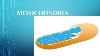
6. STRUCTURE AND FUNCTION
• Mitochondria made of two membranes. The outer membrane covers the organelle
and contains it like a skin. The inner membrane folds over many times and creates
layered structures called cristae. The fluid contained in the mitochondria is
called the matrix.
• The cristae greatly increase the total surface area of the inner membrane. ... The
membranes create two compartments.
• The inner membrane space, as implied, is the region between the inner and outer
membranes. It has an important role in the primary function of mitochondria,
which is oxidative phosphorylation (process in which ATP is formed)
7. •Its size ranges from 0.5 to 1.0 micrometer in diameter. The
structure comprises an outer membrane, an inner membrane, and
a gel-like material called the matrix.
• The outer membrane and the inner membrane are made of
proteins and phospholipid layers separated by the inner
membrane space.
•Ribosomes have two main functions — decoding the message
and the formation of peptide bonds. These two activities reside in
two large ribonucleoprotein particles (RNPs) of unequal size, the
ribosomal subunits.
•Each subunit is made of one or more ribosomal RNAs (rRNAs)
and many ribosomal proteins (r-proteins).
8.
9. • Mitochondria are known as the powerhouses of the cell.
Mitochondria are a double-membrane-bound cell organelle found
in most eukaryotic organisms.
• In all living cells, these cell organelles are found freely floating
within the cytoplasm of the cell.
• They are sites of cellular respiration.
• They uses oxygen to oxidize carbohydrates and fats present in the
cell to carbon dioxide and water. Oxidation releases energy, a
portion of which is used to form ATP.
Since mitochondria synthesizes energy rich compound ATP it is
called powerhouse of cell.
10.
11. • The heart muscle cells – with about 5,000 mitochondria per cell.
These cells need more energy, so they contain
more mitochondria than any other organ in the body.
• 5 Roles Mitochondria Play in Cells
• Production of ATP. Perhaps the most well-known role
of mitochondria is the production of ATP, the energy currency of
cells.
• Calcium Homeostasis.
• Regulation of Innate Immunity.
• Programmed Cell Death.
• Stem Cell Regulation.
12. • Mitochondria, organelles specialized to carry out aerobic
respiration, contain an inner membrane folded into cristae, which
form two separate compartments: the inner membrane space and
the matrix.
• Mitochondria convert chemical energy from the food we eat into
an energy form that the cell can use. This process is called
oxidative phosphorylation. The Krebs cycle produces a chemical
called NADH. NADH is used by enzymes embedded in the cristae
to produce ATP.
13. MITOCHONDRIA VS CHLOROPLAST
• Mitochondria and chloroplast are organelles found in a plant cell.
However, chloroplast is absent in an animal but mitochondria is found
in both. Mitochondria generates energy for the cell in the form of ATP
using oxygen and nutrients. Chloroplast is the site for photosynthesis in
a plant cell.
• Mitochondria and chloroplasts are the same size as prokaryotic cells
and divide by binary fission. Mitochondria and chloroplasts have their
own DNA which is circular, not linear. Mitochondria and
chloroplasts have their own ribosomes which have 30S and 50S
subunits, not 40S and 60S.