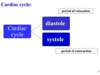
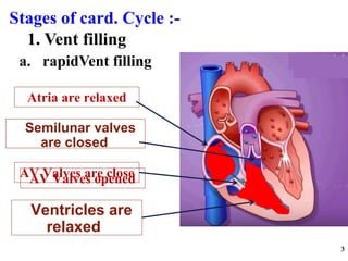
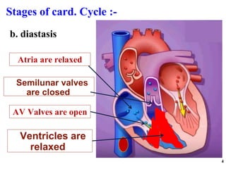
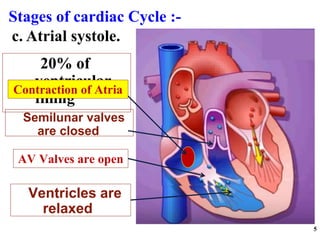
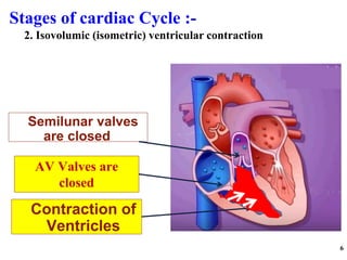
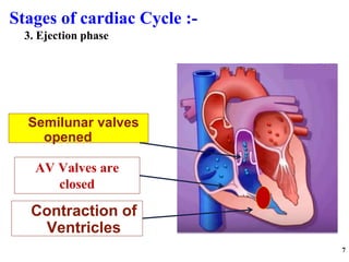
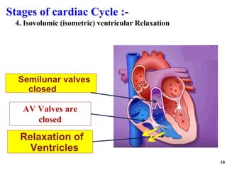
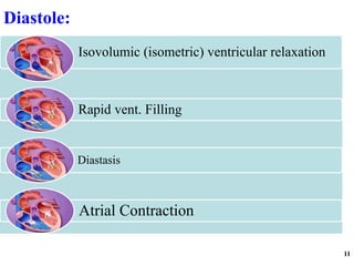
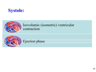
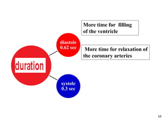
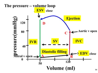
single cardiac cycle includes all of the events associated with one
heartbeat. Thus, a cardiac cycle consists of systole and diastole of the
atria plus systole and diastole of the ventricles.
The electrocardiogram (EKG) below the diagram shows the corresponding waves with each phase of the cardiac cycle. The bottom line represents the first and second heart sounds. The cardiac cycle represents the hemodynamic and electric changes that occur in systole and diastole. It has many phases.
Cardiac cycle and how the different chambers of the heart fill. We talk about the ventricular fillings and how diastole and systole work.
How pressure changes during all cycles
Medical science of cardiovascular system. It is the importance system in the human body. Blood is a specialised fruit can keep tissue which is circulated by cardiovascular system. Other system are respiratory system nervous system, gastrointestinal system . But cardiovascular system is the important system in our human body. Which involved heart
Describe events in cardiac cycle.
Describe atrial, ventricular and aortic pressure changes during cardiac cycle.
Describe the changes in ventricular volume & stroke volume during cardiac cycle.
Relate ECG changes to the phases of cardiac cycle.
Describe the functions of cardiac valves and relate their state to the production of heart sounds during cardiac cycle.
med_students0
CARDIAC CYCLE, ECG AND HEART SOUNDS.pptxthiru murugan
CARDIAC CYCLE, ECG AND HEART SOUNDS: BY Wincy Thirumurugan..
“Cardiac cycle refers to the series of events that take place when the heart beats.”
Each cycle is initiated by spontaneous contraction in the SA node and then transmit through the A-V bundle and branches into the ventricles results completion of one cycle.
EVENTS OR PHASES OF CARDIAC CYCLE: Diastolic phase (Diastole) in this phase the heart chamber are in the state of relaxation and fills with blood that receives from the veins [IVC, SVC,PULMONARY VEINS]
Systolic phase (Systole) in this the heart chambers are contracting and pumps the blood towards the periphery via the arteries. [ Pulmonary artery and aorta]
PHASES OF THE CARDIAC CYCLE
The different phases of the cardiac cycle involve:
Atrial diastole - Atrial relaxation
Atrial systole -Atrial contraction
Isovolumic relaxation -ventricular relaxation in the early phase but blood will not move and the Atrio ventricular valves will be closed
Ventricular filling - ventricular relaxation, the Atrio ventricular valves will be open allows filling blood in the ventricles
Isovolumic contraction of ventricle – ventricular systole in the early phase but no movement of the blood. The semilunar valves will be closed.
Ventricular ejection -ventricular contraction and send blood out of the ventricles through opened semilunar valves.
6. Ventricular Filling Stage: second phase. Rapid Filling, Slow Filling & Last Rapid Filling Duration of Cardiac Cycle:
In a normal person, a heartbeat is 72 beats/minute.
An Electrocardiogram (ECG) is a medical test that detects cardiac (heart) abnormalities by measuring the electrical activity generated by the heart as it. The machine that records the patient’s ECG is called an electrocardiograph.
contracts.
PLACEMENT OF ECG LEADS
ECG WAVES:
The P wave is caused by spread of depolarization through the atria, After the onset of the P wave, The QRS waves Occurs as a result of electrical depolarization of the ventricles, the ventricular T wave represents the stage of repolarization of the ventricles, The 'U' wave is a wave comes after the T wave of ventricular repolarization and may not always be observed.
HEART SOUNDS: First Heart Sound (S1)
The first heart sound results from the closing of the mitral and tricuspid valves. Second Heart Sound (S2): The second heart sound is produced by the closure of the aortic and pulmonic valves. Third Heart Sound (S3):
The third heart sound, also known as the “ventricular gallop,” occurs just after S2 when the mitral valve opens, allowing passive filling of the left ventricle. The S3 sound is actually produced by the large amount of blood striking a very compliant LV.
[Compliance heart means how easily the chamber of heart or the lumen of blood vessels expands when it is filling with the blood]
Fourth Heart Sound (S4):
The fourth heart sound, also known as the “atrial gallop,” occurs just before S1 when the atria contract to force blood into the LV.
it is my first try to make ppt. if you like than support me to make more ppt. i took help from various book thanks to ashalata anatomy and physiology and shambulingum.
This presentation describes the normal cardiac cycle referred to pressure-time curves for aorta, the left ventricle and left atrium, the electrocardiogram and the phonocardiogram.
Your heart is divided into four chambers. You have two chambers on the top (atrium, plural atria) and two on the bottom (ventricles), one on each side of the heart. Right atrium: Two large veins deliver oxygen-poor blood to your right atrium. The superior vena cava carries blood from your upper body.
Your heart is the main organ of your cardiovascular system, a network of blood vessels that pumps blood throughout your body. It also works with other body systems to control your heart rate and blood pressure. Your family history, personal health history and lifestyle all affect how well your heart works.
What is the heart?
The heart is a fist-sized organ that pumps blood throughout your body. It's the primary organ of your circulatory system.
Your heart contains four main sections (chambers) made of muscle and powered by electrical impulses. Your brain and nervous system direct your heart’s function.
What is the heart’s function?
Your heart’s main function is to move blood throughout your body. Your heart also:
Controls the rhythm and speed of your heart rate.
Maintains your blood pressure.
How does your heart work with other organs?
Your heart works with other body systems to control your heart rate and other body functions. The primary systems are:
Nervous system: Your nervous system helps control your heart rate. It sends signals that tell your heart to beat slower during rest and faster during stress.
Endocrine system: Your endocrine system sends out hormones. These hormones tell your blood vessels to constrict or relax, which affects your blood pressure. Hormones from your thyroid gland can also tell your heart to beat faster or slower.
Where is your heart located?
Your heart is located in the front of your chest. It sits slightly behind and to the left of your sternum (breastbone). Your ribcage protects your heart.
Where is your heart located?
Your heart is located in the front of your chest. It sits slightly behind and to the left of your sternum (breastbone). Your ribcage protects your heart.
What side is your heart on?
Your heart is slightly on the left side of your body. It sits between your right and left lungs. The left lung is slightly smaller to make room for the heart in your left chest.
On average, an adult’s heart weighs about 10 ounces. Your heart may weigh a little more or a little less, depending on your body size and sex.
What are the parts of the heart’s anatomy?
The parts of your heart are like the parts of a house. Your heart has:
Walls.
Chambers (rooms).
Valves (doors).
Blood vessels (plumbing).
Electrical conduction system (electricity).
Heart walls
Your heart walls are the muscles that contract (squeeze) and relax to send blood throughout your body. A layer of muscular tissue called the septum divides your heart walls into the left and right sides.
Your heart walls have three layers:
Endocardium: Inner layer.
Myocardium: Muscular middle layer.
Epicardium: Protective outer layer.
The epicardium is one layer of your
Cardiac cycle refers to a complete heartbeat from its generation to the beginning of the next beat.
Cardiac events that occur from –
beginning of one heart beat to the beginning of the next are called the cardiac cycle.
Report Back from SGO 2024: What’s the Latest in Cervical Cancer?bkling
Are you curious about what’s new in cervical cancer research or unsure what the findings mean? Join Dr. Emily Ko, a gynecologic oncologist at Penn Medicine, to learn about the latest updates from the Society of Gynecologic Oncology (SGO) 2024 Annual Meeting on Women’s Cancer. Dr. Ko will discuss what the research presented at the conference means for you and answer your questions about the new developments.
Title: Sense of Smell
Presenter: Dr. Faiza, Assistant Professor of Physiology
Qualifications:
MBBS (Best Graduate, AIMC Lahore)
FCPS Physiology
ICMT, CHPE, DHPE (STMU)
MPH (GC University, Faisalabad)
MBA (Virtual University of Pakistan)
Learning Objectives:
Describe the primary categories of smells and the concept of odor blindness.
Explain the structure and location of the olfactory membrane and mucosa, including the types and roles of cells involved in olfaction.
Describe the pathway and mechanisms of olfactory signal transmission from the olfactory receptors to the brain.
Illustrate the biochemical cascade triggered by odorant binding to olfactory receptors, including the role of G-proteins and second messengers in generating an action potential.
Identify different types of olfactory disorders such as anosmia, hyposmia, hyperosmia, and dysosmia, including their potential causes.
Key Topics:
Olfactory Genes:
3% of the human genome accounts for olfactory genes.
400 genes for odorant receptors.
Olfactory Membrane:
Located in the superior part of the nasal cavity.
Medially: Folds downward along the superior septum.
Laterally: Folds over the superior turbinate and upper surface of the middle turbinate.
Total surface area: 5-10 square centimeters.
Olfactory Mucosa:
Olfactory Cells: Bipolar nerve cells derived from the CNS (100 million), with 4-25 olfactory cilia per cell.
Sustentacular Cells: Produce mucus and maintain ionic and molecular environment.
Basal Cells: Replace worn-out olfactory cells with an average lifespan of 1-2 months.
Bowman’s Gland: Secretes mucus.
Stimulation of Olfactory Cells:
Odorant dissolves in mucus and attaches to receptors on olfactory cilia.
Involves a cascade effect through G-proteins and second messengers, leading to depolarization and action potential generation in the olfactory nerve.
Quality of a Good Odorant:
Small (3-20 Carbon atoms), volatile, water-soluble, and lipid-soluble.
Facilitated by odorant-binding proteins in mucus.
Membrane Potential and Action Potential:
Resting membrane potential: -55mV.
Action potential frequency in the olfactory nerve increases with odorant strength.
Adaptation Towards the Sense of Smell:
Rapid adaptation within the first second, with further slow adaptation.
Psychological adaptation greater than receptor adaptation, involving feedback inhibition from the central nervous system.
Primary Sensations of Smell:
Camphoraceous, Musky, Floral, Pepperminty, Ethereal, Pungent, Putrid.
Odor Detection Threshold:
Examples: Hydrogen sulfide (0.0005 ppm), Methyl-mercaptan (0.002 ppm).
Some toxic substances are odorless at lethal concentrations.
Characteristics of Smell:
Odor blindness for single substances due to lack of appropriate receptor protein.
Behavioral and emotional influences of smell.
Transmission of Olfactory Signals:
From olfactory cells to glomeruli in the olfactory bulb, involving lateral inhibition.
Primitive, less old, and new olfactory systems with different path
Lung Cancer: Artificial Intelligence, Synergetics, Complex System Analysis, S...Oleg Kshivets
RESULTS: Overall life span (LS) was 2252.1±1742.5 days and cumulative 5-year survival (5YS) reached 73.2%, 10 years – 64.8%, 20 years – 42.5%. 513 LCP lived more than 5 years (LS=3124.6±1525.6 days), 148 LCP – more than 10 years (LS=5054.4±1504.1 days).199 LCP died because of LC (LS=562.7±374.5 days). 5YS of LCP after bi/lobectomies was significantly superior in comparison with LCP after pneumonectomies (78.1% vs.63.7%, P=0.00001 by log-rank test). AT significantly improved 5YS (66.3% vs. 34.8%) (P=0.00000 by log-rank test) only for LCP with N1-2. Cox modeling displayed that 5YS of LCP significantly depended on: phase transition (PT) early-invasive LC in terms of synergetics, PT N0—N12, cell ratio factors (ratio between cancer cells- CC and blood cells subpopulations), G1-3, histology, glucose, AT, blood cell circuit, prothrombin index, heparin tolerance, recalcification time (P=0.000-0.038). Neural networks, genetic algorithm selection and bootstrap simulation revealed relationships between 5YS and PT early-invasive LC (rank=1), PT N0—N12 (rank=2), thrombocytes/CC (3), erythrocytes/CC (4), eosinophils/CC (5), healthy cells/CC (6), lymphocytes/CC (7), segmented neutrophils/CC (8), stick neutrophils/CC (9), monocytes/CC (10); leucocytes/CC (11). Correct prediction of 5YS was 100% by neural networks computing (area under ROC curve=1.0; error=0.0).
CONCLUSIONS: 5YS of LCP after radical procedures significantly depended on: 1) PT early-invasive cancer; 2) PT N0--N12; 3) cell ratio factors; 4) blood cell circuit; 5) biochemical factors; 6) hemostasis system; 7) AT; 8) LC characteristics; 9) LC cell dynamics; 10) surgery type: lobectomy/pneumonectomy; 11) anthropometric data. Optimal diagnosis and treatment strategies for LC are: 1) screening and early detection of LC; 2) availability of experienced thoracic surgeons because of complexity of radical procedures; 3) aggressive en block surgery and adequate lymph node dissection for completeness; 4) precise prediction; 5) adjuvant chemoimmunoradiotherapy for LCP with unfavorable prognosis.
New Directions in Targeted Therapeutic Approaches for Older Adults With Mantl...i3 Health
i3 Health is pleased to make the speaker slides from this activity available for use as a non-accredited self-study or teaching resource.
This slide deck presented by Dr. Kami Maddocks, Professor-Clinical in the Division of Hematology and
Associate Division Director for Ambulatory Operations
The Ohio State University Comprehensive Cancer Center, will provide insight into new directions in targeted therapeutic approaches for older adults with mantle cell lymphoma.
STATEMENT OF NEED
Mantle cell lymphoma (MCL) is a rare, aggressive B-cell non-Hodgkin lymphoma (NHL) accounting for 5% to 7% of all lymphomas. Its prognosis ranges from indolent disease that does not require treatment for years to very aggressive disease, which is associated with poor survival (Silkenstedt et al, 2021). Typically, MCL is diagnosed at advanced stage and in older patients who cannot tolerate intensive therapy (NCCN, 2022). Although recent advances have slightly increased remission rates, recurrence and relapse remain very common, leading to a median overall survival between 3 and 6 years (LLS, 2021). Though there are several effective options, progress is still needed towards establishing an accepted frontline approach for MCL (Castellino et al, 2022). Treatment selection and management of MCL are complicated by the heterogeneity of prognosis, advanced age and comorbidities of patients, and lack of an established standard approach for treatment, making it vital that clinicians be familiar with the latest research and advances in this area. In this activity chaired by Michael Wang, MD, Professor in the Department of Lymphoma & Myeloma at MD Anderson Cancer Center, expert faculty will discuss prognostic factors informing treatment, the promising results of recent trials in new therapeutic approaches, and the implications of treatment resistance in therapeutic selection for MCL.
Target Audience
Hematology/oncology fellows, attending faculty, and other health care professionals involved in the treatment of patients with mantle cell lymphoma (MCL).
Learning Objectives
1.) Identify clinical and biological prognostic factors that can guide treatment decision making for older adults with MCL
2.) Evaluate emerging data on targeted therapeutic approaches for treatment-naive and relapsed/refractory MCL and their applicability to older adults
3.) Assess mechanisms of resistance to targeted therapies for MCL and their implications for treatment selection
These simplified slides by Dr. Sidra Arshad present an overview of the non-respiratory functions of the respiratory tract.
Learning objectives:
1. Enlist the non-respiratory functions of the respiratory tract
2. Briefly explain how these functions are carried out
3. Discuss the significance of dead space
4. Differentiate between minute ventilation and alveolar ventilation
5. Describe the cough and sneeze reflexes
Study Resources:
1. Chapter 39, Guyton and Hall Textbook of Medical Physiology, 14th edition
2. Chapter 34, Ganong’s Review of Medical Physiology, 26th edition
3. Chapter 17, Human Physiology by Lauralee Sherwood, 9th edition
4. Non-respiratory functions of the lungs https://academic.oup.com/bjaed/article/13/3/98/278874
Ethanol (CH3CH2OH), or beverage alcohol, is a two-carbon alcohol
that is rapidly distributed in the body and brain. Ethanol alters many
neurochemical systems and has rewarding and addictive properties. It
is the oldest recreational drug and likely contributes to more morbidity,
mortality, and public health costs than all illicit drugs combined. The
5th edition of the Diagnostic and Statistical Manual of Mental Disorders
(DSM-5) integrates alcohol abuse and alcohol dependence into a single
disorder called alcohol use disorder (AUD), with mild, moderate,
and severe subclassifications (American Psychiatric Association, 2013).
In the DSM-5, all types of substance abuse and dependence have been
combined into a single substance use disorder (SUD) on a continuum
from mild to severe. A diagnosis of AUD requires that at least two of
the 11 DSM-5 behaviors be present within a 12-month period (mild
AUD: 2–3 criteria; moderate AUD: 4–5 criteria; severe AUD: 6–11 criteria).
The four main behavioral effects of AUD are impaired control over
drinking, negative social consequences, risky use, and altered physiological
effects (tolerance, withdrawal). This chapter presents an overview
of the prevalence and harmful consequences of AUD in the U.S.,
the systemic nature of the disease, neurocircuitry and stages of AUD,
comorbidities, fetal alcohol spectrum disorders, genetic risk factors, and
pharmacotherapies for AUD.
TEST BANK for Operations Management, 14th Edition by William J. Stevenson, Ve...kevinkariuki227
TEST BANK for Operations Management, 14th Edition by William J. Stevenson, Verified Chapters 1 - 19, Complete Newest Version.pdf
The prostate is an exocrine gland of the male mammalian reproductive system
It is a walnut-sized gland that forms part of the male reproductive system and is located in front of the rectum and just below the urinary bladder
Function is to store and secrete a clear, slightly alkaline fluid that constitutes 10-30% of the volume of the seminal fluid that along with the spermatozoa, constitutes semen
A healthy human prostate measures (4cm-vertical, by 3cm-horizontal, 2cm ant-post ).
It surrounds the urethra just below the urinary bladder. It has anterior, median, posterior and two lateral lobes
It’s work is regulated by androgens which are responsible for male sex characteristics
Generalised disease of the prostate due to hormonal derangement which leads to non malignant enlargement of the gland (increase in the number of epithelial cells and stromal tissue)to cause compression of the urethra leading to symptoms (LUTS
Title: Sense of Taste
Presenter: Dr. Faiza, Assistant Professor of Physiology
Qualifications:
MBBS (Best Graduate, AIMC Lahore)
FCPS Physiology
ICMT, CHPE, DHPE (STMU)
MPH (GC University, Faisalabad)
MBA (Virtual University of Pakistan)
Learning Objectives:
Describe the structure and function of taste buds.
Describe the relationship between the taste threshold and taste index of common substances.
Explain the chemical basis and signal transduction of taste perception for each type of primary taste sensation.
Recognize different abnormalities of taste perception and their causes.
Key Topics:
Significance of Taste Sensation:
Differentiation between pleasant and harmful food
Influence on behavior
Selection of food based on metabolic needs
Receptors of Taste:
Taste buds on the tongue
Influence of sense of smell, texture of food, and pain stimulation (e.g., by pepper)
Primary and Secondary Taste Sensations:
Primary taste sensations: Sweet, Sour, Salty, Bitter, Umami
Chemical basis and signal transduction mechanisms for each taste
Taste Threshold and Index:
Taste threshold values for Sweet (sucrose), Salty (NaCl), Sour (HCl), and Bitter (Quinine)
Taste index relationship: Inversely proportional to taste threshold
Taste Blindness:
Inability to taste certain substances, particularly thiourea compounds
Example: Phenylthiocarbamide
Structure and Function of Taste Buds:
Composition: Epithelial cells, Sustentacular/Supporting cells, Taste cells, Basal cells
Features: Taste pores, Taste hairs/microvilli, and Taste nerve fibers
Location of Taste Buds:
Found in papillae of the tongue (Fungiform, Circumvallate, Foliate)
Also present on the palate, tonsillar pillars, epiglottis, and proximal esophagus
Mechanism of Taste Stimulation:
Interaction of taste substances with receptors on microvilli
Signal transduction pathways for Umami, Sweet, Bitter, Sour, and Salty tastes
Taste Sensitivity and Adaptation:
Decrease in sensitivity with age
Rapid adaptation of taste sensation
Role of Saliva in Taste:
Dissolution of tastants to reach receptors
Washing away the stimulus
Taste Preferences and Aversions:
Mechanisms behind taste preference and aversion
Influence of receptors and neural pathways
Impact of Sensory Nerve Damage:
Degeneration of taste buds if the sensory nerve fiber is cut
Abnormalities of Taste Detection:
Conditions: Ageusia, Hypogeusia, Dysgeusia (parageusia)
Causes: Nerve damage, neurological disorders, infections, poor oral hygiene, adverse drug effects, deficiencies, aging, tobacco use, altered neurotransmitter levels
Neurotransmitters and Taste Threshold:
Effects of serotonin (5-HT) and norepinephrine (NE) on taste sensitivity
Supertasters:
25% of the population with heightened sensitivity to taste, especially bitterness
Increased number of fungiform papillae
Anti ulcer drugs and their Advance pharmacology ||
Anti-ulcer drugs are medications used to prevent and treat ulcers in the stomach and upper part of the small intestine (duodenal ulcers). These ulcers are often caused by an imbalance between stomach acid and the mucosal lining, which protects the stomach lining.
||Scope: Overview of various classes of anti-ulcer drugs, their mechanisms of action, indications, side effects, and clinical considerations.
Surat @ℂall @Girls ꧁❤8527049040❤꧂@ℂall @Girls Service Vip Top Model Safe
1599215324 (1).ppt
1. The cardiac cycle
Objectives:
1) To list the phases of the cardiac cycle in consecutive order.
2) To recognize that pressure changes determine valve action
& direction of blood flow through the heart.
3) Explain the basis of the arterial pulse and heart sounds.
4)To relate an ECG & heart sounds to events of the cardiac
cycle.
3. Stages of card. Cycle :-
3
Ventricles are
relaxed
AV Valves are close
Atria are relaxed
Semilunar valves
are closed
1. Vent filling
a. rapidVent filling
AV Valves opened
4. Stages of card. Cycle :-
4
Ventricles are
relaxed
AV Valves are open
Atria are relaxed
Semilunar valves
are closed
b. diastasis
5. 20% of
ventricular
filling
Stages of cardiac Cycle :-
5
Ventricles are
relaxed
AV Valves are open
Contraction of Atria
Semilunar valves
are closed
c. Atrial systole.
6. Stages of cardiac Cycle :-
6
Contraction of
Ventricles
AV Valves are
closed
Semilunar valves
are closed
2. Isovolumic (isometric) ventricular contraction
7. Stages of cardiac Cycle :-
7
Contraction of
Ventricles
AV Valves are
closed
Semilunar valves
opened
3. Ejection phase
8. The volume of blood in the ventricle at the end of the
systole
EDV ESV SV
8
E.D.V.
E.S.V.
S.V
The volume of blood in the ventricle at the end of the
diastole (just before ventricular systole started)
End diastolic volume
End systolic volume
The volume of blood ejected per one beat
Stroke volume
↑EDV→↑SV
↓SV → ↑ ESV
Normally about 70 ml at rest
9. 9
E.F
It is the percentage of volume of blood which is ejected
from the E.D.V.
Ejection fraction
E.F= SV ×100%
EDV
What is the ejection fraction of a left ventricle with EDV of
150ml and SV of 75 ml
E.F= SV ×100%
EDV
= 75 ×100%= 50%
150
Normally about 65%
10. Stages of cardiac Cycle :-
10
Relaxation of
Ventricles
AV Valves are
closed
Semilunar valves
closed
4. Isovolumic (isometric) ventricular Relaxation
The cardiac events that occur from the beginning of one heartbeat to the beginning of the next are called the cardiac cycle.
The cardiac cycle consists of a period of relaxation called diastole, during which the heart fills with blood, followed by a period of contraction called systole.
Filling of the Ventricles.
Rapid ventricular filling: During ventricular systole, large amounts of blood accumulate in the right and left atria because of the closed A-V valves. Therefore, as soon as systole is over and the ventricular pressures fall, the moderately increased pressures that have developed in the atria during ventricular systole immediately push the A-V valves open and allow blood to flow rapidly into the ventricles. This is called the period of rapid filling of the ventricles. The period of rapid filling lasts for about the first third of diastole.
b. Diastasis : During the middle third of diastole only a small amount of blood normally flows into the ventricles; this is blood that continues to empty into the atria from the veins and passes through the atria directly into the ventricles.
c. Atrial systole: During the last third of diastole, the atria contract and give an additional thrust to the inflow of blood
into the ventricles; this accounts for about 20 per cent of the filling of the ventricles during each heart cycle
Isovolumic (isometric) ventricular contraction:
The ventricle will contract on the same volume of blood→↑Pr. In the ventricle. When the Pr. In the left ventricle exceeds the Pr. Of the aorta (80 mmHg ) and the Pr. Of the right ventricle exceeds the Pr. of pulmonary artery (10 mmHg) Ejection phase start
Ejection phase :
The blood will be ejected to the arteries. The Pr. in the aorta =120 mmHg and Pr. in the pulmonary artery= 25 mmHg
Isovolumic or isometric ventricular relaxation.
the ventricle is going to relax on constant volume <<<<<decreases Pr. In the ventricle
Isovolumetric relaxation ends when the ventricular pressure falls below the atrial pressure
and the AV valves open, permitting the ventricles to fill
Duration of systole &diastole
Period of diastole of ventricles = 0.62 sec .
Period of systole of ventricles = 0.3 sec.
Diastolic Period is longer ALLOWING :-
1. More time for filling of the ventricle.
2. More time for relaxation of the coronary arteries→ ↑ blood supply to the ventricular muscle
The pressure – volume loop
• It is the relation between ventricular volume and pressure
It is composed of 4 phases:
Phase I: Period of vent filling.
Phase I in the volume-pressure diagram begins at a ventricular volume of about 50 milliliters and a pressure of 2 to 3 mm Hg. The amount of blood that remains in the ventricle after the previous heartbeat, 50 milliliters, is called the end-systolic volume. As venous blood flows into the ventricle from the left atrium, the ventricular volume normally increases to about 120 milliliters, called the end-diastolic volume, an increase of 70 milliliters. Therefore, the volume-pressure diagram during phase I extends along the line from point A to point B with the volume increasing to 120 milliliters and the diastolic pressure rising to about 5 to 7 mm Hg.
Phase II: Period of isovolumic contraction.
During isovolumic contraction, the volume of the ventricle does not change because all valves are closed. However, the pressure inside the ventricle increases to equal the pressure in the aorta, at a pressure value of about 80 mm Hg (point C)
Phase III: Period of ejection.
During ejection, the systolic pressure rises even higher because of still more contraction of the ventricle. At the same time, the volume of the ventricle decreases because the aortic valve has now opened and blood flows out of the ventricle into the aorta.
Phase IV: Period of isovolumic relaxation.
At the end of the period of ejection (point D) the aortic valve closes and the ventricular pressure falls back without any change in volume. Thus, the ventricle volume returns to its starting point, with about 50 milliliters of blood left in the ventricle and at an atrial pressure of 2-3 mm Hg