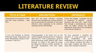
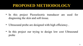
JERUSALEM COLLEGE OF ENGINEERING
DEPARTMENT OF BIOMEDICAL ENGINEERING
DESIGN OF ULTRASOUND PROBE
1
BATCH NO.:
Team Members:
1.K.Anusuya
2.T.Archana
3.J.Melita
4.S.Shruthi
Supervisor:
Ms.V.Subha Ramya
Department of Biomedical Engineering
OBJECTIVE
2
• To design a low cost and effective Ultrasound
Probe which is made with 2Mhz frequency and
mainly used for skin and soft tissue diagnosis.
Department of Biomedical Engineering
INTRODUCTION
• The father of ultrasound John J. Wild
• In the your of 1916, ultrasound was fird and for, medical proposes,
ultrasound was the brainchild of engirem Tom Brain and obstetrician lan
Donald.
• The active elemente in ultrasound transducers are reade of Special Ceranic
Crystal materials Called piezoelectrics
• Ultrasound waves are transmitted from the transduty through the gol iste the
body. These short burate of found energy hit the desired Organs and rition to
the proks as an echo.
3
Department of Biomedical Engineering
ABSTRACT
• Ultrasound technology is used high-energy sound waves to
look at tissues and Organs inside the body. Ultrasound
probes are being used for the detection. There are different
types of probes, involved in ultrasound machines which are
linear, curvilinear and phased array. Considering this in
mind our team decided to design a cost effective ultrasound
probe which involves 2Mhz frequency and are highly
efficient for skin and soft tissue diagnosis.
Department of Biomedical Engineering 4
COMPONENTS USED
• Transducer lead
• Plastic Housing
• Metal Shield
• Acoustic Insulator
• Damping Block
• Piezoelectric element
• Positive Electrode
• Ground Electrode
• Matching Layers
LITERATURE REVIEW
6
TITLE & YEAR DESCRIPTION INFERENCE
Overview of Ultrasound Imaging
Applications in Dermatology - 2021
Complete visualization of lessons is critical for
the accurate diagnosis and management of
dermatological diseases. Currently the most
readily available technologies used by
dermatologies include dermoscopy and
photography.
US is a useful non-invasive modality in
the field of dermatology that can provide
critical information to complement the
clinical examination.It can accrately assess
disease severity and activity in several
inflammatory dermatoses.
Development of Low - Cost Medical
Ultrasound Scanner Using a
monostalic Synthetic Apeature -
January 2017
The design of low-cost medical ultrasound
scanners aimmed at the detection of maternal
mortality risk factors in developing
countries.Modern Ultrasound scanners typically
employ a highh element count transducer array
with multichannel transmit & receive
electrodes.
In this paper a low cost monostatic
synthetic aperature scanner(MSAS)was
preseented with potential applications in
low resource countries for the detection of
materrnal risk facctors.This system was
designed to reduce the production costs by
replacing the multi-element piezoelectric
transducer array by asingle piezoelectric
element.
LITERATURE REVIEW
7
TITLE & YEAR DESCRIPTION INFERENCE
Ultrasound for the Evaluation of Skin
and Soft Tissue Infections - May
2015
Skin and soft tissue infcetions including
abscesses and cellulitides,are common problems
seen by physicians.The treatment of soft tissue
infections varies depending on the collection
requiring incisions and drainage
Colour flow Doppler sonogrphy will aid
in recognition of adjacent vascular
structures and mayn also detect blood
flow through a pesudoanerysm .Lymph
nodes are highly vascular and should
demonstrate strong colour flow throughout
while abcesses should not.
A Low cost Simulator to Practice
Ultrasound Image Interpretation and
Probe Manipulation: Design & firat
Evaluation - April 2o11
Ultrassonography is the lowest cost no risk
medical imaging technique.However, reading an
ultrasound US well as performing a good US
probe positioning remain difficult tasks. More
precisely we tackle in this paper the tisssue
providing acost effective realistic intefrface for
the probe manipulation with basic haptic
feedback.
We have presented a simulator for
education of young practionier to US
image interpretation and probe
manipulation .Our objective is to provide
a cost effective simulator that allows a
basic haptic feedback and which can be
easily used a standard workstation.
PROPOSED METHODOLOGY
• In this project Piezoelectric transducer are used for
diagnosing the skin and soft tissue.
• Ultrasound probe are designed with high efficiency .
• In this project our trying to design low cost Ultrasound
probe
8
REFERENCES
• https://www.ncbi.nlm.nih.gov/pmc/articles/PMC6170135/
• https://
www.researchgate.net/publication/224234134_A_low_cost_simulator_to_practice
_ultrasound_image_interpretation_and_probe_manipulation_Design_and_first_e
valuation
• https://
mail.google.com/mail/u/0?ui=2&ik=491d2e4032&attid=0.1&permmsgid=msg-a:r
6750938779666524127&th=1912e07e2b417399&view=att&disp=inline&realattid
=1912e07b791e22fedeb1
• https://mail.google.com/mail/u/0?ui=2&ik=491d2e4032&attid=0.1&permmsgid=
msg-a:r-7471925087723887928&th=1912e09063fb591c&view=att&disp=inline&r
ealattid=1912e08eadc69257c971 9
Thank you
10
Department of Biomedical Engineering

0th review template (11) (1).pptx review document template

  • 1.
    JERUSALEM COLLEGE OFENGINEERING DEPARTMENT OF BIOMEDICAL ENGINEERING DESIGN OF ULTRASOUND PROBE 1 BATCH NO.: Team Members: 1.K.Anusuya 2.T.Archana 3.J.Melita 4.S.Shruthi Supervisor: Ms.V.Subha Ramya Department of Biomedical Engineering
  • 2.
    OBJECTIVE 2 • To designa low cost and effective Ultrasound Probe which is made with 2Mhz frequency and mainly used for skin and soft tissue diagnosis. Department of Biomedical Engineering
  • 3.
    INTRODUCTION • The fatherof ultrasound John J. Wild • In the your of 1916, ultrasound was fird and for, medical proposes, ultrasound was the brainchild of engirem Tom Brain and obstetrician lan Donald. • The active elemente in ultrasound transducers are reade of Special Ceranic Crystal materials Called piezoelectrics • Ultrasound waves are transmitted from the transduty through the gol iste the body. These short burate of found energy hit the desired Organs and rition to the proks as an echo. 3 Department of Biomedical Engineering
  • 4.
    ABSTRACT • Ultrasound technologyis used high-energy sound waves to look at tissues and Organs inside the body. Ultrasound probes are being used for the detection. There are different types of probes, involved in ultrasound machines which are linear, curvilinear and phased array. Considering this in mind our team decided to design a cost effective ultrasound probe which involves 2Mhz frequency and are highly efficient for skin and soft tissue diagnosis. Department of Biomedical Engineering 4
  • 5.
    COMPONENTS USED • Transducerlead • Plastic Housing • Metal Shield • Acoustic Insulator • Damping Block • Piezoelectric element • Positive Electrode • Ground Electrode • Matching Layers
  • 6.
    LITERATURE REVIEW 6 TITLE &YEAR DESCRIPTION INFERENCE Overview of Ultrasound Imaging Applications in Dermatology - 2021 Complete visualization of lessons is critical for the accurate diagnosis and management of dermatological diseases. Currently the most readily available technologies used by dermatologies include dermoscopy and photography. US is a useful non-invasive modality in the field of dermatology that can provide critical information to complement the clinical examination.It can accrately assess disease severity and activity in several inflammatory dermatoses. Development of Low - Cost Medical Ultrasound Scanner Using a monostalic Synthetic Apeature - January 2017 The design of low-cost medical ultrasound scanners aimmed at the detection of maternal mortality risk factors in developing countries.Modern Ultrasound scanners typically employ a highh element count transducer array with multichannel transmit & receive electrodes. In this paper a low cost monostatic synthetic aperature scanner(MSAS)was preseented with potential applications in low resource countries for the detection of materrnal risk facctors.This system was designed to reduce the production costs by replacing the multi-element piezoelectric transducer array by asingle piezoelectric element.
  • 7.
    LITERATURE REVIEW 7 TITLE &YEAR DESCRIPTION INFERENCE Ultrasound for the Evaluation of Skin and Soft Tissue Infections - May 2015 Skin and soft tissue infcetions including abscesses and cellulitides,are common problems seen by physicians.The treatment of soft tissue infections varies depending on the collection requiring incisions and drainage Colour flow Doppler sonogrphy will aid in recognition of adjacent vascular structures and mayn also detect blood flow through a pesudoanerysm .Lymph nodes are highly vascular and should demonstrate strong colour flow throughout while abcesses should not. A Low cost Simulator to Practice Ultrasound Image Interpretation and Probe Manipulation: Design & firat Evaluation - April 2o11 Ultrassonography is the lowest cost no risk medical imaging technique.However, reading an ultrasound US well as performing a good US probe positioning remain difficult tasks. More precisely we tackle in this paper the tisssue providing acost effective realistic intefrface for the probe manipulation with basic haptic feedback. We have presented a simulator for education of young practionier to US image interpretation and probe manipulation .Our objective is to provide a cost effective simulator that allows a basic haptic feedback and which can be easily used a standard workstation.
  • 8.
    PROPOSED METHODOLOGY • Inthis project Piezoelectric transducer are used for diagnosing the skin and soft tissue. • Ultrasound probe are designed with high efficiency . • In this project our trying to design low cost Ultrasound probe 8
  • 9.
    REFERENCES • https://www.ncbi.nlm.nih.gov/pmc/articles/PMC6170135/ • https:// www.researchgate.net/publication/224234134_A_low_cost_simulator_to_practice _ultrasound_image_interpretation_and_probe_manipulation_Design_and_first_e valuation •https:// mail.google.com/mail/u/0?ui=2&ik=491d2e4032&attid=0.1&permmsgid=msg-a:r 6750938779666524127&th=1912e07e2b417399&view=att&disp=inline&realattid =1912e07b791e22fedeb1 • https://mail.google.com/mail/u/0?ui=2&ik=491d2e4032&attid=0.1&permmsgid= msg-a:r-7471925087723887928&th=1912e09063fb591c&view=att&disp=inline&r ealattid=1912e08eadc69257c971 9
  • 10.
    Thank you 10 Department ofBiomedical Engineering