
Recommended
More Related Content
What's hot
What's hot (20)
Similar to Cerebrum & lateral ventricle
Similar to Cerebrum & lateral ventricle (20)
More from sarikachopde
Recently uploaded
Recently uploaded (20)
Cerebrum & lateral ventricle
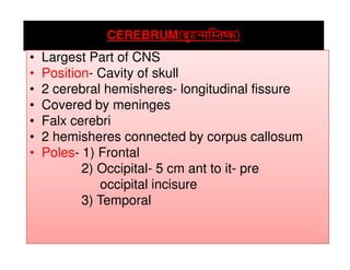
- 1. CEREBRUM((((oÉëÑWûlqÉÎxiÉwMüoÉëÑWûlqÉÎxiÉwMüoÉëÑWûlqÉÎxiÉwMüoÉëÑWûlqÉÎxiÉwMü)))) • Largest Part of CNS• Largest Part of CNS • Position- Cavity of skull • 2 cerebral hemisheres- longitudinal fissure • Covered by meninges • Falx cerebri • 2 hemisheres connected by corpus callosum• 2 hemisheres connected by corpus callosum • Poles- 1) Frontal 2) Occipital- 5 cm ant to it- pre occipital incisure 3) Temporal
- 2. Sulci & Gyri Types of Sulci Limiting Axial OperculatedComplete Separating Develop Third areaPresent onSeparating 2 functional Areas e.g.Central Develop along rapidly developing Brain axis Calcarine Third area Does not Appear on Surface Lunate Present on Surface, Project on lat.ventricle calcarine
- 3. CEREBRUM Rt.Cerebral Hemisphere Lt.Cerebral Hemisphere Longitudinal Cerebral Fissure(Falx cerebri Rt.Cerebral Hemisphere
- 4. Surfaces & Borders Surfaces Superolat. Medial Inferior Orbital TentorialTentorial Borders Inferolat.Superomedial Median Occipital Median Orbital
- 8. Lat.Sulcus Stem Starts from ant perforated substance Lesser wing Rami Ant.ascending Post Ant. Ant.ascending Post Pars orbitalis Pars triangularis Pars opercularis
- 9. Stem of lat sucus Transmits middle cerebral vessels
- 10. Lesser wing
- 11. Sulci on frontal lobe • Frontal lobe boundaries 1) Sup- Superomed. Border 2) Inf- stem of lat. Sulcus/superciliary border 3) Post- Central sulcus CENTRAL SULCUSCENTRAL SULCUS • Starts behind the midpoint of frontal & occipital poles • Ends above the post ramus • Marks the junction of motor & sensory area PRECENTRAL SULCUS • Ant to central sulcus
- 12. Sulci on frontal lobe 1) Sup frontal 2) Inf frontal Gyri on frontal lobe 1. Precentral- coticonuclear & corticospinal tracts 2. Sup. Frontal 3. Middle frontal 4. Inf frontal- Broca’s area- pars
- 20. Sulci on Parietal lobe • Parietal lobe boundaries 1) Sup- Superomed. Border 2) Inf- Post ramus 3) Post- Parieto occipital sulcus 4) Ant- Central sulcus Post central • Parallel & post.to central• Parallel & post.to central Intraparietal • Starts from postcentral. Passes horizontally to occipital lobe Gyri on parietal lobe 1) Postcentral 2) Sup parietal lobule 3) Inf. Parietal lobule- a) Supramarginal b) Angular- Visual c) Posterior
- 22. Sulci on Occipital lobe Transverse occipital • Descends from superomed border Lat occipital • Short horizontal Lunate In front of occipital poleIn front of occipital pole GYRI ON OCCIPITAL LOBE 1) Arcus parieto occipitalis 2) Sup occipital 3) Inf occipital 4) Gyrus descendes
- 24. Sulci on Temporal lobe Sup temporal • Begins near temporal pole runs horizontally Inf temporal • Below sup temoporal GYRI ON TEMPORAL LOBE 1) Sup temporal1) Sup temporal 2) Middle temporal 3) Inf temporal
- 25. INSULA • Considered as 5th lobe • Deep in the floor of lat sulcus • Surrounded by circular sulcus • Area of cerebral cortex overlapping insula- Opercula • Apex of insula- Limen insulae • Surface divided- sulcus centralis insulae Frontal operculum Bet- ant horizontal & ascending Frontoparietal operculum Bet- ant ascending & post Temporal operculum Below post ramus
- 27. Medial surface • Corpus callosum • Septum pellucidum • Lateral ventricle • Sulci & gyri- Sulci & GyriSulci & Gyri 1. Cingulate sulcus- Above & parallel to corpus callosum. Ends at the superomedial border 2. Parieto- occipital sulcus- starts on superomed border meets calcarine 3. Calcarine sulcus- Starts from occipital pole. Passes towards corpus callosum
- 28. Medial surface Med frontal Lower limb movements
- 29. Medial surface Gyri 1. Cingulate gyrus – Below the cingulate sulcus 2. Medial frontal- Above cingulate- anteriorly 3. Paracentral lobule- Above cingulate- posteriorly.Lower limb movements 4. Precuneus- Bet. Parieto occipital & cingulate 5. Cuneus- Bet. Parieto occipital & calcarine 6. Lingual- Below calcarine
- 30. Corpus callosum •Connects the 2 cerebral hemispheres • Length- 10 cm • Ant end- 4 cm away from frontal pole • Post end- 6 cm away from occipital pole Relations of genu- Post- septum pellucidum - Ant – Ant cerebral artery- Ant – Ant cerebral artery Relations of rostrum- Ant – Ant cerebral artery Relations of splenium- Inf- Pineal gland -Ant – Thalamus
- 31. Relations of trunk- Inf- septum pellucidum/ Lateral ventricle -Sup – Covered by Indusium gresium. Related to falx cerebri Ant cerebral vessels
- 33. Relations Of body & splenium Thalamus Pineal body Fornix
- 34. Relations Of Genu & trunk Genu Anterior cerebral artery Genu
- 35. Relations Of corpus callosum
- 36. Communicating Fibres Forceps minor Fibres of genu connecting frontal lobes Tapetum Forceps Major Fibres of splenium Tapetum Fibres of trunk & splenium
- 38. Cerebrum Inf. Surface-Rt. Lat. orbital Gyrus Rectus Orbital Olfactory Sulcus Orbital Sulcus
- 39. Cerebrum Inf. Surface-Rt. Lat. Tentorial
- 40. Sulci 1) Collateral 2) Occipitotemporal Gyri- Cerebrum Inf. Surface-Rt. Lat. Tentorial 1) Lingual 2) Parahippocampal 3) Uncus 4) Med.occipitotemporal 5) Lat. occipitotemporal
- 41. Limbic Lobe & Olfactory Pathways Associated with visceral olfactory,social somatic,emotional factors 1. Olfactory Bulb- Oval ,reddish grey(1. Olfactory Bulb- Oval ,reddish grey( cribriform ) 2. Olfactory tract- orbital surface (frontal) 3. Amygdala- Resembling Almond a) Amygdaloid body b) Amygdaloid nuclear complex
- 42. 4. Stria terminalis- Fine mylinated fibres from amygdaloid complex 5. Ant. Commissure- Crossing midline ant to columns of fornix 6. Mammilary bodies- Memory Structure of cerebrum Outer Grey mater Inner white mater- Red dots( Blood escape)
- 43. Nuclei Of cerebrum 1. Amygadaloid 2. Claustrum 3. Caudate 4. Lentiform
- 44. Fornix C shaped bundle of fibres
- 45. Fibres of cerebrum Commissural Connect 2 Hemispheres Association Diff. cortical Areas on same Projection Cerebral cortex toHemispheres Corpus callosum Areas on same Side e.g.Cingulum cortex to Brain stem & Sp.cord e.g.Corona radiata
- 47. Blood supply- Ant. Cerebral Middle cerebral post cerebral Venous drain- Sup.& Inf.cerebral Middle cerebralMiddle cerebral Ant.cerebral Lymphatics – No lymphatics in CNS Applied- 1.Tumour 2.Infarct 3.Infection
- 51. Lateral Ventricle • Irregular cavities in lower & med. Parts of cerebrum • Both sides separated by septum pellucidum • Communicates- 3rd ventricle- Interventricular foramen/ Monro • Lined with ependyma. • Secrets & contains CSF• Secrets & contains CSF • Structure Body Cornu/Horns Ant Post Inf
- 52. Lateral ventricle Post horn Body Ant horn Inf .horn Post horn
- 54. Central part/body • From interventricular foramen to splenium • Relations- 1) Roof- Corpus callosum 2) Floor- In lateromed. Order- a) Caudate nucleus- Narrow post, /long axis directed posteromed. b) Stria terminalis- Small bundle of white fibres occupyingb) Stria terminalis- Small bundle of white fibres occupying narrow groove med to caudate nucleus c) Thalamostriate vein d) Thalamus- Sup surface. Almost hidden by choroid plexus. 3) Medial wall- Septum pellucidum
- 58. Relations Of Ant. Cornu Passes in the frontal lobe Appears triangular in coronal section Relations- 1.Sup.- Ant part of corpus callosum 2. Ant- Post. surface of genu 3. Inf.- Caudate nucleus(head) 4. Med.- Septum pellucidum
- 61. May be absent In occipital lobe Relations- 1. Roof- Tapetum 2. Lat- Tapetum 3. Med.- Forceps major. Form bulb of post. Horn Relations Of Post. Cornu of post. Horn Below this- Elevation- Calcar axis( Calcarine sulcus)
- 62. Post .Horn
- 64. • Largest of all • In the temporal lobe • Curves around the post end of thalamus • Position indicated by sup. temporal sulcus Relations- 1.Roof- Tapetum, caudate nucleus Relations Of Inf. Cornu Relations- 1.Roof- Tapetum, caudate nucleus 2. Floor- Collateral eminence, hippo campus