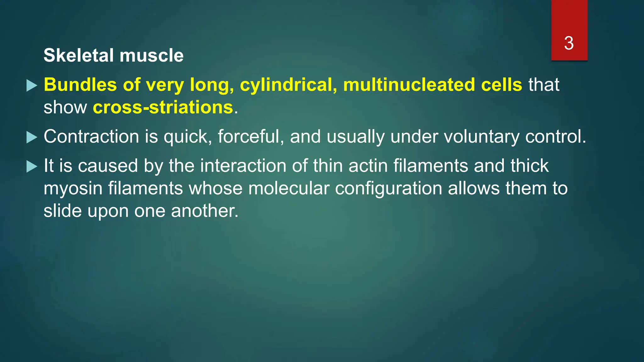
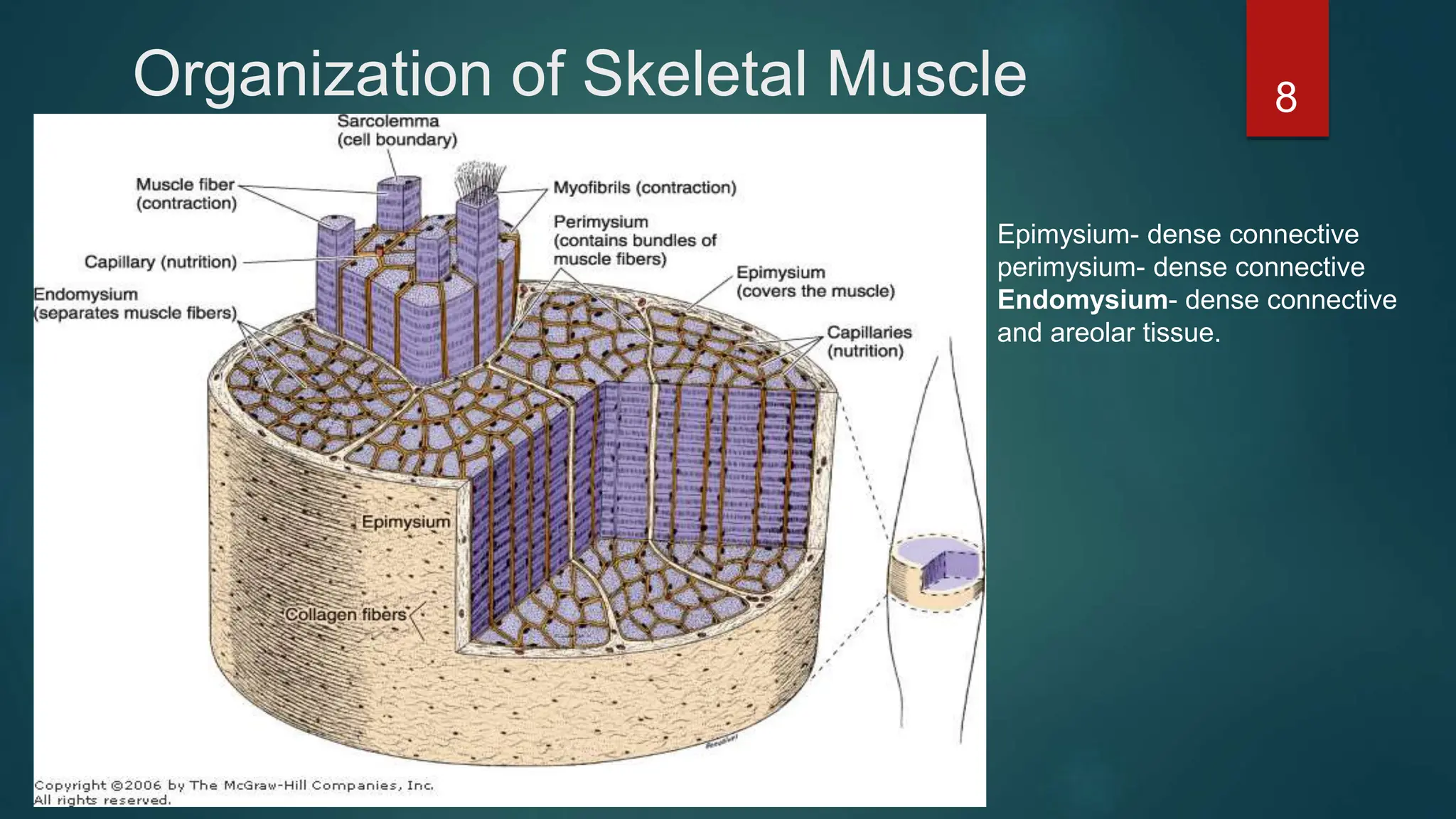
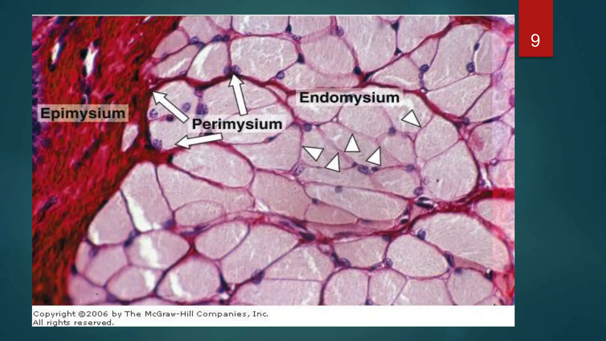
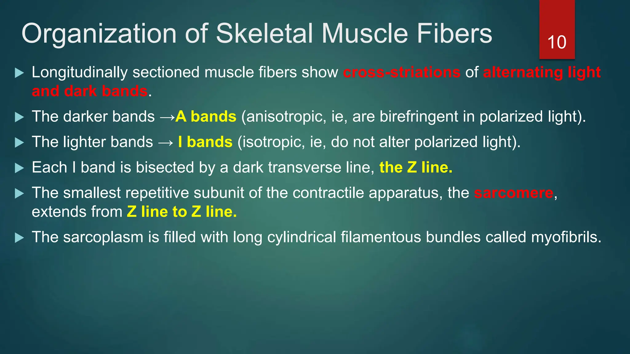
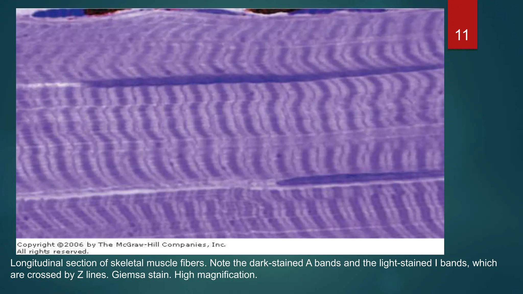
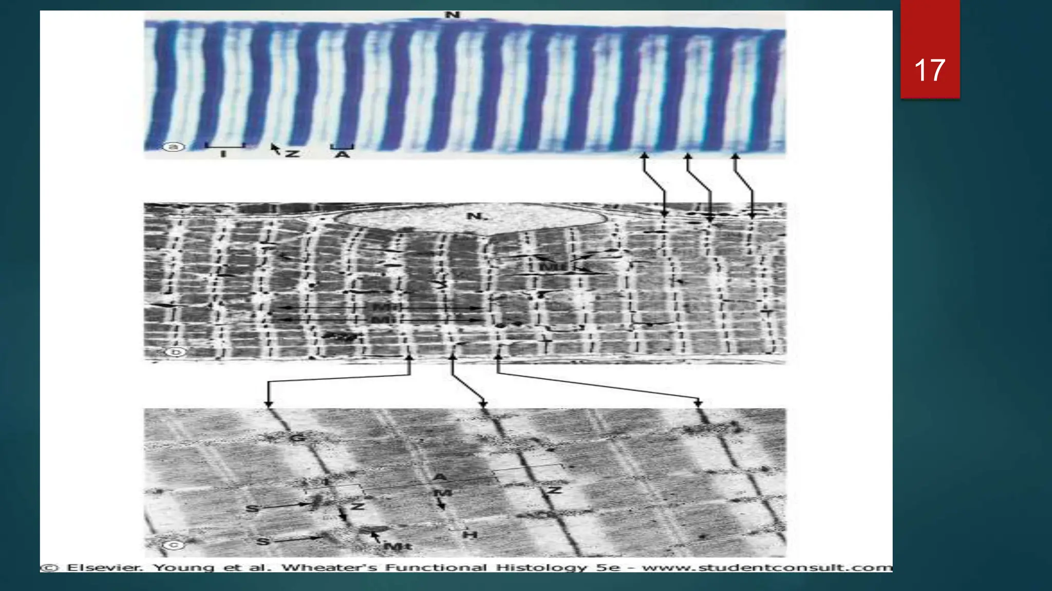
2. Introduction
Muscles are multicellular contractile units of contractile cells.
Cells containing contractile proteins.
Structural biology of these proteins generates the forces
necessary for cellular contraction, which drives movement within
certain organs and the body as a whole
On the basis of morphological and functional characteristics
1. Skeletal muscles.
2. Cardiac muscles.
3. Smooth muscle.
2
3. Skeletal muscle
Bundles of very long, cylindrical, multinucleated cells that
show cross-striations.
Contraction is quick, forceful, and usually under voluntary control.
It is caused by the interaction of thin actin filaments and thick
myosin filaments whose molecular configuration allows them to
slide upon one another.
3
4. Cardiac muscle
has cross-striations and is composed of elongated, branched
individual cells that lie parallel to each other.
At sites of end-to-end contact are the intercalated disks,
structures found only in cardiac muscle.
Contraction of cardiac muscle is involuntary, vigorous, and
rhythmic.
Smooth muscle
consists of collections of fusiform cells that do not show cross-
striations.
Their contraction process is slow and not subject to voluntary
control.
4
5. PLEASE NOTE
Some muscle cell organelles have names that differ from
their counterparts in other cells.
The cell membrane of a muscle is called sarcolemma or
plasmalemma.
The cytoplasm is called sarcoplasm
The smooth endoplasmic reticulum is called
sarcoplasmic reticulum.
5
7. Skeletal Muscle
Consists of muscle fibers, bundles of very long (up to
30 cm) cylindrical multinucleated cells.
Multinucleation results from the fusion of embryonic
mononucleated myoblasts (muscle cell precursors).
The oval nuclei are usually found at the periphery
of the cell under the cell membrane.
7
8. Organization of Skeletal Muscle 8
Epimysium- dense connective
perimysium- dense connective
Endomysium- dense connective
and areolar tissue.
10. Organization of Skeletal Muscle Fibers
Longitudinally sectioned muscle fibers show cross-striations of alternating light
and dark bands.
The darker bands →A bands (anisotropic, ie, are birefringent in polarized light).
The lighter bands → I bands (isotropic, ie, do not alter polarized light).
Each I band is bisected by a dark transverse line, the Z line.
The smallest repetitive subunit of the contractile apparatus, the sarcomere,
extends from Z line to Z line.
The sarcoplasm is filled with long cylindrical filamentous bundles called myofibrils.
10
11. 11
Longitudinal section of skeletal muscle fibers. Note the dark-stained A bands and the light-stained I bands, which
are crossed by Z lines. Giemsa stain. High magnification.
13. The Sarcomere
The smallest repetitive subunit of the contractile apparatus, the
sarcomere, extends from Z line to Z line.
This sarcomere pattern is due mainly to the presence of two types of
filaments thick and thin.
The thick filaments occupy the A band.
The thin filaments run between and parallel to the thick filaments and
have one end attached to the Z line.
13
15. The Sarcomere
A band shows the presence of a lighter zone in its center, the
H band, that corresponds to a region consisting only of the
rodlike portions of the myosin molecule.
Bisecting the H band is the M line, a region at which lateral
connections are made between adjacent thick filaments.
15
18. Striated muscle proteins;
The four main proteins are actin, tropomyosin, troponin, and
myosin.
Thin filaments are composed of actin, tropomyosin, troponin.
Thick filaments consist primarily of myosin.
18
19. Cardiac Muscle
Mature cardiac muscle exhibit a cross-striated banding pattern identical to
that of skeletal muscle.
Each cardiac muscle cell possesses one or two centrally located pale-
staining nuclei.
Surrounding the muscle cells is a delicate sheath of endomysial
connective tissue containing a rich capillary network.
A unique and distinguishing characteristic of cardiac muscle is the presence
of dark-staining transverse lines that cross the chains of cardiac cells at
irregular intervals.
19
20. Intercalated disc
Junctional complexes found at the interface between adjacent cardiac muscle cells.
Appear as straight lines or may exhibit a steplike pattern.
It has two regions a transverse portion,and a lateral portion.
There are three main junctional specializations within the disk.
1. Zonulae adherentes which serve as anchoring sites for actin filaments of the terminal
sarcomeres.
2. Maculae adherentes (desmosomes) bind the cardiac cells together, preventing them from pulling
apart under constant contractile activity.
3. Gap junctions provide ionic continuity between adjacent cells.
The significance of ionic coupling is that chains of individual cells act as a syncytium, allowing the
signal to contract to pass in a wave from cell to cell.
20
21. 21
Drawing of a section of heart muscle, showing central nuclei, cross-striation, and intercalated disks.
24. Smooth Muscle
Smooth muscle is composed of elongated, nonstriated cells, each of
which is enclosed by a basal lamina and a network of reticular fibers .
The basal lamina and a network of reticular fibers serve to combine the force
generated by each smooth muscle fiber into a concerted action, eg,
peristalsis in the intestine.
cells are fusiform.
Cells have a single nucleus located in the center of the broadest part of the
cell.
The borders of the cell become scalloped when smooth muscle contracts,
and the nucleus becomes folded or has the appearance of a corkscrew.
24
25. Regeneration of Muscle Tissue
The three types of adult muscle have different potentials for regeneration after injury.
Cardiac muscle
no regenerative capacity beyond early childhood.
Defects or damage (eg, infarcts) in heart muscle are generally replaced by the
proliferation of connective tissue, forming myocardial scars.
skeletal muscle
undergo limited regeneration.
The source of regenerating cells is believed to be the satellite cells.
They are considered to be inactive myoblasts that persist after muscle differentiation.
Smooth muscle
an active regenerative response.
After injury, viable mononucleated smooth muscle cells and pericytes from blood
vessels undergo mitosis and provide for the replacement of the damaged tissue.
25
However, this is often difficult to see with light microscopy due to the irregular branching shape of the cells and their myofibrils.
The latter are a sparse population of mononucleated spindle-shaped cells that lies within the basal lamina surrounding each mature muscle fiber. Because of their intimate apposition with the surface of the muscle fiber, they can be identified only with the electron microscope.