- Mycobacterium leprae is the causative bacteria of leprosy (Hansen's disease), which was first recognized in ancient times and described by Hippocrates. The bacteria was discovered in 1873 and causes a chronic granulomatous disease primarily affecting the skin, nerves, and respiratory tract.
- Leprosy has a long incubation period of 5-7 years on average and can be classified based on clinical presentation and bacterial load as tuberculoid, borderline, or lepromatous. Effective treatment involves multidrug therapy with rifampicin, dapsone, and clofazimine for 6-12 months depending on classification.
- Without treatment, le
Russian Call Girls In Pune 👉 Just CALL ME: 9352988975 ✅❤️💯low cost unlimited ...
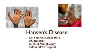
Hansens disease
1.
2. INTRODUCTION
• Mycobacterium leprae is the causative agent of Hansen’s Disease; a disease of antiquity,
having been recognized since long time such as:
o Vedic times in India (described as Kushta Roga in Sushruta Sambita, 600 BC)
o Biblical times in the Middle East
o Hippocrates, 460 BC.
• The credit of discovery of lepra bacilli goes to GH Armauer Hansen (1873) in Norway.
• Although, M. leprae was the first bacterial pathogen of humans to be described, still it
remains one of the least understood organisms probably because it is not cultivable.
(Exception of KOCH’S Postulates)
• However, Shepard (1960) had done a breakthrough by multiplying the lepra bacilli in the
footpads of mice kept at a low temperature (20°C).
3. CLINICAL MANIFESTRATION
• Leprosy is a chronic granulomatous disease of humans.
• Primarily involving cooler parts of the body (skin, peripheral nerves, upper
respiratory tract, eyes, and testes, etc.)
• But are capable of affecting any tissue or organs causing bony deformities and
disfigurements in untreated cases.
• Incubation period: Leprosy has a long incubation period, an average of 5-7years
(vary between 2 and 40 years)
o Lepra bacilli:12-13 days
o Tubercle bacillus: 14hr
o Coliform: 20min
4. CLASSIFICATION
• Leprosy can be classified into various categories based on clinical, bacteriological,
immunological and histological status of the patients.
• There are three classification schemes.
• Following initiation of treatment or alteration of host immunity, the leprosy category of
patients changes from one type to another type.
RIDLEY-JOPLING CLASSIFICATION
(1966)
MADRID CLASSIFICATION
(1953)
INDIAN CLASIFICATION (1981)
[By Leprosy Association of India]
Lepromatous Leprosy (LL) Lepromatous type Lepromatous type
Borderline Lepromatous Leprosy (BL) Borderline Borderline
Borderline Leprosy (BB) Interminate type Interminate type
Borderline Tuberculoid Leprosy (BT) Tuberculoid type Pure Nuritic type
Tuberculoid Leprosy (TT) Tuberculoid type
5. Clinical Classification
Based on the number of skin lesions, presence of nerve involvement and identification of
bacilli on slit skin smear, leprosy can be classified into two categories.
Paucibacillary (PB) leprosy: A case of leprosy which fulfills all the criteria
• 1 to 5 skin lesions
• No nerve involvement
• Slit-skin smear negative for lepra bacilli
Multibacillary (MB) leprosy: A case of leprosy fulfills any one of the criteria
• >5 skin lesions
• Nerve involvement (neuritis); or
• Slits positive for lepra bacilli.
6. CHARACTER LEPROMATOUS LEPROSY (LL) TUBERCULOID LEPROSY (TT)
Bacillary load Multibacillary (MB) Paucibacillary (PB)
Skin lesion Many, Symmetrical, Irregular
margin appear as multiple
nodules (Lepromata)
Few, Asymmetrical, Sharpe
margin, Hypopigmented,
Annular macule with
elevated border
(Anaesthetic patches)
Nerve lesion Very late Early, Thick & Enlarged,
Leads to deformity
CMI Low Normal
Lepromin test Negative Positive
7. • Borderline type: It is seen in patients possessing characteristics in between
tuberculoid and lepromatous types. They may shift to either TT or LL type
depending on chemotherapy or alterations in the host resistance
• Indeterminate type: This denotes those early unstable cases with one or two
hypopigmented macules and definite sensory impairment, Lesions are
bacteriologically negative
• Pure neuritic type: These patients develop neural involvement without any skin
lesion. Cases are bacteriologically negative.
8. IMMUNE RESPONSE
Immune response to the lepra bacilli is the most important factor that determines the
outcome of the infection.
Innate immunity: People show high degree of innate immunity to lepra bacilli so that only a
minority of those infected develop clinical disease.
Cell-mediated immune response: CMI plays a vital role in the control of the disease. The
category of leprosy develops is determined by the CMI status of the individual
• People with low CMI usually develop LL type of lesions
• People with intact CMI develop TT type lesions.
Humoral immune response: Antibodies have a minor role in disease control as M. leprae is
intracellular.
9. PATHOGENESIS
Source of infection:
• Multibacillary (LL and BL) cases are the most important sources of infection.
• Tuberculoid leprosy cases do not transmit infection efficiently
Mode of transmission:
M. leprae has multiple routes or transmission. Portal of entry is either nose or skin.
• Nasal droplet infection is the most common mode.
• Direct contact from person to person
• Indirect contact with infected soil, fomites such as clothes.
Communicability: Leprosy is not highly communicable. Intimate and prolonged contact is
necessary for transmission.
10. DIAGNOSIS
Smear microscopy is done to demonstrate the acid-fast bacilli in the lesion.
Specimen Collection
• Total samples are collected; 4 from skin (forehead. cheek, c chin and buttock), 1 from ear
lobe and nasal mucosa by nasal blow/scraping
• Slit skin smear is the technique followed to collect the skin and ear lobe specimens
• Biopsy from the thickened nerves and nodular lesions maybe necessary in some cases.
11. Appearance
• M. leprae is less acid-fast compared
to tubercle bacilli, therefore the
smears are stained by Ziehl-Neelsen
technique by using 5% sulfuric acid
for decolorization.
• Under oil immersion objective, red
acid-fast bacilli are seen, arranged
singly or in groups (cigar like
bundles), bound together by lipid-
like substance, the glia to form
globi.
12. LEPROMIN TEST
• Lepromin test is discovered by Mitsuda (1919).
• It demonstrates the delayed hypersensitivity reaction against the lepra antigen.
• It also indicates an intact host's CMI.
• However, it is not used for diagnosis of active infection but classifying lesions of leprosy
and also used as a prognostic indicator.
Procedure: Lepromin antigen is injected intradermally to forearm and reading is taken at
two occasions.
• At 48hr (Early or Fernandez reaction): Induration >10 mm produced at the site of
inoculation indicates past exposure lepra bacilli
• At 21 days (Late or Mitsuda reaction): A nodule >5mm size is formed at the site of
inoculation which subsequently ulcerates
o If positive, indicates that the patient's CMI is intact and good prognosis
o If negative, indicates absence of CMI and poor prognosis.
13. TREATMENT
Because of risk of development of drug resistance to single drug WHO recommends
multidrug therapy (MDT) for treatment leprosy.
WHO Regimen (2018)
• 3-drug regimen: Rifampicin, Dapsone and Clofazimine for all leprosy patients.
o Dapsone (100 mg) is given daily, self-administered
o Rifampicin (600 mg) is given once a month under super vision
o Clofazimine (300 mg) is given once a month under supervision, and by 50 mg daily,
self-administered
• Duration of treatment: 6 months for paucibacillary leprosy and 12 months for
multibacillary leprosy
Follow-up: Annually for 2 years for paucibacillary leprosy and for 5 years for multibacillary
leprosy.
14. COMPLICATION
Complications in leprosy patients may be of 2 types
Deformities
About 25% of untreated cases develop deformities in due course of time which may arise
due to
• Nerve injury leading to muscle weakness or paralysis, or
• Disease process (facial deformities or loss of eyebrow), or
• Infection or injury (ulcers).
• Common deformities include
• Face: Leonine facies, sagging face, loss of eyebrow/eye lashes, saddle nose and
corneal opacity and ulcers
• Hands: Claw hand and wrist drop
• Feet: Foot drop, clawing of toes, inversion of foot, and plantar ulcers.
Lepra Reactions
Though leprosy runs as a chronic disease, several allergic tvpe of acute exacerbations occur
throughout its course called lepra reactions.
15. PREVENTION
Active case finding and effective treatment of cases is the most important measure to
control leprosy.
There is no effective vaccine available so far. Only trials are done.
BCG vaccine: Trials were done using BCG vaccine alone or in combination with killed
lepra bacilli, ICRC bacillus
MIP vaccine: A killed leprosy vaccine has been di developed in India in 2018, using
Mycobacterium pranii (MIP).