Giardia and Trichomonas.ppt
•Download as PPT, PDF•
1 like•626 views

Protozoa of this group possess one or more whip like flagella as their organs of locomotion Classification According to their habitat Lumen dwelling flagellates Alimentary canal – Intestinal flagellates Urogenital tract – Genital flagellattes 2.Hemoflagellates - flagellates found in blood and tissues Pathogenic: Intestinal flagellates - Giardia lamblia Duodenum, Jejunum -Diarrhoea. Genital flagellates - Trichomonas vaginalis Vagina, Urethra -Vaginitis , Urethritis Non pathogenic: Trichomonas tenax ( Mouth) Trichomonas hominis ( Caecum). Enteromonas hominis ( Colon) Dientamoeba fragilis( Colon)

Recommended
More Related Content
What's hot
What's hot (20)
Similar to Giardia and Trichomonas.ppt
Similar to Giardia and Trichomonas.ppt (20)
More from NCRIMS, Meerut
More from NCRIMS, Meerut (20)
Recently uploaded
Recently uploaded (20)
Giardia and Trichomonas.ppt
- 1. FLAGELLATES
- 2. Protozoa of this group possess one or more whip like flagella as their organs of locomotion
- 3. Classification According to their habitat 1. Lumen dwelling flagellates • Alimentary canal – Intestinal flagellates • Urogenital tract – Genital flagellattes 2. Hemoflagellates - flagellates found in blood and tissues
- 4. Pathogenic: Intestinal flagellates - Giardia lamblia Duodenum, Jejunum -Diarrhoea. Genital flagellates - Trichomonas vaginalis Vagina, Urethra -Vaginitis , Urethritis Non pathogenic: • Trichomonas tenax ( Mouth) • Trichomonas hominis ( Caecum). • Enteromonas hominis ( Colon) • Dientamoeba fragilis( Colon)
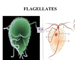
- 5. Giardiasis
- 6. Scanning electron micrograph (SEM) depicting a Giardia lamblia protozoan undergoing binary fission, creating what appears to be a microscopic "heart"
- 7. Life cycle
- 8. Leewenhoek discovered Giardia in his own stools
- 9. • World-wide in distribution • caused by - Giardia intestinalis / Giardia lamblia.
- 10. • Habitat: - Duodenum & the upper part of jejunum. • Morphology: (exists in 2 forms) - Trophozoite. - Cyst.
- 11. TROPHOZOITE OF GIARDIA LAMBLIA Shape: Viewed from front – Tennis racket shaped Viewed from side – Longitudinally split pear
- 12. TROPHOZOITE OF GIARDIA LAMBLIA Shape: Viewed from front – Tennis racket shaped Viewed from side – Longitudinally split pear
- 13. TROPHOZOITE OF GIARDIA LAMBLIA Shape: Viewed from front – Tennis racket shaped Viewed from side – Longitudinally split pear
- 14. TROPHOZOITE OF GIARDIA LAMBLIA Shape: Viewed from front – Tennis racket shaped Viewed from side – Longitudinally split pear
- 15. TROPHOZOITE OF GIARDIA LAMBLIA Shape: Viewed from front – Tennis racket shaped Viewed from side – Longitudinally split pear
- 16. TROPHOZOITE OF GIARDIA LAMBLIA Shape: Viewed from front – Tennis racket shaped Viewed from side – Longitudinally split pear
- 17. Trophozoite • 14 u long x 7 u broad • Bilaterally symmetrical • 2 nuclei • 4 pairs of flagella • 2 axostyles • Motility: falling-leaf
- 18. • The dorsal surface is convex & the ventral surface is concave with a sucking disc. • Concave sucking disc ventrally-used for attachment to surface of intestinal epithelial cells.
- 19. Cyst • Shape : oval • Size: 12µm x 7µm • Four nuclei displaced to one pole • axostyles lie diagonally across long axis forming a dividing line within cyst wall. • Remnants of flagella • Surrounded by tough hyaline cyst wall.
- 20. Cyst • Shape : oval • Size: 12µm x 7µm • Four nuclei displaced to one pole • axostyles lie diagonally across long axis forming a dividing line within cyst wall. • Remnants of flagella • Surrounded by tough hyaline cyst wall.
- 21. Cyst • Shape : oval • Size: 12µm x 7µm • Four nuclei displaced to one pole • axostyles lie diagonally across long axis forming a dividing line within cyst wall. • Remnants of flagella • Surrounded by tough hyaline cyst wall.
- 22. Cyst • Shape : oval • Size: 12µm x 7µm • Four nuclei displaced to one pole • axostyles lie diagonally across long axis forming a dividing line within cyst wall. • Remnants of flagella • Surrounded by tough hyaline cyst wall.
- 23. LIFE CYCLE • in one host • Mature cyst is the infective form • Mode of infection – Faeco-oral route • Within 30 min excystation occurs in the duodenum • One cyst forms two trophozoites, which multiply by binary fission and colonises the duodenum and upper part of jejunum • To avoid acidity of duodenum they may localises in biliary tract • By means of concavity of ventral surface it attaches to mucosal surfaces of duodenum and jejunum. • Encystation occurs while transit down the colon
- 24. Trophozoite in the intestine of man Multiply by binary fission During unfavourable conditions, encystment occurs in large intestine Infection in man occurs by ingestion of cyst Each cyst hatchesout two trophozoites Trophozoites multiply and localises in duodenum Due to high acidity of duodenum, trophozoites localises in billiary tract
- 25. Cyst • Shape : oval • Size: 12µm x 7µm • Four nuclei displaced to one pole • axostyles lie diagonally across long axis forming a dividing line within cyst wall. • Remnants of flagella • Surrounded by tough hyaline cyst wall.
- 27. Pathogenesis • Does not penetrate mucosa or invade tissue. • remains tightly attached to surface epithelial cells of duodenum and jejunum by sucking disc. • structure and function of villi of intestines is disturbed causing disturbance of intestinal function, leading to 1. malabsorption of lipids and lipid soluble vitamins 2. diarrhoea
- 28. Clinical disease • watery diarrhoea • Foul smelling stools, no pus or blood • Flatulence • Abdominal distention • Mal absorption
- 29. PATHOGENESIS AND SYMPTOMS • It is present within the glandular crypts of duodenal and jejunal mucosa. • It does not invade the tissues • Duodenitis and jejunitis
- 30. GIARDIASIS PRESENTATION • Incubation: one to two weeks • Giardia may cause symptoms by creating a brush border enzyme deficiency. • Onset: gradual • Symptoms: nausea, vomiting, malaise, flatulence, bloating, cramping, mild diarrhea, steatorrhea • The stool is voluminous,foul smelling,large amount of mucus anfd fat but no blood. • Sequelae: malabsorbtion, lactase deficiency, weight loss, fatigue, depression and rarely reactive arthritis or urticaria
- 31. 2. Pathogenesis and symptoms G. lamblia inhabits in the duodenum and upper jejunum Trophozoites are attached to the mucosa surface by sucker, reproduced by binary fission Histology: shortening of microvilli, elongation of crypts, and damaging the brush border of the absorptive cells Diarrhea, abdominal pain, bloating, nausea, and vomiting Mechanical blockage of the intestinal mucosa, competition for nutrients, inflammation
- 32. LABORARORY DIAGNOSIS • Identification of cyst in formed stools and trophozoites in diarrhoeal stool. • As the parasites are attached firmly to the mucosa by means of sucking disc, even 5 or 6 samples may not show any parasite. • Trophozoites may also be detected in duodenal aspirates. • Useful method for obtaining duodenal aspirate is ENTEROTEST
- 33. Prevention and Control • Public Health: – Sewage treatment – Treated drinking water – Medicating infected members of the public – Monitoring and treatment of food handlers • Personal: – Boiling stream water for minimum of 1 minute – Iodine tablets – Avoid contaminated water – Keeping hands clean and dry – Thoroughly drying utensils and dishes – Treat any pets showing signs of infection
- 34. Lab Diagnosis Microscopic examination of freshly passed stools.
- 35. Trichomoniasis
- 36. TRICHOMONADS • 3-5 anterior flagella • one undulating membrane • axostyle • hydrogensome (EM) Human Trichomonas Species T. tenax oral cavity T. hominis* intestine T. vaginalis uro-genital *aka: Pentatrichomonas
- 37. TRICHOMONAS VAGINALIS Normal inhabitant of vagina, prostate and urinary tract Obligate parasite Morphology – pear shaped trophozoite,10-30um in length and 5-20um broad 4 ant flagella and cystostome, ant end is rounded Post end pointed Undulating membrane up to middle, an axostyle is also present Motile by jerky movement, divide by binary fission No cyst form
- 38. Caused by Trichomonas vaginalis Habitat In females – urethra and vagina In males – urethra and prostate gland
- 39. Morphology Occurs only as trophozoite form. No cystic form.
- 40. • Ovoid or pear shaped • 13 x7 u • Short undulating membrane reaching upto the middle of body • Single nucleus • Prominent axostyle projects posteriorly • 4 anterior flagella and fifth runs along outer margin of undulating membrane • Motility – rapid, jerky
- 41. • Sexually transmitted disease • In women – leukorrhea, vaginitis. • In men, usually asymptomatic. May develop urethritis, prostatitis, cystitis.
- 42. Lab diagnosis Microscopy of : • Vaginal or urethral discharge • Urine sediment Direct wet mount – actively motile trophozoites seen.
- 43. DIAGNOSIS • demonstration of parasite • direct observation or in vitro culture • vaginal discharge • urine sediment • prostatic secretion TREATMENT • metronidazole (Flagyl) • 250 mg (3/d) for 5-7 days • single 2 g dose • simultaneous treatment of partner! (85-90% cure rate) PREVENTION • limit # of sexual partners • condoms
- 44. Key Features of Cysts • oval shape • 11-14 x 6-10 m • distinct cell wall set apart from cytoplasm • 4 nuclei at anterior end • large karyosome, no peripheral chromatin • fibrils (axonemes) evident • median bodies
- 47. • pear shape • 12-15 x 5-10 x 2-4 m • 2 nuclei • large karyosome, no peripheral chromatin • fibrils (axonemes) evident • bilateral symmetry • pair of median bodies • adhesive disk (not always evident) • 4 pair flagella • motility likened to falling leaf Key Features of Trophozoites
- 49. Other Flagellates Found in Human Feces • Dientamoeba fragilis • no flagella (discuss with amebas) • Pentatrichomonas hominis • formerly called Trichomonas hominis • Chilomastix mesnili • Enteromonas hominis • Retortamonas intestinalis
- 50. Non-Pathogenic Intestinal Flagellates • 7-15 mm trophozoite • no cyst • single nucleus • axostyle • 4 free flagella + undulating membrane • costa Trichomonas hominis
- 51. Non-Pathogenic Intestinal Flagellates Chilomastix mesnili • 10-20 mm trophozoite • 6-20 mm cyst • single nucleus • 4 flagella • cytostome
- 53. THANK YOU