
Recommended
More Related Content
Similar to Balantidium coli.pdf
Similar to Balantidium coli.pdf (20)
More from OsmanHassan35
More from OsmanHassan35 (20)
Recently uploaded
Recently uploaded (20)
Balantidium coli.pdf
- 2. INTRODUCTION • It is largest protozoan • Only ciliated parasites of humans • Causes Balantidiasis Taxonomy: belongs to Phylum Ciliophora Class: Litostomatea Order: Vestibuliferida Family: Balantidiidae Habitat: large intestines of man, pig (main reservoir) and other animals.
- 3. MORPHOLOGY • Two forms: a. Trophozoite – in dysenteric stool b. Cyst: - in carriers and chronic cases Both forms: binucleated - large macronucleus and small micronucleus
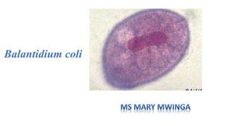
- 4. TROPHOZOITE • Found in active stage of disease – invasive form • shape: oval • Size: 30-300 µm long x 30-100 µm breadth • Whole body covered with a row of tiny delicate cilia – organ of locomotion • Cilia present near the mouth part – longer called “adoral cilia” • Anterior end- narrow - Bears a groove (peristome) that leads to a mouth (cytostome) - followed by a short funnel shaped gullet (cytopharynx) extending up to one-third of the body. • Posterior end- broad, round - Bears an excretory opening (cytopyge)
- 5. TROPHOZOITE • No anus • Cytoplasm- outer clear ectoplasm and inner granular endoplasm Endoplasm - Contains two nuclei: large kidney shaped macronucleus in center and a small micronucleus in the concavity of the macronucleus - Two contractile vacuoles: lie side by side or one above the other maintain the proper osmotic pressure inside cell - Numerous food vacuole: contains food particles like debris from host gut, bacteria, starch grains, fat droplets and occasional RBCs, etc. Where digestion of food particles takes place.
- 7. CYST • Shape: round • Size: 40-60 µm • Immobile and dominant • Surrounded by a thick transparent cyst wall allows the cysts to resist degradation in the acidic environment of the stomach and the basic environment of the small intestine - Contains two nuclei- macronucleus and micronucleus and vacuoles - Cilia- seen in younger cyst but is absorbed on maturity movement ceases
- 8. Development in large intestine- Life cycle • Mode of transmission: faecal-oral route • Virulence factor: Hyaluronidase- help to penetrate intestinal mucosa • Excystation: occurs in small intestine- when trophozoites are produced from cysts - Multiplication in large intestine - Single trophozoite forms from each cyst - trophozoite- is the feeding stage of the parasite multiply either in gut lumen or enter the sub mucosa of large intestine Cell division Asexual reproduction Sexual reproduction
- 10. Asexual reproduction • Division by binary fission • Micronucleus divide first followed by macronucleus • A transverse septum forms – separates the cytoplasm into halves. Sexual reproduction • Replicate sexually (Syngamy) by conjugation • Two trophozoites come in contact with each other at their anterior ends • Exchange the nuclear material for few moments then they detach • No increase in number of trophozoites • Both trophozoite and cyst are excreted in faeces • Trophozoites disintegrates, cysts are resistant and are infective to man and pig
- 11. Life cycle
- 12. Risk factors • Pig’s faeces carrying vast volumes of Balantidium coli contaminates water sources • Humans who work with pigs exposed to Balantidium coli.
- 13. Clinical features Asymptomatic carriers • Results from majority of infection • Harbours the cyst and spread the infection Acute disease • Similar to acute amoebic dysentery • Trophozoites invade gut sub mucosa- form multiple tiny superficial ulcers - Ulcers with necrotic base and undermined edge • Microscopically- cluster of trophozoites are found in sub mucosa with inflammatory cells (lymphocytic) • Patients present have frequent diarrhoea with profuse mucus and blood. • Other features- fever, nausea, vomiting and abdominal pain • Haemorrhage- may lead to shock and death
- 14. Clinical features Chronic disease • Have periods of increased bowel movements (mucous or rarely bloody) • Alternate periods of constipations • Organism load is less and requires repeated stool examination Complications • Seen in immunocompromised and malnourished people • Perforation of large intestine, involvement of appendix, peritonitis, severe dehydration leading to renal failure • Extra intestinal manifestations- rare: liver abscess, pleuritis and pneumonia
- 15. EPIDEMIOLOGY Balantidiasis: • Distributed worldwide- in tropical and subtropical countries with increased pig to human contact • Highest prevalence- 20% reported from mountain districts of West Irian (Indonesia) • Human outbreak reported from Pacific Island of Truk in 1973 • India: rare
- 16. LABORATORY DIAGNOSIS • Stool examination-detects trophozoites and cysts • Histopathology • Culture • serology
- 17. STOOL MICROSCOPY • Trophozoites- detected in acute disease (dysenteric stool) -easy to identify by its rotatory motility, large kidney shaped macronucleus and presence of cilia • Cysts- seen in chronic cases or carriers - round, 40-60 µm in size, surrounded by a cyst wall and presence of two nuclei
- 18. HISTOPATHOLOGY • Histopathological staining of biopsy tissue or scrapping of the ulcers taken by sigmoidoscopy -reveals clusters of trophozoites, cysts and lymphocytic infiltration found in sub mucosa
- 19. CULTURE • Media used: Boeck and Drbohlav egg serum media and Balamuth’s media • Culture rarely necessary as parasites are easily detected by stool microscopy or histopathology Serology • scrapings of colonic and ceacal mucosa can be stained with H&E.
- 20. PREVENTION • Treatment of carriers shedding the cysts • Hygienic rearing of pigs and prevention of pig to human contact • Prevention of contamination of food or water with pig and human faeces
- 21. TREATMENT • DOC: Tetracycline- 500 mg four times a day for 10 days • Alternatively Metronidazole- 750 mg three times a day for 5-7 days • Treatment of carriers- preventing spread of the disease. • No relapse or drug resistance reported
- 22. Things to remember Trophozoite • Shape Oval, pointed at anterior end • Size 30-300 µm long x 30-100 µm wide • Surface Covered in cilia • Not infective • Reproduction By binary fission or conjugation • Nuclei: Macronucleus and micronucleus
- 23. Cyst • Shape Spherical • Size 40-60 m across • Surface Covered with thick, hard cyst wall • with cilia • Infectious: Infective • Reproduction Non-reproductive • Nuclei: macronucleus and micronucleus