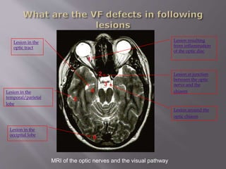
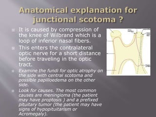
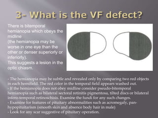
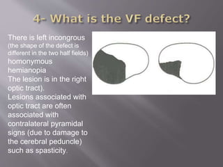
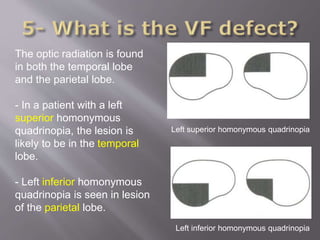
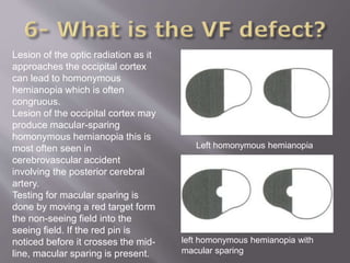
Dr. Riyad Banayot
 Most common field defects and their
underlying conditions are:
 Tunnel vision in advanced retinitis pigmentosa
and advanced glaucoma
 Altitudinal field defect in ischaemic optic
neuropathy and branch retinal artery
occlusion
 Arcuate scotoma in glaucoma
MRI of the optic nerves and the visual pathway
Lesion at junction
between the optic
nerve and the
chiasm
Lesion around the
optic chiasm
Lesion in the
optic tract
Lesion in the
temporal/parietal
lobe
Lesion in the
occipital lobe
Lesion resulting
from inflammation
of the optic disc
 Remember to
describe the visual
field as the patient
sees it.
 Mention if the field
defect is congruous
(behind the lateral
geniculate body) or
incongruous (in the
optic tract).
 Look for other
neurological signs
such as hemiplegia
from cerebrovascular
accident.
The peripheral visual
fields are normal in both
eyes.
There is a right central
scotoma (revealed by
testing the central field with a
red pin).
Examine the patient's
fundus for any evidence
of papillitis or optic
atrophy.
Check for RAPD.
The patient has a right
central scotoma and
superior temporal field
defect in the left eye.
The findings suggest a
lesion in the junction
between the optic
nerve and the chiasm
on the side with the
central scotoma.
 It is caused by compression of
the knee of Wilbrand which is a
loop of inferior nasal fibers.
 This enters the contralateral
optic nerve for a short distance
before traveling in the optic
tract.
 Examine the fundi for optic atrophy on
the side with central scotoma and
possible papilloedema on the other
side.
 Look for causes. The most common
causes are meningioma (the patient
may have proptosis ) and a prefixed
pituitary tumor (the patient may have
signs of hypopituitarism or
Acromegaly).
There is bitemporal
hemianopia which obeys the
midline
(the hemianopia may be
worse in one eye than the
other or denser superiorly or
inferiorly).
This suggests a lesion in the
optic chiasm.
- The hemianopia may be subtle and revealed only by comparing two red objects
in each hemifield. The red color in the temporal field appears washed out.
- If the hemianopia does not obey midline consider pseudo-bitemporal
hemianopia such as bilateral sectoral retinitis pigmentosa, tilted discs or bilateral
infero-temporal retinoschisis. Examine the fundi for any such changes.
- Examine for features of pituitary abnormalities such as acromegaly, pan-
hypopituitarism (smooth skin and absence body hair in male)
- Look for any scar suggestive of pituitary operation.
There is left incongrous
(the shape of the defect is
different in the two half fields)
homonymous
hemianopia
The lesion is in the right
optic tract).
Lesions associated with
optic tract are often
associated with
contralateral pyramidal
signs (due to damage to
the cerebral peduncle)
such as spasticity.
Left superior homonymous quadrinopia
Left inferior homonymous quadrinopia
The optic radiation is found
in both the temporal lobe
and the parietal lobe.
- In a patient with a left
superior homonymous
quadrinopia, the lesion is
likely to be in the temporal
lobe.
- Left inferior homonymous
quadrinopia is seen in lesion
of the parietal lobe.
 Test the patient for Gertmann's
syndrome ?
 Gertmann's syndrome which is seen in
lesion of the dominant parietal lobe
which is usually the left in right handed
person
 Failure of patient's ability to calculate,
name fingers and tell left from right
 Perform the opto-kinetic drum test for
evidence of parietal lobe lesion ?
 There is impaired pursuit movement to
the side of the lesion.
 Astereognosis (failure to tell an object
through touch)
 Joint position sense loss
 Loss of two point discrimination
 Alternatively touching the patient's right
then his left hand and then touching the
two hand together (with patient's eyes
closed).
 If the patient has sensory inattention, he
would notice only one side being touched
when you touch both his hands
simultaneously.
Normal opticokinetic nystagmus
with smooth pursuit movement.
Abnormal opticokinetic
nystagmus with small saccadic
movement replacing the
smooth pursuit movement.
Left homonymous hemianopia
left homonymous hemianopia with
macular sparing
Lesion of the optic radiation as it
approaches the occipital cortex
can lead to homonymous
hemianopia which is often
congruous.
Lesion of the occipital cortex may
produce macular-sparing
homonymous hemianopia this is
most often seen in
cerebrovascular accident
involving the posterior cerebral
artery.
Testing for macular sparing is
done by moving a red target form
the non-seeing field into the
seeing field. If the red pin is
noticed before it crosses the mid-
line, macular sparing is present.
 History:
 Acute (vascular disorder) or
 Chronic (space-occupying lesion)
 History of CVD, DM and smoking
 Examination:
 Cardiovascular examination for hypertension, atrial
fibrillation, cardiac disorders and carotid artery disease.
 Neurological examination for raised ICP
 Blood tests for full blood count for hyperviscosity
conditions such as polycythemia, ESR (? Vasculitis)
 CT scan for CVA, space occupying lesion.
 Chest X-ray if there is evidence of cerebral space-
occupying lesion.

Common Cases: Neurological Visual Field defects

  • 1.
  • 2.
     Most commonfield defects and their underlying conditions are:  Tunnel vision in advanced retinitis pigmentosa and advanced glaucoma  Altitudinal field defect in ischaemic optic neuropathy and branch retinal artery occlusion  Arcuate scotoma in glaucoma
  • 3.
    MRI of theoptic nerves and the visual pathway Lesion at junction between the optic nerve and the chiasm Lesion around the optic chiasm Lesion in the optic tract Lesion in the temporal/parietal lobe Lesion in the occipital lobe Lesion resulting from inflammation of the optic disc
  • 4.
     Remember to describethe visual field as the patient sees it.  Mention if the field defect is congruous (behind the lateral geniculate body) or incongruous (in the optic tract).  Look for other neurological signs such as hemiplegia from cerebrovascular accident.
  • 5.
    The peripheral visual fieldsare normal in both eyes. There is a right central scotoma (revealed by testing the central field with a red pin). Examine the patient's fundus for any evidence of papillitis or optic atrophy. Check for RAPD.
  • 6.
    The patient hasa right central scotoma and superior temporal field defect in the left eye. The findings suggest a lesion in the junction between the optic nerve and the chiasm on the side with the central scotoma.
  • 7.
     It iscaused by compression of the knee of Wilbrand which is a loop of inferior nasal fibers.  This enters the contralateral optic nerve for a short distance before traveling in the optic tract.  Examine the fundi for optic atrophy on the side with central scotoma and possible papilloedema on the other side.  Look for causes. The most common causes are meningioma (the patient may have proptosis ) and a prefixed pituitary tumor (the patient may have signs of hypopituitarism or Acromegaly).
  • 8.
    There is bitemporal hemianopiawhich obeys the midline (the hemianopia may be worse in one eye than the other or denser superiorly or inferiorly). This suggests a lesion in the optic chiasm. - The hemianopia may be subtle and revealed only by comparing two red objects in each hemifield. The red color in the temporal field appears washed out. - If the hemianopia does not obey midline consider pseudo-bitemporal hemianopia such as bilateral sectoral retinitis pigmentosa, tilted discs or bilateral infero-temporal retinoschisis. Examine the fundi for any such changes. - Examine for features of pituitary abnormalities such as acromegaly, pan- hypopituitarism (smooth skin and absence body hair in male) - Look for any scar suggestive of pituitary operation.
  • 9.
    There is leftincongrous (the shape of the defect is different in the two half fields) homonymous hemianopia The lesion is in the right optic tract). Lesions associated with optic tract are often associated with contralateral pyramidal signs (due to damage to the cerebral peduncle) such as spasticity.
  • 10.
    Left superior homonymousquadrinopia Left inferior homonymous quadrinopia The optic radiation is found in both the temporal lobe and the parietal lobe. - In a patient with a left superior homonymous quadrinopia, the lesion is likely to be in the temporal lobe. - Left inferior homonymous quadrinopia is seen in lesion of the parietal lobe.
  • 11.
     Test thepatient for Gertmann's syndrome ?  Gertmann's syndrome which is seen in lesion of the dominant parietal lobe which is usually the left in right handed person  Failure of patient's ability to calculate, name fingers and tell left from right
  • 12.
     Perform theopto-kinetic drum test for evidence of parietal lobe lesion ?  There is impaired pursuit movement to the side of the lesion.
  • 13.
     Astereognosis (failureto tell an object through touch)  Joint position sense loss  Loss of two point discrimination
  • 14.
     Alternatively touchingthe patient's right then his left hand and then touching the two hand together (with patient's eyes closed).  If the patient has sensory inattention, he would notice only one side being touched when you touch both his hands simultaneously.
  • 15.
    Normal opticokinetic nystagmus withsmooth pursuit movement. Abnormal opticokinetic nystagmus with small saccadic movement replacing the smooth pursuit movement.
  • 16.
    Left homonymous hemianopia lefthomonymous hemianopia with macular sparing Lesion of the optic radiation as it approaches the occipital cortex can lead to homonymous hemianopia which is often congruous. Lesion of the occipital cortex may produce macular-sparing homonymous hemianopia this is most often seen in cerebrovascular accident involving the posterior cerebral artery. Testing for macular sparing is done by moving a red target form the non-seeing field into the seeing field. If the red pin is noticed before it crosses the mid- line, macular sparing is present.
  • 17.
     History:  Acute(vascular disorder) or  Chronic (space-occupying lesion)  History of CVD, DM and smoking  Examination:  Cardiovascular examination for hypertension, atrial fibrillation, cardiac disorders and carotid artery disease.  Neurological examination for raised ICP  Blood tests for full blood count for hyperviscosity conditions such as polycythemia, ESR (? Vasculitis)  CT scan for CVA, space occupying lesion.  Chest X-ray if there is evidence of cerebral space- occupying lesion.