Vesicular stomatitis in Cattle, Horse and pigs
•Download as PPTX, PDF•
4 likes•1,466 views

Caused by Rhabdoviridae family, Vesiculovirus. Development of vesicles on the mouth and feet. The virus, an arbovirus, is spread to cattle, horses, and pigs primarily by sandflies and blackflies. The mechanism of injury in vesicular stomatitis is cell dysfunction and lysis leading to intercellular edema with vesiculation, erosion, and ulceration of mucosae and skin. Pathogenesis of the disease is by the bite of the flies, entry of virus, viral replication in the cell and rupture of the cell, which form intercellular space that is fluid filled to form vesicles. Rupture of this vesicles leads to erosion/ulceration of overlying mucosa or skin.

Recommended
More Related Content
What's hot
What's hot (20)
Similar to Vesicular stomatitis in Cattle, Horse and pigs
Similar to Vesicular stomatitis in Cattle, Horse and pigs (20)
Recently uploaded
Recently uploaded (20)
Vesicular stomatitis in Cattle, Horse and pigs
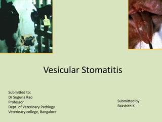
- 1. Vesicular Stomatitis Submitted by: Rakshith K Submitted to: Dr Suguna Rao Professor Dept. of Veterinary Pathlogy Veterinary college, Bangalore
- 2. Vesicular Stomatitis • Rhabdoviridae, Vesiculovirus, Arbo virus. • Enveloped, single stranded, Negative sense RNA. • Vesicular stomatitis is an infectious disease caused by a vesiculovirus, and characterized clinically by the development of vesicles on the mouth and feet.
- 3. • Clinical signs of vesicular stomatitis are identical to those of FMD. • The mechanism of injury in vesicular stomatitis is cell dysfunction and lysis leading to intercellular edema with vesiculation, erosion, and ulceration of mucosae and skin.
- 4. • The virus, an arbovirus, is spread to cattle, horses, and pigs primarily by sandflies and blackflies. • Vesicular lesions occur near the sites of insect bites, suggesting that the virus infects target cells locally and there is no systemic spread of virus. • Squamous epithelial cells of mucosae and skin are the primary target cells. • Langerhans cells (dendritic cells) and cells of the monocyte macrophage system are also target cells – Rare.
- 5. Host range: • Primary disease- Horse, Cattle, and Pigs. • Goat and sheep- Resistant.
- 6. Morbidity/ Mortality • Morbidity – Range: 5 to 90% – Most animals seroconvert • Mortality – Higher in adults – Death rare in cattle and horses
- 7. Spread of disease: • Insects- Principle vectors of transmission. • Mosquitoes, black flies, sand flies, gnats, and midges. Sand fly Black fly
- 8. Sand flies & Black flies carrying virus bites Cattle (Arbo virus) Injures blood vessels & deposit virus direct into plasma of BV or Interstitial fluid Squamous epithelial cells of mucosa and skin are the primary target cells for viral infection (Stratum basale and/or Stratum spinosum—Target layers) Viral envelope glycoprotein-G binds to LDL receptors on epi. cell enter the cells via endocytosis Replicate in the cytoplasm, and escape from the cell by rupture of cells Formation of intercellular spaces that fill with fluid which form vesicles Trauma ruptures the vesicles and leads to erosion/ulceration of the overlying mucosa or skin Pathogenesis
- 9. Clinical signs: • Mild fever • Development of vesicles on the dorsum of the tongue, dental pad, lips and the buccal mucosa • The vesicles rupture, and the resulting irritation causes profuse salivation and anorexia • Lesions may also occur on the teats and the coronary band.
- 11. Microscopic lesions • There is intercellular oedema in the stratum spinosum. • This leads to cell dissociation and necrosis. • Neutrophils and macrophages infiltrate the necrotic tissue, which sloughs, leaving erosions. • The intra-epithelial oedema result in a vesicle.
- 12. Clinical Diagnosis • Clinical signs: Salivation and lameness • Laboratory Diagnosis • Virus isolation • Viral antigen detection – Vesicular fluid or epithelium – ELISA, complement fixation, virus neutralization • Antibody tests – Paired serum samples – ELISA, complement fixation, virus neutralization
- 13. Differential diagnosis Vesicular lesions • FMD Foot lesions • Dermatophilosis • Foot-rot • MCF - Chemical Irritants Ulcerative lesions • BVD/MD • MCF • LSD- Microscopically • IBR • Rinderpest • Blue tongue
- 14. Treatment • No specific treatment available • Supportive care – Fresh, clean water • Electrolytes if necessary – Soft feeds • Antibiotics for secondary infection • Good prognosis • Production animals may suffer losses
- 15. Prevention • Avoid grazing at peak insect feeding hours • Segregation and isolation necessary for controlling spread • Sanitation: Good sanitation practices (cleaning out feed twice daily, disinfect feed bunks and water troughs) • Insect control programs • Easily inactivated – Area must be free of organic matter – Contact time of at least 10 minutes • Disinfectants – Phenolic, halogen-based disinfectants – Chlorine dioxide, 1% chlorine bleach