SlideShare a Scribd company logo
1 of 55
The skin
www.medrockets.com
Fb:Medrockets
The skin (the interface between humans and their
environment) is the largest organ in the body. It weighs an
average of 4 kg and covers an area of 2 m2.
The skin has two layers:
1.The epidermis, outer epithelial layer.
2.The dermis, inner connective tissue.
Beneath the dermis is, the subcutis/hypodermis which usually
contains abundant fat.
The epidermis adheres to the dermis partly by the interlocking
of its downward projections (epidermal ridges or pegs) with
upward projections of the dermis (dermal papillae).
The Integument
Is the largest system of the body
 16% of body weight,
 1.5 to 2m2
in area,
 The integument is made up of two parts:
1. Cutaneous membrane
a. Epidermis– Superficial epithelium
b. Dermis – underlying CT with blood supply
2. Accessory structures
a. Hair
b. Nails
c. Exocrine Glands
Protection
 First line of defense against
 Bacteria
 Viruses
 Protects underlying structures from
 Ultraviolet (UV) radiation
 Dehydration
 Vitamin D production
 Needed for calcium absorption
 Sensation
 Sensory receptors
Body temperature regulation
If too hot
 Dermal blood vessels dilate
 Vessels carry more blood to surface so heat can escape
If too cold
 Dermal blood vessels constrict
 Prevents heat from escaping
Excretion
Small amounts of waste products are lost through
perspiration
www.medrockets.com
Fb:Medrockets
Understanding how the skin can function in these many
ways starts with understanding the structure of the 3 layers
of skin
The Epidermis
Epithelial tissue
Dermis
Dense connective tissue proper – irregular
Hypodermis
Subcutaneous tissue- loose connective tissue proper
and adipose tissue
www.medrockets.com
Fb:Medrockets
Epidermis
Dermis
Hypodermis
or
subcutaneous layer
www.medrockets.com
Fb:Medrockets
Epidermis:
The epidermis is formed from many layers of closely packed cells
called keratinocytes:
1.Basal layer (stratum germinativum(.
2.Prickle cell layer (stratum spinosum(.
3.Granular layer (stratum ranulosum(.
4.Horny layer (stratum corneum(
On the palms and soles a pale or pink layer, the stratum lucidum,
is noted just above the granular layer.
The epidermis varies in thickness from less than 0.1 mm on the
eyelids to nearly 1 mm on the palms and soles.
Structures of the Epidermis
 The five strata of keratinocytes in thick skin
 From basal lamina to free surface
1. Stratum basale
2. Stratum spinosum
3. Stratum granulosum
4. Stratum lucidum
5. Stratum corneum
www.medrockets.com
Fb:Medrockets
11.The basal layer:
Is the deepest layer, rests on basement membrane,
which attaches it to the dermis.
This layer generate cells of the epidermis.
It is a single layer of columnar cells, whose basal
surfaces sprout many fine processes and
hemidesmosomes, anchoring them to the lamina densa
of the basement membrane.
www.medrockets.com
Fb:Medrockets
22..The spinous or prickle cell layer:
Composed of differentiating cells, contain some tonofibrils
and kertohyalin granules, which synthesize keratins.
They are larger than basal cells.
Keratinocytes are firmly attached to each other by small
interlocking cytoplasmic processes, and by abundant
desmosomes.
Under the light microscope, the desmosomes look like
‘prickles’, they are specialized attachment plaques.
3.Granular layer:
Consists of two or three layers of cells that are flatter than
those in the spinous layer, and have more tonofibrils.
As the name of the layer implies, these cells contain large
irregular basophilic granules of keratohyalin, which merge
with tonofibrils.
As keratinocytes migrate out through the outermost layers,
their keratohyalin granules break up and their contents are
dispersed throughout the cytoplasm, leading to keratinization
and the formation of a thick and tough peripheral protein
coating called the horny envelope.
Stratum Lucidum — the “clear layer”
Found only in thick skin
Covers stratum granulosum
Stratum Corneum — the “horn layer”
Exposed surface of skin
15 to 30 layers of keratinized cells
Water resistant
Shed and replaced every 2 weeks
www.medrockets.com
Fb:Medrockets
Other cells of the epidermis:
1. Melanocytes.
2. Langerhans cells.
3. Merckle cells
www.medrockets.com
Fb:Medrockets
Melanocytes are the only cells that can synthesize
melanin.
They migrate from the neural crest into the
basal layer of the ectoderm where, in human embryos,
they are seen as early as the eighth week of gestation.
They are also found in hair bulbs, the retina and pia
arachnoid.
Melanocytes
www.medrockets.com
Fb:Medrockets
Each dendritic melanocyte associates with a number
of keratinocytes, forming an ‘epidermal
melanin unit.
The dendritic processes of melanocytes wind between
the epidermal cells and end as discs in contact with
them.
Their cytoplasm contains discrete organelles, the
melanosomes, containing varying amounts of the
pigment melanin.
www.medrockets.com
Fb:Medrockets
The Langerhans cell is a dendritic cell like the
melanocyte.
It also lacks desmosomes and tonofibrils, but has a
lobulated nucleus.
The specific granules within the cell look like a tennis
racket when seen in two dimensions in an electron
micrograph. They are plate-like, with a rounded bleb
protruding from the surface.
Langerhans cells
www.medrockets.com
Fb:Medrockets
Langerhans cells come from precursors originating in the bone
marrow.
There are approximately 800 Langerhans cells per mm2.
Langerhans cells are alone among epidermal cells in
possessing surface receptors for C3b and the Fc portions of
IgG and IgE, and in bearing major histocompatibility complex
(MHC) Class II antigens (HLA-DR, -DP and -DQ).
They are best thought of as specialized macrophages.
Langerhans cells have a key role in many immune reactions.
They take up exogenous antigen, process
it and present it to T lymphocytes either in the skin
or in the local lymph nodes.
They probably play a part in immuno-surveillance for viral
and tumor antigens.
In this way, ultraviolet radiation can induce skin tumors both
by causing mutations in the epidermal cells, and by decreasing
the number of epidermal Langerhans cells.
Topical or systemic glucocorticoids also reduce the density of
epidermal Langerhans cells.
Merkel cells are found in normal epidermis.
Act as transducers for fine touch.
They are nondendritic cells.
Lying in or near the basal layer.
Are of the same size as keratinocytes.
They are concentrated in localized thickenings of the
epidermis near hair follicles (hair discs).
Contain membrane bound spherical granules, 80–100 nm in
diameter.
Sparse desmosomes connect these cells to neighbouring
keratinocytes.
Fine unmyelinated nerve endings are often associated with
Merkel cells.
Merkel cells
Epidermis—a stratified epithelium
www.medrockets.com
Fb:Medrockets
The basement membrane lies at the interface between the
epidermis and dermis.
With light microscopy it can be highlighted using a periodic
acid–Schiff (PAS) stain, because of its abundance of neutral
mucopolysaccharides..
The dermo-epidermal junction
Electron microscopy shows that the basement membrane has 4
componenets:
1.The plasma membrane of basal cells which has hemidesmosomes
(containing bullous pemphigoid antigens, collagen XVII and á6 â4
integrin).
2.Electron-lucent area, the lamina lucida which separate lamina densa
from the basal cells. The lamina lucida contains laminin-1, laminin-5 and
entactin.
3.Lamina densa (rich in type IV collagen).
4.Anchoring fibrils (of type VII collagen), dermal microfibril bundles
and single small collagen fibres (types I and III), extend from the
papillary dermis to the deep part of the lamina densa.
The structures within the dermo-epidermal junction provide
mechanical support, encouraging the adhesion, growth,
differentiation and migration of the overlying basal cells, and
also act as a semipermeable filter that regulates the transfer of
nutrients and cells from dermis to epidermis.
www.medrockets.com
Fb:Medrockets
Dermis:
The dermis lies between the epidermis and the subcutaneous fat. It is tow
parts:
1.Upper papillary dermis.
2.Lower reticular dermis.
Its thickness varies, being greatest in the palms and soles and least in the
eyelids and penis.
In old age, the dermis thins and loses its elasticity.
The dermis interdigitates with the epidermis so that upward projections
of the dermis, the dermal papillae, interlock with downward ridges of the
epidermis, the rete pegs.
This interdigitation is responsible for the ridges seen most readily on the
fingertips (as fingerprints).
The dermis has three components:
cells, fibers and amorphous ground substance.
Cells of the dermis:
The main cells of the dermis are fibroblasts, but there
are also small numbers of resident and transitory mononuclear
phagocytes, lymphocytes, Langerhans cells and mast cells.
Other blood cells, e.g. polymorphs, are seen during
inflammation.
Fibres of the dermis:
The dermis is largely made up of interwoven fibresThe dermis is largely made up of interwoven fibres,,
principally of collagen, packed in bundlesprincipally of collagen, packed in bundles..
Those in the papillary dermis are finer than those in the deeperThose in the papillary dermis are finer than those in the deeper
reticular dermisreticular dermis..
When the skin is stretched, collagen, with its high tensileWhen the skin is stretched, collagen, with its high tensile
strength, prevents tearing, and the elastic fibres, intermingledstrength, prevents tearing, and the elastic fibres, intermingled
with the collagen, later return it to the unstretched statewith the collagen, later return it to the unstretched state..
www.medrockets.com
Fb:Medrockets
Collagen fibers:
Makes up to 70–80% of the dry weight of the dermis.
Its fibres are composed of thinner fibrils, which are in turn made up of
microfibrils built from individual collagen molecules. These molecules
consist of three polypeptide chains forming a triple helix with a non-
helical segment at both ends.
Collagen is an unusual protein as it contains a high proportion of proline
and hydroxyproline and many glycine residues.
Defects in the enzymes needed for collagen synthesis are responsible for
some skin diseases, including the Ehlers–Danlos syndrome and
osteogenesis imperfecta (fragility of bones(.
Elastic fibers:
Account for about 2% of the dry weight of adult dermis.
They have two distinct protein components:
an amorphous elastin core and a surrounding ‘elastic tissue microfibrillar
component’.
Abnormalities in the elastic tissue cause cutis laxa (sagging inelastic
skin( and pseudoxanthoma elasticum.
Reticulin fibres::
Are fine collagen fibers, seen in fetal skin and around the blood vessels
and appendages of adult skin.
The ground substance of the dermis consists largely of two
glycosaminoglycans (hyaluronic acid and dermatan sulphate( with
smaller amounts of heparan sulphate and chondroitin sulphate.
The ground substance has several important functions:
•it binds water, allowing nutrients, hormones and waste products to pass
through the dermis;
•it acts as a lubricant between the collagen and elastic fibre networks
during skin movement; and
•it provides bulk, allowing the dermis to act as a shock absorber.
Ground substance of the dermis:
Both smooth and striated muscle are found in the skin.
The smooth arrector pili muscles are used by animals to raise their fur
and so protect them from the cold.
They are vestigial in humans, but may help to express sebum. Smooth
muscle is also responsible for ‘goose pimples’ (bumps( from cold, nipple
erection, and the raising of the scrotum by the dartos muscle.
Striated fibres (e.g. the platysma( and some of the muscles of facial
expression, are also found in the dermis.
Muscles:
www.medrockets.com
Fb:Medrockets
Although the skin consumes little oxygen, its abundant blood supply
regulates body temperature.
The blood vessels lie in two main horizontal layers:
1.The deep plexus is just above the subcutaneous fat, and its arterioles
supply the sweat glands and hair papillae.
2.The superficial plexus is in the papillary dermis and arterioles from it
become capillary loops in the dermal papillae.
The blood vessels in the skin are important in thermoregulation.
Blood vessels:
:
Afferent lymphatics begin as blind-ended capillaries in the dermal
papilla and pass to a superficial lymphatic plexus in the papillary dermis.
There are also two deeper horizontal plexuses, and collecting lymphatics
from the deeper one run with the veins in the superficial fascia.
Cutaneous lymphatics
www.medrockets.com
Fb:Medrockets
The skin is supplied with an estimated one million nerve fibres.
Most are found in the face and extremities.
Their cell bodies lie in the dorsal root ganglia.
Both myelinated and non-myelinated fibres exist, with the latter making
up an increasing proportion peripherally.
Most free sensory nerves end in the dermis; however, a few non-
myelinated nerve endings penetrate into the epidermis. Some of these are
associated with Merkel cells.
Nerves:
Free nerve endings detect stimuli of heat and pain (nocioceptors(, while
specialized end organs in the dermis, Pacinian and Meissner corpuscles,
detect pressure (mechanoreceptors(, vibration and touch.
Autonomic nerves supply the blood vessels, sweat glands and arrector
pili muscles.
Itching is an important feature of many skin diseases. It follows the
stimulation of fine free nerve endings lying close to the dermo-epidermal
junction.
Impulses from these free endings pass centrally in two ways:
quickly along myelinated A fibres, and more slowly along non-
myelinated C fibres.
In itchy skin diseases, pruritogenic chemicals such as histamine and
proteolytic enzymes are liberated close to the dermoepidermal junction.
The hard keratin of the nail plate is formed in the nail matrix,
which lies in an invagination of the epidermis (the nail fold(
on the back of the terminal phalanx of each digit.
The matrix runs from the proximal end of the floor of the nail
fold to the distal margin of the lunula.
From this area the nail plate grows forward over the nail bed,
ending in a free margin at the tip of the digit.
The nail:
The nail bed is capable of producing small amounts of keratin
which contribute to the nail and which are responsible for the
‘false nail’ formed when the nail matrix is obliterated by
surgery or injury.
The cuticle acts as a seal to protect the potential space of the
nail fold from chemicals and from infection.
The nails provide strength and protection for the terminal
phalanx.
Their presence helps with fine touch and with the handling of
small objects.
The rate at which nails grow varies from person to person:
fingernails average between 0.5 and 1.2 mm per week, while
toenails grow more slowly.
Nails grow faster in the summer, if they are bitten, and in
youth.
They change with ageing from the thin, occasionally spooned
nails of early childhood to the duller, paler and more opaque
nails of the very old.
Cosists of 2 parts:
1. Hair follicle, include: inner root sheath and outer rot sheath
2. Hair shaft, consists of: hair cuticle, cortex and medulla.
Hair shaft and hair follicle are produced by the matrix portion of hair
bulb.
Along one side, the sebaceous gland open to the upper part of the hair
follicle, arrector pili muscle attach to the lower part.
Apocrine gland also open to the hair follicle from the opposite side.
From the surface opening of the hair follicle to the enterance of the
sebaceous duct is called infundibular segment.
The portion between the sebaceous duct and insertion of arrector pili
muscle is isthmus.
The lowest portion is hair bulb.
Hair unit:
1.Lanugo hairs. Fine long hairs covering the fetus, but shed
about 1 month before birth.
2.Vellus hairs. Fine short unmedullated hairs covering much
of the body surface. They replace thelanugo hairs just before
birth.
3.Terminal hairs. Long coarse medullated hairs seen,
for example, in the scalp or pubic regions. Their growth
is often influenced by circulating androgen levels.
Classification of hairs:
There are three phases of follicular activity:
1. Anagen. The active phase of hair production.
2. Catagen. A short phase of conversion from
active growth to the resting phase. Growth stops, and the
end of the hair becomes club-shaped.
3. Telogen. A resting phase at the end of which the
club hair is shed.
The hair cycle
The scalp contain an average of 100 000 hairs,
anagen lasts for up to 5 years,
catagen for about 2 weeks,
and telogen for about 3 months.
As many as 100 hairs may be shed from the normal
scalp every day as a normal consequence of cycling.
On the scalp, about 85% are normally in anagen and
15% in the telogen phase.
www.medrockets.com
Fb:Medrockets
Sebaceous glands:
They are associated with hair follicle, lie in the obtuse angle between
the follicle and the epidermis.
They are multilobed and contain cells full of lipid, which are shed whole
(holocrene secretion) during secretion into the upper part of the hair
follicle.
Sebum contains a mixture of triglycerides, fatty acids, waxy esters,
squalene and cholesterol.
It lubricates and waterproofs the skin and protects it from drying.
Free sebaceous glands may be found in the eye lids (meibomian glands),
mucous membranes (Fordyce spots), nipples, peri-anal region and
genetalia.
Androgen hormones, especially dihydrotestosterone stimulate sebaceous
gland activity.
There are 2-3 million sweat glands distributed all over the body surface
but they are most numerous on the palms, soles and axillae.
The tightly coiled glands lie deep in the dermis, and the emerging duct
passes to the surface by penetrating the epidermis in a corkscrew
fashion.
Initially sweat is isotonic like plasma but, under normal conditions, it
becomes hypotonic by the time it discharged at the surface, after the
tubular resorption of electrolytes and water under the influence of
aldosterone and antidiuretic hormones.
Eccrine sweat glands:
The PH of sweat is between 4.0 and 6.8; it contains sodium, potassium
chloride, lactate, urea and ammonia.
Sweat glands have an important role in temperature control, the skin
surface being cooled by evaporation. Up to 10 L/day of sweat can be
excreted.
The sweat glands are innervated by cholinergic fibers of the sympathetic
nervous system. Sweating can therefore be induced by cholinergic and
blocked by anticholenergic drugs.
Central control of sweating resides in the preoptic hypothalamic sweat
centre.
Apocrine sweat glands:
They are limited to the axillae, nipples, peri-umbilical area, perineum
and genitalia.
The coiled tubular glands (larger than sweat glands) lie deep in the
dermis, and during sweating the luminal part of their cells is lost
(decapitation secretion).
Apocrine sweat passes via the duct into the mid-portion of the hair
follicle.
The action of bacteria on apocrine sweat is responsible for body odour.
The glands are innervated by adrenergic fibres of the sympathetic
nervous system.
Thank you
www.medrockets.com
Fb:Medrockets

More Related Content

What's hot (20)

Structure of blood vessels
Structure of blood vesselsStructure of blood vessels
Structure of blood vessels
 
Artery and veins, capillaries, arteriole and venules, systemic circulation an...
Artery and veins, capillaries, arteriole and venules, systemic circulation an...Artery and veins, capillaries, arteriole and venules, systemic circulation an...
Artery and veins, capillaries, arteriole and venules, systemic circulation an...
 
Cardiovascular system
Cardiovascular systemCardiovascular system
Cardiovascular system
 
The Myocardium
The MyocardiumThe Myocardium
The Myocardium
 
Integumentary system
Integumentary systemIntegumentary system
Integumentary system
 
Histology of cardiovascular system
Histology of cardiovascular systemHistology of cardiovascular system
Histology of cardiovascular system
 
Integumentary system
Integumentary systemIntegumentary system
Integumentary system
 
Integumentary System
Integumentary SystemIntegumentary System
Integumentary System
 
Blood vessels
Blood vesselsBlood vessels
Blood vessels
 
Cardiovascular system
Cardiovascular systemCardiovascular system
Cardiovascular system
 
Lymphatic system
Lymphatic systemLymphatic system
Lymphatic system
 
Blood vessels.2.
Blood vessels.2.Blood vessels.2.
Blood vessels.2.
 
skin ppt
skin pptskin ppt
skin ppt
 
Anatomy Of The Heart
Anatomy Of The HeartAnatomy Of The Heart
Anatomy Of The Heart
 
Function of arterial & venous system
Function of arterial & venous systemFunction of arterial & venous system
Function of arterial & venous system
 
The skin
The skinThe skin
The skin
 
Skin
SkinSkin
Skin
 
Anatomy of skin
Anatomy of skinAnatomy of skin
Anatomy of skin
 
Muscle tissues
Muscle tissuesMuscle tissues
Muscle tissues
 
Cardiovascular System
Cardiovascular SystemCardiovascular System
Cardiovascular System
 

Similar to Skin

dermatology.Skin anatomy.(dr.darseem)
dermatology.Skin anatomy.(dr.darseem)dermatology.Skin anatomy.(dr.darseem)
dermatology.Skin anatomy.(dr.darseem)student
 
184715 integuments
184715 integuments184715 integuments
184715 integumentsYoAmoNYC
 
184715 integuments
184715 integuments184715 integuments
184715 integumentsabctutor
 
Skin structures and functions ppt Dr.Mumux
Skin structures and functions ppt Dr.MumuxSkin structures and functions ppt Dr.Mumux
Skin structures and functions ppt Dr.MumuxMumux Mirani
 
INTEGUMENTARY SYSTEM (1).pdf
INTEGUMENTARY SYSTEM (1).pdfINTEGUMENTARY SYSTEM (1).pdf
INTEGUMENTARY SYSTEM (1).pdfSaqibShaik2
 
Skin Tissue - Histology and Embryrology.
Skin Tissue - Histology and Embryrology.Skin Tissue - Histology and Embryrology.
Skin Tissue - Histology and Embryrology.Denizasmaz
 
ANATOMY AND PHYSIOLOGY OF SKIN
ANATOMY AND PHYSIOLOGY OF SKINANATOMY AND PHYSIOLOGY OF SKIN
ANATOMY AND PHYSIOLOGY OF SKINAlmasMajeeth
 
Skin and its diseases. Dermatology description
Skin and its diseases. Dermatology descriptionSkin and its diseases. Dermatology description
Skin and its diseases. Dermatology descriptionKeshav177622
 
Structure of skin
Structure of skin Structure of skin
Structure of skin docjikisha
 
Introduction to dermatovenerology.pptx
Introduction to dermatovenerology.pptxIntroduction to dermatovenerology.pptx
Introduction to dermatovenerology.pptxssuser48e933
 
SKIN : Excretory system in Human
SKIN : Excretory system in HumanSKIN : Excretory system in Human
SKIN : Excretory system in Humannurul ikhsani
 
Introduction of Human skin
Introduction of Human skin Introduction of Human skin
Introduction of Human skin ShivanshuGangwar
 

Similar to Skin (20)

dermatology.Skin anatomy.(dr.darseem)
dermatology.Skin anatomy.(dr.darseem)dermatology.Skin anatomy.(dr.darseem)
dermatology.Skin anatomy.(dr.darseem)
 
184715 integuments
184715 integuments184715 integuments
184715 integuments
 
184715 integuments
184715 integuments184715 integuments
184715 integuments
 
The Skin
The SkinThe Skin
The Skin
 
Skin structure and development
Skin structure and developmentSkin structure and development
Skin structure and development
 
Skin structures and functions ppt Dr.Mumux
Skin structures and functions ppt Dr.MumuxSkin structures and functions ppt Dr.Mumux
Skin structures and functions ppt Dr.Mumux
 
INTEGUMENTARY SYSTEM (1).pdf
INTEGUMENTARY SYSTEM (1).pdfINTEGUMENTARY SYSTEM (1).pdf
INTEGUMENTARY SYSTEM (1).pdf
 
TAM1
TAM1TAM1
TAM1
 
Skin Tissue - Histology and Embryrology.
Skin Tissue - Histology and Embryrology.Skin Tissue - Histology and Embryrology.
Skin Tissue - Histology and Embryrology.
 
ANATOMY AND PHYSIOLOGY OF SKIN
ANATOMY AND PHYSIOLOGY OF SKINANATOMY AND PHYSIOLOGY OF SKIN
ANATOMY AND PHYSIOLOGY OF SKIN
 
Skin and its diseases. Dermatology description
Skin and its diseases. Dermatology descriptionSkin and its diseases. Dermatology description
Skin and its diseases. Dermatology description
 
skin
skinskin
skin
 
Dermatology 5th year, 1st lecture (Dr. Kazhan)
Dermatology 5th year, 1st lecture (Dr. Kazhan)Dermatology 5th year, 1st lecture (Dr. Kazhan)
Dermatology 5th year, 1st lecture (Dr. Kazhan)
 
Structure of skin
Structure of skin Structure of skin
Structure of skin
 
Integumentary System
Integumentary SystemIntegumentary System
Integumentary System
 
Integumentary System
Integumentary SystemIntegumentary System
Integumentary System
 
Introduction to dermatovenerology.pptx
Introduction to dermatovenerology.pptxIntroduction to dermatovenerology.pptx
Introduction to dermatovenerology.pptx
 
intoduction to skin
intoduction to skinintoduction to skin
intoduction to skin
 
SKIN : Excretory system in Human
SKIN : Excretory system in HumanSKIN : Excretory system in Human
SKIN : Excretory system in Human
 
Introduction of Human skin
Introduction of Human skin Introduction of Human skin
Introduction of Human skin
 

More from rahulverma1194

More from rahulverma1194 (20)

Spinal anesthesia
Spinal anesthesiaSpinal anesthesia
Spinal anesthesia
 
Regional anesthesia
Regional anesthesiaRegional anesthesia
Regional anesthesia
 
Postanesthetic care
Postanesthetic carePostanesthetic care
Postanesthetic care
 
Local anaesthesia
Local anaesthesiaLocal anaesthesia
Local anaesthesia
 
General anesthesia
General anesthesiaGeneral anesthesia
General anesthesia
 
Epidural anesthesia
Epidural anesthesiaEpidural anesthesia
Epidural anesthesia
 
Anesthesiology
AnesthesiologyAnesthesiology
Anesthesiology
 
Anaesthetic management of the surgical patient
Anaesthetic management of the  surgical patientAnaesthetic management of the  surgical patient
Anaesthetic management of the surgical patient
 
Presentation1
Presentation1 Presentation1
Presentation1
 
Copd
Copd Copd
Copd
 
Infertility
Infertility Infertility
Infertility
 
Head injuries
Head injuriesHead injuries
Head injuries
 
Upper git bleeding
Upper git bleedingUpper git bleeding
Upper git bleeding
 
Tracheostomy a
Tracheostomy a Tracheostomy a
Tracheostomy a
 
Parathyroid disorders
Parathyroid disorders Parathyroid disorders
Parathyroid disorders
 
Diabetic coma clinical_features
Diabetic coma clinical_features Diabetic coma clinical_features
Diabetic coma clinical_features
 
Investigations in cardiology
Investigations in cardiology Investigations in cardiology
Investigations in cardiology
 
Prostatic cancer
Prostatic cancerProstatic cancer
Prostatic cancer
 
Benign prostatic hypertrophy
Benign prostatic hypertrophyBenign prostatic hypertrophy
Benign prostatic hypertrophy
 
Thyroid neoplasms
Thyroid neoplasmsThyroid neoplasms
Thyroid neoplasms
 

Recently uploaded

Kesar Bagh Call Girl Price 9548273370 , Lucknow Call Girls Service
Kesar Bagh Call Girl Price 9548273370 , Lucknow Call Girls ServiceKesar Bagh Call Girl Price 9548273370 , Lucknow Call Girls Service
Kesar Bagh Call Girl Price 9548273370 , Lucknow Call Girls Servicemakika9823
 
Call Girls Thane Just Call 9910780858 Get High Class Call Girls Service
Call Girls Thane Just Call 9910780858 Get High Class Call Girls ServiceCall Girls Thane Just Call 9910780858 Get High Class Call Girls Service
Call Girls Thane Just Call 9910780858 Get High Class Call Girls Servicesonalikaur4
 
VIP Call Girls Mumbai Arpita 9910780858 Independent Escort Service Mumbai
VIP Call Girls Mumbai Arpita 9910780858 Independent Escort Service MumbaiVIP Call Girls Mumbai Arpita 9910780858 Independent Escort Service Mumbai
VIP Call Girls Mumbai Arpita 9910780858 Independent Escort Service Mumbaisonalikaur4
 
Call Girls Jayanagar Just Call 7001305949 Top Class Call Girl Service Available
Call Girls Jayanagar Just Call 7001305949 Top Class Call Girl Service AvailableCall Girls Jayanagar Just Call 7001305949 Top Class Call Girl Service Available
Call Girls Jayanagar Just Call 7001305949 Top Class Call Girl Service Availablenarwatsonia7
 
Call Girls In Andheri East Call 9920874524 Book Hot And Sexy Girls
Call Girls In Andheri East Call 9920874524 Book Hot And Sexy GirlsCall Girls In Andheri East Call 9920874524 Book Hot And Sexy Girls
Call Girls In Andheri East Call 9920874524 Book Hot And Sexy Girlsnehamumbai
 
Mumbai Call Girls Service 9910780858 Real Russian Girls Looking Models
Mumbai Call Girls Service 9910780858 Real Russian Girls Looking ModelsMumbai Call Girls Service 9910780858 Real Russian Girls Looking Models
Mumbai Call Girls Service 9910780858 Real Russian Girls Looking Modelssonalikaur4
 
Call Girls Frazer Town Just Call 7001305949 Top Class Call Girl Service Avail...
Call Girls Frazer Town Just Call 7001305949 Top Class Call Girl Service Avail...Call Girls Frazer Town Just Call 7001305949 Top Class Call Girl Service Avail...
Call Girls Frazer Town Just Call 7001305949 Top Class Call Girl Service Avail...narwatsonia7
 
Call Girls Jp Nagar Just Call 7001305949 Top Class Call Girl Service Available
Call Girls Jp Nagar Just Call 7001305949 Top Class Call Girl Service AvailableCall Girls Jp Nagar Just Call 7001305949 Top Class Call Girl Service Available
Call Girls Jp Nagar Just Call 7001305949 Top Class Call Girl Service Availablenarwatsonia7
 
Bangalore Call Girls Majestic 📞 9907093804 High Profile Service 100% Safe
Bangalore Call Girls Majestic 📞 9907093804 High Profile Service 100% SafeBangalore Call Girls Majestic 📞 9907093804 High Profile Service 100% Safe
Bangalore Call Girls Majestic 📞 9907093804 High Profile Service 100% Safenarwatsonia7
 
Call Girls Whitefield Just Call 7001305949 Top Class Call Girl Service Available
Call Girls Whitefield Just Call 7001305949 Top Class Call Girl Service AvailableCall Girls Whitefield Just Call 7001305949 Top Class Call Girl Service Available
Call Girls Whitefield Just Call 7001305949 Top Class Call Girl Service Availablenarwatsonia7
 
Asthma Review - GINA guidelines summary 2024
Asthma Review - GINA guidelines summary 2024Asthma Review - GINA guidelines summary 2024
Asthma Review - GINA guidelines summary 2024Gabriel Guevara MD
 
College Call Girls Vyasarpadi Whatsapp 7001305949 Independent Escort Service
College Call Girls Vyasarpadi Whatsapp 7001305949 Independent Escort ServiceCollege Call Girls Vyasarpadi Whatsapp 7001305949 Independent Escort Service
College Call Girls Vyasarpadi Whatsapp 7001305949 Independent Escort ServiceNehru place Escorts
 
Vip Call Girls Anna Salai Chennai 👉 8250192130 ❣️💯 Top Class Girls Available
Vip Call Girls Anna Salai Chennai 👉 8250192130 ❣️💯 Top Class Girls AvailableVip Call Girls Anna Salai Chennai 👉 8250192130 ❣️💯 Top Class Girls Available
Vip Call Girls Anna Salai Chennai 👉 8250192130 ❣️💯 Top Class Girls AvailableNehru place Escorts
 
Call Girls Hebbal Just Call 7001305949 Top Class Call Girl Service Available
Call Girls Hebbal Just Call 7001305949 Top Class Call Girl Service AvailableCall Girls Hebbal Just Call 7001305949 Top Class Call Girl Service Available
Call Girls Hebbal Just Call 7001305949 Top Class Call Girl Service Availablenarwatsonia7
 
Aspirin presentation slides by Dr. Rewas Ali
Aspirin presentation slides by Dr. Rewas AliAspirin presentation slides by Dr. Rewas Ali
Aspirin presentation slides by Dr. Rewas AliRewAs ALI
 
Hi,Fi Call Girl In Mysore Road - 7001305949 | 24x7 Service Available Near Me
Hi,Fi Call Girl In Mysore Road - 7001305949 | 24x7 Service Available Near MeHi,Fi Call Girl In Mysore Road - 7001305949 | 24x7 Service Available Near Me
Hi,Fi Call Girl In Mysore Road - 7001305949 | 24x7 Service Available Near Menarwatsonia7
 
Call Girl Bangalore Nandini 7001305949 Independent Escort Service Bangalore
Call Girl Bangalore Nandini 7001305949 Independent Escort Service BangaloreCall Girl Bangalore Nandini 7001305949 Independent Escort Service Bangalore
Call Girl Bangalore Nandini 7001305949 Independent Escort Service Bangalorenarwatsonia7
 
Call Girl Koramangala | 7001305949 At Low Cost Cash Payment Booking
Call Girl Koramangala | 7001305949 At Low Cost Cash Payment BookingCall Girl Koramangala | 7001305949 At Low Cost Cash Payment Booking
Call Girl Koramangala | 7001305949 At Low Cost Cash Payment Bookingnarwatsonia7
 

Recently uploaded (20)

Kesar Bagh Call Girl Price 9548273370 , Lucknow Call Girls Service
Kesar Bagh Call Girl Price 9548273370 , Lucknow Call Girls ServiceKesar Bagh Call Girl Price 9548273370 , Lucknow Call Girls Service
Kesar Bagh Call Girl Price 9548273370 , Lucknow Call Girls Service
 
Call Girls Thane Just Call 9910780858 Get High Class Call Girls Service
Call Girls Thane Just Call 9910780858 Get High Class Call Girls ServiceCall Girls Thane Just Call 9910780858 Get High Class Call Girls Service
Call Girls Thane Just Call 9910780858 Get High Class Call Girls Service
 
VIP Call Girls Mumbai Arpita 9910780858 Independent Escort Service Mumbai
VIP Call Girls Mumbai Arpita 9910780858 Independent Escort Service MumbaiVIP Call Girls Mumbai Arpita 9910780858 Independent Escort Service Mumbai
VIP Call Girls Mumbai Arpita 9910780858 Independent Escort Service Mumbai
 
Call Girls Jayanagar Just Call 7001305949 Top Class Call Girl Service Available
Call Girls Jayanagar Just Call 7001305949 Top Class Call Girl Service AvailableCall Girls Jayanagar Just Call 7001305949 Top Class Call Girl Service Available
Call Girls Jayanagar Just Call 7001305949 Top Class Call Girl Service Available
 
Call Girls In Andheri East Call 9920874524 Book Hot And Sexy Girls
Call Girls In Andheri East Call 9920874524 Book Hot And Sexy GirlsCall Girls In Andheri East Call 9920874524 Book Hot And Sexy Girls
Call Girls In Andheri East Call 9920874524 Book Hot And Sexy Girls
 
Mumbai Call Girls Service 9910780858 Real Russian Girls Looking Models
Mumbai Call Girls Service 9910780858 Real Russian Girls Looking ModelsMumbai Call Girls Service 9910780858 Real Russian Girls Looking Models
Mumbai Call Girls Service 9910780858 Real Russian Girls Looking Models
 
Call Girls Frazer Town Just Call 7001305949 Top Class Call Girl Service Avail...
Call Girls Frazer Town Just Call 7001305949 Top Class Call Girl Service Avail...Call Girls Frazer Town Just Call 7001305949 Top Class Call Girl Service Avail...
Call Girls Frazer Town Just Call 7001305949 Top Class Call Girl Service Avail...
 
sauth delhi call girls in Bhajanpura 🔝 9953056974 🔝 escort Service
sauth delhi call girls in Bhajanpura 🔝 9953056974 🔝 escort Servicesauth delhi call girls in Bhajanpura 🔝 9953056974 🔝 escort Service
sauth delhi call girls in Bhajanpura 🔝 9953056974 🔝 escort Service
 
Call Girls Jp Nagar Just Call 7001305949 Top Class Call Girl Service Available
Call Girls Jp Nagar Just Call 7001305949 Top Class Call Girl Service AvailableCall Girls Jp Nagar Just Call 7001305949 Top Class Call Girl Service Available
Call Girls Jp Nagar Just Call 7001305949 Top Class Call Girl Service Available
 
Bangalore Call Girls Majestic 📞 9907093804 High Profile Service 100% Safe
Bangalore Call Girls Majestic 📞 9907093804 High Profile Service 100% SafeBangalore Call Girls Majestic 📞 9907093804 High Profile Service 100% Safe
Bangalore Call Girls Majestic 📞 9907093804 High Profile Service 100% Safe
 
Call Girls Whitefield Just Call 7001305949 Top Class Call Girl Service Available
Call Girls Whitefield Just Call 7001305949 Top Class Call Girl Service AvailableCall Girls Whitefield Just Call 7001305949 Top Class Call Girl Service Available
Call Girls Whitefield Just Call 7001305949 Top Class Call Girl Service Available
 
Asthma Review - GINA guidelines summary 2024
Asthma Review - GINA guidelines summary 2024Asthma Review - GINA guidelines summary 2024
Asthma Review - GINA guidelines summary 2024
 
College Call Girls Vyasarpadi Whatsapp 7001305949 Independent Escort Service
College Call Girls Vyasarpadi Whatsapp 7001305949 Independent Escort ServiceCollege Call Girls Vyasarpadi Whatsapp 7001305949 Independent Escort Service
College Call Girls Vyasarpadi Whatsapp 7001305949 Independent Escort Service
 
Vip Call Girls Anna Salai Chennai 👉 8250192130 ❣️💯 Top Class Girls Available
Vip Call Girls Anna Salai Chennai 👉 8250192130 ❣️💯 Top Class Girls AvailableVip Call Girls Anna Salai Chennai 👉 8250192130 ❣️💯 Top Class Girls Available
Vip Call Girls Anna Salai Chennai 👉 8250192130 ❣️💯 Top Class Girls Available
 
Call Girls Hebbal Just Call 7001305949 Top Class Call Girl Service Available
Call Girls Hebbal Just Call 7001305949 Top Class Call Girl Service AvailableCall Girls Hebbal Just Call 7001305949 Top Class Call Girl Service Available
Call Girls Hebbal Just Call 7001305949 Top Class Call Girl Service Available
 
Aspirin presentation slides by Dr. Rewas Ali
Aspirin presentation slides by Dr. Rewas AliAspirin presentation slides by Dr. Rewas Ali
Aspirin presentation slides by Dr. Rewas Ali
 
Hi,Fi Call Girl In Mysore Road - 7001305949 | 24x7 Service Available Near Me
Hi,Fi Call Girl In Mysore Road - 7001305949 | 24x7 Service Available Near MeHi,Fi Call Girl In Mysore Road - 7001305949 | 24x7 Service Available Near Me
Hi,Fi Call Girl In Mysore Road - 7001305949 | 24x7 Service Available Near Me
 
Escort Service Call Girls In Sarita Vihar,, 99530°56974 Delhi NCR
Escort Service Call Girls In Sarita Vihar,, 99530°56974 Delhi NCREscort Service Call Girls In Sarita Vihar,, 99530°56974 Delhi NCR
Escort Service Call Girls In Sarita Vihar,, 99530°56974 Delhi NCR
 
Call Girl Bangalore Nandini 7001305949 Independent Escort Service Bangalore
Call Girl Bangalore Nandini 7001305949 Independent Escort Service BangaloreCall Girl Bangalore Nandini 7001305949 Independent Escort Service Bangalore
Call Girl Bangalore Nandini 7001305949 Independent Escort Service Bangalore
 
Call Girl Koramangala | 7001305949 At Low Cost Cash Payment Booking
Call Girl Koramangala | 7001305949 At Low Cost Cash Payment BookingCall Girl Koramangala | 7001305949 At Low Cost Cash Payment Booking
Call Girl Koramangala | 7001305949 At Low Cost Cash Payment Booking
 

Skin

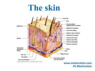
  • 2. The skin (the interface between humans and their environment) is the largest organ in the body. It weighs an average of 4 kg and covers an area of 2 m2. The skin has two layers: 1.The epidermis, outer epithelial layer. 2.The dermis, inner connective tissue. Beneath the dermis is, the subcutis/hypodermis which usually contains abundant fat. The epidermis adheres to the dermis partly by the interlocking of its downward projections (epidermal ridges or pegs) with upward projections of the dermis (dermal papillae).
  • 3.
  • 4. The Integument Is the largest system of the body  16% of body weight,  1.5 to 2m2 in area,  The integument is made up of two parts: 1. Cutaneous membrane a. Epidermis– Superficial epithelium b. Dermis – underlying CT with blood supply 2. Accessory structures a. Hair b. Nails c. Exocrine Glands
  • 5. Protection  First line of defense against  Bacteria  Viruses  Protects underlying structures from  Ultraviolet (UV) radiation  Dehydration  Vitamin D production  Needed for calcium absorption  Sensation  Sensory receptors
  • 6. Body temperature regulation If too hot  Dermal blood vessels dilate  Vessels carry more blood to surface so heat can escape If too cold  Dermal blood vessels constrict  Prevents heat from escaping Excretion Small amounts of waste products are lost through perspiration www.medrockets.com Fb:Medrockets
  • 7. Understanding how the skin can function in these many ways starts with understanding the structure of the 3 layers of skin The Epidermis Epithelial tissue Dermis Dense connective tissue proper – irregular Hypodermis Subcutaneous tissue- loose connective tissue proper and adipose tissue www.medrockets.com Fb:Medrockets
  • 9. Epidermis: The epidermis is formed from many layers of closely packed cells called keratinocytes: 1.Basal layer (stratum germinativum(. 2.Prickle cell layer (stratum spinosum(. 3.Granular layer (stratum ranulosum(. 4.Horny layer (stratum corneum( On the palms and soles a pale or pink layer, the stratum lucidum, is noted just above the granular layer. The epidermis varies in thickness from less than 0.1 mm on the eyelids to nearly 1 mm on the palms and soles.
  • 10. Structures of the Epidermis  The five strata of keratinocytes in thick skin  From basal lamina to free surface 1. Stratum basale 2. Stratum spinosum 3. Stratum granulosum 4. Stratum lucidum 5. Stratum corneum www.medrockets.com Fb:Medrockets
  • 11. 11.The basal layer: Is the deepest layer, rests on basement membrane, which attaches it to the dermis. This layer generate cells of the epidermis. It is a single layer of columnar cells, whose basal surfaces sprout many fine processes and hemidesmosomes, anchoring them to the lamina densa of the basement membrane. www.medrockets.com Fb:Medrockets
  • 12. 22..The spinous or prickle cell layer: Composed of differentiating cells, contain some tonofibrils and kertohyalin granules, which synthesize keratins. They are larger than basal cells. Keratinocytes are firmly attached to each other by small interlocking cytoplasmic processes, and by abundant desmosomes. Under the light microscope, the desmosomes look like ‘prickles’, they are specialized attachment plaques.
  • 13. 3.Granular layer: Consists of two or three layers of cells that are flatter than those in the spinous layer, and have more tonofibrils. As the name of the layer implies, these cells contain large irregular basophilic granules of keratohyalin, which merge with tonofibrils. As keratinocytes migrate out through the outermost layers, their keratohyalin granules break up and their contents are dispersed throughout the cytoplasm, leading to keratinization and the formation of a thick and tough peripheral protein coating called the horny envelope.
  • 14. Stratum Lucidum — the “clear layer” Found only in thick skin Covers stratum granulosum Stratum Corneum — the “horn layer” Exposed surface of skin 15 to 30 layers of keratinized cells Water resistant Shed and replaced every 2 weeks www.medrockets.com Fb:Medrockets
  • 15. Other cells of the epidermis: 1. Melanocytes. 2. Langerhans cells. 3. Merckle cells www.medrockets.com Fb:Medrockets
  • 16. Melanocytes are the only cells that can synthesize melanin. They migrate from the neural crest into the basal layer of the ectoderm where, in human embryos, they are seen as early as the eighth week of gestation. They are also found in hair bulbs, the retina and pia arachnoid. Melanocytes www.medrockets.com Fb:Medrockets
  • 17. Each dendritic melanocyte associates with a number of keratinocytes, forming an ‘epidermal melanin unit. The dendritic processes of melanocytes wind between the epidermal cells and end as discs in contact with them. Their cytoplasm contains discrete organelles, the melanosomes, containing varying amounts of the pigment melanin. www.medrockets.com Fb:Medrockets
  • 18. The Langerhans cell is a dendritic cell like the melanocyte. It also lacks desmosomes and tonofibrils, but has a lobulated nucleus. The specific granules within the cell look like a tennis racket when seen in two dimensions in an electron micrograph. They are plate-like, with a rounded bleb protruding from the surface. Langerhans cells www.medrockets.com Fb:Medrockets
  • 19. Langerhans cells come from precursors originating in the bone marrow. There are approximately 800 Langerhans cells per mm2. Langerhans cells are alone among epidermal cells in possessing surface receptors for C3b and the Fc portions of IgG and IgE, and in bearing major histocompatibility complex (MHC) Class II antigens (HLA-DR, -DP and -DQ). They are best thought of as specialized macrophages.
  • 20. Langerhans cells have a key role in many immune reactions. They take up exogenous antigen, process it and present it to T lymphocytes either in the skin or in the local lymph nodes. They probably play a part in immuno-surveillance for viral and tumor antigens. In this way, ultraviolet radiation can induce skin tumors both by causing mutations in the epidermal cells, and by decreasing the number of epidermal Langerhans cells. Topical or systemic glucocorticoids also reduce the density of epidermal Langerhans cells.
  • 21. Merkel cells are found in normal epidermis. Act as transducers for fine touch. They are nondendritic cells. Lying in or near the basal layer. Are of the same size as keratinocytes. They are concentrated in localized thickenings of the epidermis near hair follicles (hair discs). Contain membrane bound spherical granules, 80–100 nm in diameter. Sparse desmosomes connect these cells to neighbouring keratinocytes. Fine unmyelinated nerve endings are often associated with Merkel cells. Merkel cells
  • 23. The basement membrane lies at the interface between the epidermis and dermis. With light microscopy it can be highlighted using a periodic acid–Schiff (PAS) stain, because of its abundance of neutral mucopolysaccharides.. The dermo-epidermal junction
  • 24. Electron microscopy shows that the basement membrane has 4 componenets: 1.The plasma membrane of basal cells which has hemidesmosomes (containing bullous pemphigoid antigens, collagen XVII and á6 â4 integrin). 2.Electron-lucent area, the lamina lucida which separate lamina densa from the basal cells. The lamina lucida contains laminin-1, laminin-5 and entactin. 3.Lamina densa (rich in type IV collagen). 4.Anchoring fibrils (of type VII collagen), dermal microfibril bundles and single small collagen fibres (types I and III), extend from the papillary dermis to the deep part of the lamina densa.
  • 25.
  • 26. The structures within the dermo-epidermal junction provide mechanical support, encouraging the adhesion, growth, differentiation and migration of the overlying basal cells, and also act as a semipermeable filter that regulates the transfer of nutrients and cells from dermis to epidermis. www.medrockets.com Fb:Medrockets
  • 27. Dermis: The dermis lies between the epidermis and the subcutaneous fat. It is tow parts: 1.Upper papillary dermis. 2.Lower reticular dermis. Its thickness varies, being greatest in the palms and soles and least in the eyelids and penis. In old age, the dermis thins and loses its elasticity. The dermis interdigitates with the epidermis so that upward projections of the dermis, the dermal papillae, interlock with downward ridges of the epidermis, the rete pegs. This interdigitation is responsible for the ridges seen most readily on the fingertips (as fingerprints). The dermis has three components: cells, fibers and amorphous ground substance.
  • 28. Cells of the dermis: The main cells of the dermis are fibroblasts, but there are also small numbers of resident and transitory mononuclear phagocytes, lymphocytes, Langerhans cells and mast cells. Other blood cells, e.g. polymorphs, are seen during inflammation.
  • 29. Fibres of the dermis: The dermis is largely made up of interwoven fibresThe dermis is largely made up of interwoven fibres,, principally of collagen, packed in bundlesprincipally of collagen, packed in bundles.. Those in the papillary dermis are finer than those in the deeperThose in the papillary dermis are finer than those in the deeper reticular dermisreticular dermis.. When the skin is stretched, collagen, with its high tensileWhen the skin is stretched, collagen, with its high tensile strength, prevents tearing, and the elastic fibres, intermingledstrength, prevents tearing, and the elastic fibres, intermingled with the collagen, later return it to the unstretched statewith the collagen, later return it to the unstretched state.. www.medrockets.com Fb:Medrockets
  • 30. Collagen fibers: Makes up to 70–80% of the dry weight of the dermis. Its fibres are composed of thinner fibrils, which are in turn made up of microfibrils built from individual collagen molecules. These molecules consist of three polypeptide chains forming a triple helix with a non- helical segment at both ends. Collagen is an unusual protein as it contains a high proportion of proline and hydroxyproline and many glycine residues. Defects in the enzymes needed for collagen synthesis are responsible for some skin diseases, including the Ehlers–Danlos syndrome and osteogenesis imperfecta (fragility of bones(.
  • 31. Elastic fibers: Account for about 2% of the dry weight of adult dermis. They have two distinct protein components: an amorphous elastin core and a surrounding ‘elastic tissue microfibrillar component’. Abnormalities in the elastic tissue cause cutis laxa (sagging inelastic skin( and pseudoxanthoma elasticum. Reticulin fibres:: Are fine collagen fibers, seen in fetal skin and around the blood vessels and appendages of adult skin.
  • 32. The ground substance of the dermis consists largely of two glycosaminoglycans (hyaluronic acid and dermatan sulphate( with smaller amounts of heparan sulphate and chondroitin sulphate. The ground substance has several important functions: •it binds water, allowing nutrients, hormones and waste products to pass through the dermis; •it acts as a lubricant between the collagen and elastic fibre networks during skin movement; and •it provides bulk, allowing the dermis to act as a shock absorber. Ground substance of the dermis:
  • 33. Both smooth and striated muscle are found in the skin. The smooth arrector pili muscles are used by animals to raise their fur and so protect them from the cold. They are vestigial in humans, but may help to express sebum. Smooth muscle is also responsible for ‘goose pimples’ (bumps( from cold, nipple erection, and the raising of the scrotum by the dartos muscle. Striated fibres (e.g. the platysma( and some of the muscles of facial expression, are also found in the dermis. Muscles: www.medrockets.com Fb:Medrockets
  • 34. Although the skin consumes little oxygen, its abundant blood supply regulates body temperature. The blood vessels lie in two main horizontal layers: 1.The deep plexus is just above the subcutaneous fat, and its arterioles supply the sweat glands and hair papillae. 2.The superficial plexus is in the papillary dermis and arterioles from it become capillary loops in the dermal papillae. The blood vessels in the skin are important in thermoregulation. Blood vessels:
  • 35. : Afferent lymphatics begin as blind-ended capillaries in the dermal papilla and pass to a superficial lymphatic plexus in the papillary dermis. There are also two deeper horizontal plexuses, and collecting lymphatics from the deeper one run with the veins in the superficial fascia. Cutaneous lymphatics www.medrockets.com Fb:Medrockets
  • 36.
  • 37. The skin is supplied with an estimated one million nerve fibres. Most are found in the face and extremities. Their cell bodies lie in the dorsal root ganglia. Both myelinated and non-myelinated fibres exist, with the latter making up an increasing proportion peripherally. Most free sensory nerves end in the dermis; however, a few non- myelinated nerve endings penetrate into the epidermis. Some of these are associated with Merkel cells. Nerves:
  • 38. Free nerve endings detect stimuli of heat and pain (nocioceptors(, while specialized end organs in the dermis, Pacinian and Meissner corpuscles, detect pressure (mechanoreceptors(, vibration and touch. Autonomic nerves supply the blood vessels, sweat glands and arrector pili muscles. Itching is an important feature of many skin diseases. It follows the stimulation of fine free nerve endings lying close to the dermo-epidermal junction. Impulses from these free endings pass centrally in two ways: quickly along myelinated A fibres, and more slowly along non- myelinated C fibres. In itchy skin diseases, pruritogenic chemicals such as histamine and proteolytic enzymes are liberated close to the dermoepidermal junction.
  • 39. The hard keratin of the nail plate is formed in the nail matrix, which lies in an invagination of the epidermis (the nail fold( on the back of the terminal phalanx of each digit. The matrix runs from the proximal end of the floor of the nail fold to the distal margin of the lunula. From this area the nail plate grows forward over the nail bed, ending in a free margin at the tip of the digit. The nail:
  • 40. The nail bed is capable of producing small amounts of keratin which contribute to the nail and which are responsible for the ‘false nail’ formed when the nail matrix is obliterated by surgery or injury. The cuticle acts as a seal to protect the potential space of the nail fold from chemicals and from infection. The nails provide strength and protection for the terminal phalanx. Their presence helps with fine touch and with the handling of small objects.
  • 41. The rate at which nails grow varies from person to person: fingernails average between 0.5 and 1.2 mm per week, while toenails grow more slowly. Nails grow faster in the summer, if they are bitten, and in youth. They change with ageing from the thin, occasionally spooned nails of early childhood to the duller, paler and more opaque nails of the very old.
  • 42.
  • 43.
  • 44. Cosists of 2 parts: 1. Hair follicle, include: inner root sheath and outer rot sheath 2. Hair shaft, consists of: hair cuticle, cortex and medulla. Hair shaft and hair follicle are produced by the matrix portion of hair bulb. Along one side, the sebaceous gland open to the upper part of the hair follicle, arrector pili muscle attach to the lower part. Apocrine gland also open to the hair follicle from the opposite side. From the surface opening of the hair follicle to the enterance of the sebaceous duct is called infundibular segment. The portion between the sebaceous duct and insertion of arrector pili muscle is isthmus. The lowest portion is hair bulb. Hair unit:
  • 45.
  • 46. 1.Lanugo hairs. Fine long hairs covering the fetus, but shed about 1 month before birth. 2.Vellus hairs. Fine short unmedullated hairs covering much of the body surface. They replace thelanugo hairs just before birth. 3.Terminal hairs. Long coarse medullated hairs seen, for example, in the scalp or pubic regions. Their growth is often influenced by circulating androgen levels. Classification of hairs:
  • 47. There are three phases of follicular activity: 1. Anagen. The active phase of hair production. 2. Catagen. A short phase of conversion from active growth to the resting phase. Growth stops, and the end of the hair becomes club-shaped. 3. Telogen. A resting phase at the end of which the club hair is shed. The hair cycle
  • 48. The scalp contain an average of 100 000 hairs, anagen lasts for up to 5 years, catagen for about 2 weeks, and telogen for about 3 months. As many as 100 hairs may be shed from the normal scalp every day as a normal consequence of cycling. On the scalp, about 85% are normally in anagen and 15% in the telogen phase. www.medrockets.com Fb:Medrockets
  • 49.
  • 50.
  • 51. Sebaceous glands: They are associated with hair follicle, lie in the obtuse angle between the follicle and the epidermis. They are multilobed and contain cells full of lipid, which are shed whole (holocrene secretion) during secretion into the upper part of the hair follicle. Sebum contains a mixture of triglycerides, fatty acids, waxy esters, squalene and cholesterol. It lubricates and waterproofs the skin and protects it from drying. Free sebaceous glands may be found in the eye lids (meibomian glands), mucous membranes (Fordyce spots), nipples, peri-anal region and genetalia. Androgen hormones, especially dihydrotestosterone stimulate sebaceous gland activity.
  • 52. There are 2-3 million sweat glands distributed all over the body surface but they are most numerous on the palms, soles and axillae. The tightly coiled glands lie deep in the dermis, and the emerging duct passes to the surface by penetrating the epidermis in a corkscrew fashion. Initially sweat is isotonic like plasma but, under normal conditions, it becomes hypotonic by the time it discharged at the surface, after the tubular resorption of electrolytes and water under the influence of aldosterone and antidiuretic hormones. Eccrine sweat glands:
  • 53. The PH of sweat is between 4.0 and 6.8; it contains sodium, potassium chloride, lactate, urea and ammonia. Sweat glands have an important role in temperature control, the skin surface being cooled by evaporation. Up to 10 L/day of sweat can be excreted. The sweat glands are innervated by cholinergic fibers of the sympathetic nervous system. Sweating can therefore be induced by cholinergic and blocked by anticholenergic drugs. Central control of sweating resides in the preoptic hypothalamic sweat centre.
  • 54. Apocrine sweat glands: They are limited to the axillae, nipples, peri-umbilical area, perineum and genitalia. The coiled tubular glands (larger than sweat glands) lie deep in the dermis, and during sweating the luminal part of their cells is lost (decapitation secretion). Apocrine sweat passes via the duct into the mid-portion of the hair follicle. The action of bacteria on apocrine sweat is responsible for body odour. The glands are innervated by adrenergic fibres of the sympathetic nervous system.