
Recommended
More Related Content
What's hot
What's hot (20)
Similar to Microsporogenesis
Similar to Microsporogenesis (20)
More from Pushpinderjeet Kaur
Recently uploaded
Recently uploaded (20)
Microsporogenesis
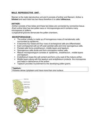
- 1. MALE REPRODUCTIVE UNIT : Stamen is the male reproductive unit and it consists of anther and filament. Anther is bilobed and each lobe has two theca therefore it is called dithecous. ANTHER- -Anther consists of two lobes and these two lobes are connected by connective tissue. -Each anther lobe has two pollen sacs or microsporangia and it contains many microspores or pollens. -Longitudinal grooves demarcate the pollen chambers. MICROPORNAGIUM – The anther initially is made up of homogenous mass of meristematic cells surrounded by epidermis. It becomes four lobed and four rows of archesporial cells are differentiated. Each archesporial cell cut off outer parietal cells and inner sporogenous cells. Parietal cells forms endothecium, middle layers and tapetum. Sporogenous cells divide and form microspore mother cells. Mature microsporangium consists of epidermis , endothecium , middle layers and tapetum. Endotheicum loses the cell content and form a dry coat of the mature anther. Middle layers along with the tapetum and endpthesium protects the microspores and helps in dehiscence of the anther. Tapetum provides nourishment to the developing pollen grains. Tapetum- Possess dense cytoplasm and have more than one nucleus.
- 2. MICROSPOROGENSIS- The process of the formation and differentiation of microspores (pollen grains) from microspore mother cells (MMC) by reductional division is called microsporogenesis. Sporogenous cells undergo differemtiation to form microspore mother cell. Microspore mother cells undergo meiosis and give rise to formation of four haploid nuclei and followed by the cell wall formation. These are called microspore tetrad. As the anther mature and dehydrate, the microspores dissociate from each other and develop pollen grains. POLLEN GRAIN Pollen grain represents the male gametophytes. Pollen grains are made of 2 layered Wall, 1. Exine :- Made of sporopollenin - most resistant organic matter known. It can withstand high temperatures and strong acids and alkali. No enzyme can degrade sporopollenin 2. Intine :- -Thin and continuous layer – Made of cellulose and pectin 3. Germ pores – apertures on exine where sporopollenin is absent – forms pollen tube. 4. A plasma membrane surrounds cytoplasm of pollen grain. MATURE POLLEN While still in anther lobe, pollen grains begin to germinate. The nuclei undergo mitotic division. At this stage pollen grain contains two nuclei- a large vegetative nuclei and a small generative nucleus. Both these nuclei lie freely in the cytoplasm. A mature pollen consist of 2 cells with nucleus (Vegetative and Generative). VEGETATIVE CELL Bigger Abundant food reserve Large irregular nucleus Responsible for the development of pollen grain GENERATIVE CELL Small Involves in syngamy (as it undergoes mitotic division to form two male gametes one of which fuse with an egg) Dense cytoplasm and nucleus
- 3. In 60 % of angiosperms pollen grain are shed at 2 celled stage – which contains generative and vegetative cells In remaining 40 % angiosperms pollen grains are shed at 3 celled stage – which contain vegetative cells and two male gametes. ( generative cells undergo mitotic division to form two male gametes. Pollen grains of many species e.g Parthenium cause severe allergies and bronchial diseases in some people and leads to chronic respiratory disorders– asthma, bronchitis, etc. • Pollen grains are rich in nutrients and are used as pollen tablets as food supplements. • Viability of pollen grain varies with species to species and should land on stigma before this period to germinate. Pollen grains of large number of species are stored in liquid nitrogen at temperature – 1960 C, can be stored as pollen bank. This technique is called CRYOPRESERVATION. Cryopreservation – Cryo is a greek word (krayos – frost ) It means preservation in frozen state. Principle – to bring plant tissue or cell to a zero metabolism or non - dividing state by reducing the temperature in the presence of cryopreservant. It can be liquid nitrogen at -196 C. The Pistil, Megasporangium (Ovule) and Embryo sac Monocarpellary - Gynoecium may consists of single pistil. Multicarpellary – if gynoceium consists of more than one pistil. Syncarpous – If pistils are fused it is called syncarpous. Apocarpous – If pistils are free it is called apocarpous. e.g Multicarpellary and syncarpous pistil- Papaver Multicarpellary and apocarpous pistil- Michelia
- 4. • Each pistil has three parts the stigma, style and ovary. Inside the ovary is ovarian cavity (locule). The placenta is located inside the ovarian cavity. Megasporangia (ovules) arise from placenta. Megasporangium (ovule) Ovule is a small structure attached to placenta. Funicle – stalk by which ovule is attached to placenta Hilum- junction between ovule and funicle Integuments- protective envelops Micropyle- small opening at the tip of ovule into where pollen tube enters Chalaza- basal part of ovule Nucellus (2n)-mass of cells enclosed in integuments. Has abundant food reserve. Megasporogenesis- The process of formation of megaspore from megaspore mother cell by meiotic division is known as megasporogenesis. This process takes place in ovule. Ovule differentiates a single megaspore mother cell (MMC) in the micropylar region of nucellus. MMC undergoes meiotic division that results into the production of four megaspores. • In most of the flowering plants three megaspores degenerate. 1megaspore develops into female gametophyte (embryo sac). Therefore it is also called monosporic division.