Excretory system 2
•Download as PPTX, PDF•
32 likes•20,659 views

COMPLETE DESCRIPTION OF ENDOCRINE SYSTEM

Recommended
More Related Content
What's hot
What's hot (20)
Viewers also liked
Viewers also liked (20)
Similar to Excretory system 2
Similar to Excretory system 2 (20)
More from pramod kumar
More from pramod kumar (20)
Recently uploaded
Recently uploaded (20)
Excretory system 2
- 2. OBJECTIVES OF THE CLASS At the end of the presentation, all of you should able to: Enlist the organs of excretory system. Explain about function of liver. Describe the functions of large intestine. Explain about the skin. Enlist the structures of urinary system
- 3. OBJECTIVES CONTINUE….. Explain about kidneys. Explain the organs associated with kidneys. Discuss the gross structure of kidney. Explain microscopic structure of the kidney. Illustrate functions of kidneys. Describe renal blood supply of kidney
- 5. INTRODUCTION OF EXCRETORY SYSTEM The human excretory system functions to remove waste from the human body, the bodily process of discharging wastes.
- 6. EXCRETORY SYSTEM Excretory system consist of following : Lungs Skin Large intestine Liver Urinary system
- 7. LIVER It is a largest gland of the body. Detoxifies and breaks down chemicals. The liver also produces bile.
- 8. LUNGS Filter out carbon dioxide, from the blood.
- 9. LARGE INTESTINE The large intestine collects waste from throughout the body. It extracts any remaining usable water and then removes solid waste.
- 10. SKIN Skin excretes sweat through sweat glands throughout the body. This helps to remove additional wastes, such as excess urine. Helps to keep the body cool when it is warm.
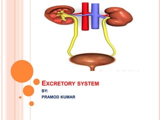
- 11. URINARY SYSTEM It consists of following structures: Kidneys Ureters Urinary bladder Urethra
- 12. KIDNEY
- 13. KIDNEY- LOCATION Lies in the abdominal cavity below diaphragm. On each side of the vertebral column. Extend from the level of 12th thoracic vertebra to the 3rd lumbar vertebra. The right kidney is usually slightly lower than the left. About 11cm long, 6cm wide, 3cm thick and weight 150 gm.
- 14. KIDNEYS CONTD.
- 15. ORGANS ASSOCIATED WITH THE KIDNEY RIGHT KIDNEY Superiorly- the right adrenal gland Anteriorly - the right lobe of the liver, duodenum and the hepatic flexure of the colon Posteriorly - the diaphragm and muscles of the posterior abdominal wall.
- 16. ORGANS ASSOCIATED WITH THE KIDNEY LEFT KIDNEY Superiorly- the left adrenal gland. Anteriorly- the spleen, stomach, jejunum and splenic flexure of the colon. Posteriorly - the diaphragm and muscles of the posterior abdominal wall.
- 17. GROSS STRUCTURE OF THE KIDNEY Fibrous capsule- surrounding the kidney. Cortex- a reddish brown layer of tissue. Medulla- Innermost layer, consisting of pale conical – shaped striations is called renal pyramids.
- 18. ORGANS ASSOCIATED WITH KIDNEY
- 19. GROSS STRUCTURE OF THE KIDNEY Hilum- Is the concave medial border of the kidney where the kidney where the renal blood and lymph vessels, the ureter and nerves enter. It has a number of distal branches called calyces. Calyces are of two types. Major calyx Minor calyx
- 20. MICROSCOPIC STRUCTURE OF KIDNEY The kidney is composed of about 1 million functional units called Nephrons and a smaller number of collecting tubes.
- 21. FUNCTIONS OF KIDNEY Formation of urine. Simple filtration. Selective reabsorption Secretion Water balance and urine output Electrolyte balance
- 22. BLOOD SUPPLY OF KIDNEY Kidneys need to have constant supply in order to control the composition of body fluids. Renal artery a branch from aorta enters the kidney at hilum. supplies blood rich in nitrogenous waste, oxygen and nutrients.
- 23. BLOOD FLOW
- 24. BLOOD SUPPLY CONTD. Renal vein: carries purified deoxygenated blood to the inferior vena cava and then to the heart.
- 25. NEPHRON Is the basic structural and functional unit of the kidney. Its chief function is- To regulate the concentration of water and soluble substances Reabsorbing what is needed and excreting the rest as urine
- 26. NEPHRON A nephron eliminates wastes from the body regulates blood volume and blood pressure control levels of electrolytes and metabolites and regulates blood pH.
- 27. NEPHRON CONTD. Each nephron consists of the following parts: 1) Glomerulus ; 2) Bowman’s capsule ; 3) Proximal tubule ; 4) loop of Henle ; 5) Distal tubule ; 6) Collecting duct.
- 28. NEPHRON
- 30. PARTS OF NEPHRON: Bowman’s Capsule Glomerulus Proximal Convoluted Tubule Loop of henle Distal Convoluted Tubule Collecting tubules Bowman’s capsule: is the initial dilated part of nephron. Glomerulus: is formed of a tuft of capillaries into the bowman’s capsule.
- 31. Proximal convoluted tubule: PCT lumen is continuous with that of Bowman’s capsule. Loop of henle: it consists of a descending limb which continues into the thin segment from which arises the thick ascending limb. Distal convoluted Tubule: Thick ascending limb is continued with the distal convoluted tubule. Collecting tubules: DCT join to form collecting tubules.
- 32. Composition of urine About 96% of the urine is water. 1.5% is salts, mainly sodium chloride. Urea make up 2%. About 1.5L urine is produced daily.
- 33. URETERS The ureter is the tube that carries urine from the kidney to the urinary bladder. In humans there are two ureters, one attached to each kidney.
- 34. URETERS CONTINUE……… The upper half of the ureter is located in the abdomen and the lower half is located in the pelvic area. The ureter is approximately 12 inches long. The tube has thick walls composed of a fibrous, a muscular, and a mucus coat, which are able to contract.
- 35. URINARY BLADDER The urinary bladder is a muscular sac in the pelvis, just above and behind the pubic bone. When empty, the bladder is about the size and shape of a pear.
- 36. URINARY BLADDER CONTINUE….. Urine is made in the kidneys, and travels down two tubes called ureters to the bladder. The bladder stores urine, allowing urination to be infrequent and voluntary. The bladder is lined by layers of muscle tissue that stretch to accommodate urine. The normal capacity of the bladder is 400 to 600 mL
- 37. URETHRA In both genders, the urethra works as a tube connecting the urinary bladder to the genitals.
- 38. URETHRA CONTINUE……. The bladder collects and stores urine until when it is ready to be discharged through the urethra. While the function remains the same for both genders, slight differences exist due to differences between male and female genitals.
- 39. PATHOLOGY RELATED TO EXCRETORY SYSTEM: Glomerulonephritis
- 41. URINARY TRACT INFECTIONS: A urinary tract infection (UTI) (also known as acute cystitis or bladder infection) is an infection that affects part of the urinary tract. When it affects the lower urinary tract it is known as a simple cystitis (a bladder infection) and when it affects the upper urinary tract it is known as pyelonephritis (a kidney infection).
- 42. KIDNEY STONES A kidney stone, also known as a renal calculus (from the Latin rēnēs, "kidneys," and calculus, "pebble"), is a solid concretion orcrystal aggregation formed in the kidneys from minerals in the urine.
- 43. RENAL FAILURE Renal failure (also kidney failure or renal insufficiency) is a medical condition in which the kidneys fail to adequately filter waste products from the blood.
- 44. HYDRONEPHROSIS
- 45. URETHRITIS
- 47. CONCLUSION The main function of the Excretory System is to get rid of bodily waste. It is one of the most important systems in the body because the waste it gets rid of is poisonous, and if they build up in your body for too long they can kill you.
- 48. SUMMARY The kidneys regulate the amount of water, salts and other substances in the blood. The kidneys are fist-sized, bean shaped structures that remove nitrogenous wastes (urine) and excess salts from the blood. The ureters are tubes that carry urine from the pelvis of the kidneys to the urinary bladder. The urinary bladder temporarily stores urine until it is released from the body. The urethra is the tube that carries urine from the urinary bladder to the outside of the body. The outer end of the urethra is controlled by a circular muscle called a sphincter.
- 49. RECAPTUALIZATION Enlist the organs of excretory system? Enlist the structures of urinary system? explain the organs associated with kidneys? Illustrate functions of kidneys? Enlist parts of nephron? Explain about urinary bladder? Explain about urethra?