2. (carbohydrates)
Phospholipids fatty acids membranes
Nucleic Acids
(DNA/RNA)
nucleotides DNA: nucleoid (chromosome), plasmids
rRNA: ribosomes; mRNA, tRNA:
cytoplasm
Procaryotic Cell Architecture
At one time it was thought that bacteria and other procaryotes were essentially "bags of enzymes" with no
inherent cellular architecture. The development of the electron microscope in the 1950s revealed the distinct
anatomical features of bacteria and confirmed the suspicion that they lacked a nuclear
membrane.Procaryotes are cells of relatively simple construction, especially if compared to eucaryotes.
Whereas eucaryotic cells have a preponderance of organelles with separate cellular functions, procaryotes
carry out all cellular functions as individual units.
A procaryotic cell has five essential structural components: a nucleoid (DNA),ribosomes, cell
membrane, cell wall, and some sort of surface layer, which may or may not be an inherent part of the
wall.
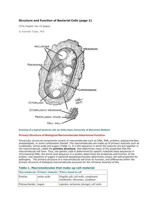
Structurally, there are three architectural regions: appendages (attachments to the cell surface) in the
form of flagella and pili (or fimbriae); a cell envelopeconsisting of a capsule, cell wall and plasma
membrane; and a cytoplasmic region that contains the cell chromosome (DNA) and ribosomes and
various sorts of inclusions (Figure 1).
Figure 1. Cutaway drawing of a typical bacterial cell illustrating structural components. See Table 2 below for
chemical composition and function of the labeled components.
Table 2. Summary of characteristics of typical bacterial cell structures
3. Structure
Flagella
Function(s)
Swimming movement
Predominant chemical composition
Protein
Pili
Sex pilus
Stabilizes mating bacteria during DNA transfer by
conjugation
Protein
Common pili or
fimbriae
Attachment to surfaces; protection against
phagotrophic engulfment
Protein
Capsules (includes
"slime layers" and
glycocalyx)
Attachment to surfaces; protection against
phagocytic engulfment, occasionally killing or
digestion; reserve of nutrients or protection
against desiccation
Usually polysaccharide;
occasionally polypeptide
Cell wall
Gram-positive
bacteria
Prevents osmotic lysis of cell protoplast and
confers rigidity and shape on cells
Peptidoglycan (murein) complexed
with teichoic acids
Gram-negative
bacteria
Peptidoglycan prevents osmotic lysis and confers
rigidity and shape; outer membrane is
permeability barrier; associated LPS and proteins
have various functions
Peptidoglycan (murein) surrounded
by phospholipid protein-
lipopolysaccharide "outer
membrane"
Plasma membrane
Permeability barrier; transport of solutes; energy
generation; location of numerous enzyme systems
Phospholipid and protein
Ribosomes Sites of translation (protein synthesis) RNA and protein
Inclusions
Often reserves of nutrients; additional specialized
functions
Highly variable; carbohydrate, lipid,
protein or inorganic
Chromosome Genetic material of cell DNA
Plasmid Extrachromosomal genetic material DNA
4. Figure 2 . Electron micrograph of an ultra-thin section of a dividing pair of group A streptococci (20,000X).
The cell surface fimbriae (fibrils) are evident. The bacterial cell wall is seen as the light staining region
between the fibrils and the dark staining cell interior. Cell division in progress is indicated by the new septum
formed between the two cells and by the indentation of the cell wall near the cell equator. The streptococcal
cell diameter is equal to approximately one micron. Electron micrograph of Streptococcus pyogenes by Maria
Fazio and Vincent A. Fischetti, Ph.D. with permission. The Laboratory of Bacterial Pathogenesis and
Immunology, Rockefeller University.