
Recommended
More Related Content
What's hot
What's hot (20)
Similar to |HAP-II| Unit-3: Respiratory System. |Complete Notes||
Similar to |HAP-II| Unit-3: Respiratory System. |Complete Notes|| (20)
Recently uploaded
Recently uploaded (20)
|HAP-II| Unit-3: Respiratory System. |Complete Notes||
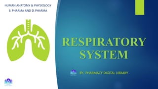
- 1. RESPIRATORY SYSTEM BY- PHARMACY DIGITAL LIBRARY HUMAN ANATOMY & PHYSIOLOGY B. PHARMA AND D. PHARMA
- 2. Contents Introduction Classification of the Respiratory system Upper Respiratory System Lower Respiratory system Physiology of Respiration Transport of O2 and Co2 Pulmonary volume and capacities Disorders of the Respiratory System IMP questions 2
- 3. Introduction The respiratory system is a biological system consisting of specific organs and structures used for the respiration process in an organism. It is involved in the intake and exchange of oxygen and carbon dioxide between an organism and the environment. The branch of medicine that deals with the diagnosis and treatment of diseases of the ears, nose, and throat is called Otorhinolaryngology. RESPIRATION- The oxidative process occurring within living cells by which the chemical energy of organic molecules is released in a series of metabolic steps involving the consumption of oxygen and the liberation of carbon dioxide and water is called as respiration. 3
- 4. The process of respiration is divided into 3 different processes Pulmonary ventilation: It is the inspiration (inflow) and expiration (outflow) of air between the atmosphere and the lungs. External respiration: It is the exchange of gases between the lungs and the blood. Internal respiration: It is the exchange of gases between the blood and cells. 4
- 5. Classification of The Respiratory System Anatomically it is divided into Respiratory System Upper respiratory system (Nose, pharynx, and associated structure) Lower respiratory system (larynx, trachea, bronchi, and lungs) 5
- 6. Functionally it is divided into Respiratory System Conducting System Associated with the conduction of gases it consists of the nose, pharynx, larynx, trachea, bronchi, and terminal bronchioles Respiration System Associated with the exchange of gases. It consists of respiration bronchioles, alveolar ducts, and alveoli 6
- 8. Upper Respiratory System Nose The nose is a structure of the face made of cartilage, bone, muscle, and skin that supports and protects the anterior portion of the nasal cavity. It is the external portion of the respiratory system and opens through the nostrils. The nasal cavity is divided by a septum into left and right portions. The nasal cavity is a hollow space within the nose and skull that is lined with hair and mucus membrane. The nose is lined by vascular ciliated columnar epithelium containing mucus- secreting goblet cells 8
- 9. Functions The nasal cavity prepares the air entering the body for the lungs by warming it, moistening it, and filtering it. Hair and mucus lining the nasal cavity help trap dust, mold, pollen, and other environmental contaminants before they can reach the inner portions of the body. Air exiting the body through the nose returns moisture and heat to the nasal cavity before being exhaled into the environment. The nose is the organ of the sense of smell. The nerve endings that detect and smell are are in the roof of the nose. 9
- 10. Pharynx The pharynx or throat is a funnel-shaped tube about 13 cm long. Both the mouth and nose open into the pharynx. It lies just posterior to the nasal cavity, oral cavity, and larynx and just interior to the cervical vertebrae Its wall is composed of skeletal muscles and lined with mucus membranes. Broadly pharynx is divided into 3 parts: 1. Nasopharynx: The uppermost part of the pharynx that lies just posterior to the nasal cavity. 2. Oropharynx: The middle part of the pharynx that lies just posterior to the oral cavity. 3. Laryngopharynx: The lowest portion of the pharynx that lies just superior to the larynx. 10
- 11. Pharynx 11
- 12. Functions- Passageway for air and food Pharynx is an organ involved in both the respiratory and digestive systems. Air passes through nasal and oral sections and food passes through oral and laryngeal sections. Warming and humidifying the air By the same methods as in the nose, the air is further warmed and moistened as it passes through the pharynx. Taste The olfactory nerve endings of the sense of taste are located in the epithelium of the oral and pharyngeal parts. Protection The lymphatic tissue of the pharyngeal and laryngeal tonsils produces antibodies in response to antigens. Speech The pharynx functions in speech by acting as a resonating chamber for sound. It helps to give the voice its characteristics. 12
- 13. Lower Respiratory System Larynx It is also called a voice box, It is a short passage that connects the pharynx with the trachea. The larynx is composed of irregularly shaped cartilage attached by ligaments and membranes. The main cartilages are 1, Thyroid cartilage 1 Cricoid cartilage 2 Arytenoid cartilage 1 Epiglottis: Elastic cartilage Lower Respiratory System 13
- 14. Thyroid cartilage (Adam’s apple) It consists of two fused plates of hyaline cartilage that form the anterior wall of the larynx. which gives it a triangular shape. It is larger in males than in females. Cricoid cartilage This lies below the thyroid cartilage. It is a ring of hyaline cartilage, attached to the 1 ring of trachea. It forms the inferior wall of the larynx. Arytenoid Cartilage These are the triangular pieces of hyaline cartilage It is located at the posterior of the cricoid cartilage. They provide attachment to the vocal cords. 14
- 15. Epiglottis It is a large leaf-shaped piece of elastic cartilage. The stem of the epiglottis is attached to the thyroid cartilage, but the leaf portion is unattached and free to move up and down like a trap door. If the larynx is the voice box, then the epiglottis acts as a lid, it closes the larynx during swallowing and protects the lungs from entry of foreign objects. Pharynx 15
- 16. Voice Production The Vocal Cords are two folds of a mucus membrane with cord-like free edges from thyroid cartilage anteriorly and arytenoid cartilage posteriorly. When the muscles controlling the vocal cords are relaxed the vocal cords open and the passageway for air through the larynx is clear, the vocal cords are called abducted (open vocal cords). The pitch of the sound produced by vibrating the vocal cords in this portion is low. When the muscle controlling the vocal cords contracts, the vocal cords are closed, and the vocal cords are called as adducted (closed vocal cords). The pitch of the sound produced by such a vocal cord is high. When the vocal cords are stretched to this extent and are vibrated by air passing through them from the lungs, the sound produced is high-pitched. The pitch of the voice is determined by the tension applied to the vocal cords through an appropriate set of muscles. If they have been pulled taut by the muscles, they vibrate more rapidly, and a higher pitch results. 16
- 17. Epiglottis 17
- 18. Functions Production of sound Protection of the lower respiratory tract by ensuring that food passes into the esophagus and not into the lower respiratory tract. Acts as a passageway for air. Larynx produces humidification, filtration, and warming of air as it travels through the larynx. 18
- 19. Trachea (Windpipe) It is a tubular passageway for air. It is 12 cm long and 2.5 cm in diameter. It extends from the larynx to the 5th thoracic vertebrae where it divides into left and right primary bronchi. The trachea is located anterior to the esophagus and has C-shaped cartilaginous bands within the wall. The trachea is made up of 4 main layers. Adventitia (outer layer of areolar connective tissue) The hyaline cartilage The submucosa The mucosa The mucosa is made up of pseudo-stratified ciliated columnar epithelium containing ciliated columnar cells, goblet cells, and basal cells. The cilia move in a single direction thereby keeping the tract free of dust and particles. The single C-shaped cartilage ring provides rigid support to the tracheal wall. At the point where the trachea divides into the right and left primary bronchi, there is an internal ridge called a carina. 19
- 20. Trachea 20
- 21. Functions Mucociliary escalator: Mucus secreted by the goblet cells of mucosa moistens the air and traps the dust particles. The cilia move the dust particles to the pharynx where they can be eliminated from the respiratory tract by expectoration {spitting). Cough reflex: A nerve ending in the larynx, trachea, and bronchi are sensitive to the irritation which generates nerve impulses conducted by the Vagus nerve to the respiratory center in the brain stem. Warming, humidifying, and filtering: Trachea produces humidification, filtration, and warming of air as it travels through the trachea. 21
- 22. Lungs The lungs are a pair of spongy, air-filled cone-shaped organs located on either side of the chest (thorax). The lungs extend laterally from the heart to the ribs on both sides of the chest and continue posteriorly toward the spine. The superior end of the lung forms the cone, and the inferior end forms the base. The left lung is slightly smaller than the right lung The right lung is thicker and broader than the left lung The lungs are divided into Apex: The narrow superior portion of the lung Base: The broad inferior portion of the lung Coastal surface: The surface of the lung lying against the ribs 22
- 23. Anatomy of the Lungs 23
- 24. Medial surface: It forms the lateral boundary of the mediastinum. The medial surface of each lung contains a region the hilus through which bronchi, pulmonary blood vessels, lymphatic vessels, and nerves enter and exit. The lung is surrounded by two layers of delicate serous membrane called as a pleural membrane. The inner membrane which covers the lungs is called as visceral pleura and the outer layer which is attached to the wall of the thoracic cavity is called as parietal pleura. The space between these two layers, the pleural cavity, contains the lubricating fluid secreted by membranes. This fluid reduces friction between the membranes and allows them to move easily on one another during breathing. 24
- 25. Lobes of lungs The right lung is subdivided into 3 lobes: Superior lobe Middle lobe Inferior lobe The left lung is subdivided into 2 lobes: Superior lobe Inferior lobe Fissures of lungs Fissures are double-fold visceral pleura that either completely or incompletely invaginate lung parenchyma to form the lung lobes. Right lung: It has two fissures: • Oblique fissure: It separates the upper lobes from the lower lobes. • Horizontal fissure: It separates the right upper and middle lobes. Lungs: Lobes of the Lungs 25
- 26. Left lung: It has one fissure: • Oblique fissure: It separates the superior lobe and inferior lobe. Bronchi • At the sternal angle, the trachea divides into the right pulmonary bronchus which goes into the right lung, and the left pulmonary bronchus which goes into the left lung. • Bronchi are large, hollow tubes made of hyaline cartilage lined with ciliated pseudostratified epithelium. Right bronchus • It is wider, shorter, and more vertical than the left bronchus. • It is 2.5 cm long. • After entering the right lung at the hilum, it is (the primary bronchi) divided into secondary bronchi, one for each lobe of lung. 26
- 27. Left bronchus • It is 5 cm long and narrower than the right bronchus. • After entering the left lung, it divides into 2 secondary bronchi, one for each lobe of the lung. • The secondary bronchi further divide into smaller bronchi that divide into bronchioles. bronchi called tertiary • Bronchioles further divides into smaller bronchioles to form tube called terminal bronchioles. • Terminal bronchioles further divide into respiratory bronchioles. Functions • The primary function is the control of air entering the lungs. • The diameter of the respiratory system is altered by contraction or relaxation, thus regulating the volume of air entering the lungs. 27
- 28. Lobules • Each lobe of the lung is divided into many small compartments called lobules. • Each lobule is covered by elastic connective tissue and contains lymphatic vessels, an arteriole, a venule, and a branch from a terminal bronchiole. • Terminal bronchioles are subdivided into microscopic branches called respiratory bronchioles. • Respiratory bronchioles in turn are subdivided into several (2-11) alveolar ducts. Bronchioles, Alveolar ducts and Alveoli 28
- 29. • The alveolar ducts are surrounded by alveoli and alveoli sacs. • The alveoli are cup-shaped structures surrounded by a capillary network. • The alveolar wall consists of two types of cells. Type 1 alveolar cells: it forms a lining of the alveolar wall. Type 2 alveolar cells: it secretes an alveolar fluid (a complex mixture of phospholipids is lipoproteins), which keeps the alveolar cells moist. Alveoli 29
- 30. Physiology of Respiration Respiration is divided into two phases, external and internal respiration. External Respiration (Pulmonary gas exchange) It is the exchange of gases between the air spaces of the lungs and blood in pulmonary capillaries. In this process, pulmonary capillary blood gains O2 and loses CO2. Right ventricle: It pumps deoxygenated blood to the lungs for purification. Left ventricle: It pumps oxygenated blood to all other parts of the body. External respiration or pulmonary gas exchange is the diffusion of O2 from the air in the alveoli of the lungs to blood in pulmonary capillaries and the diffusion of CO2 in the opposite direction. External respiration in the lungs converts deoxygenated blood coming from the right side of the heart into oxygenated blood that returns to the left side of the heart. As blood flows through the pulmonary capillaries, it picks up O2 from alveolar air and unloads CO2 into alveolar air. This process is called an exchange of gases; each gas diffuses independently from the area where partial pressure is low. 30
- 31. O2 diffuses from alveolar air, where its partial pressure is 105 mm Hg into the blood in pulmonary capillaries, where Pco2 is only 40 mm Hg in resting person. In a person who is exercising the Po2 will be even lower because contracting muscle fibers are using more O2. Diffusion continues until the Po2 of pulmonary capillary blood increases to match the Po2 of alveolar air, 105 mm Hg. While O2 is diffusing from alveolar air into deoxygenated blood, CO2, is diffusing in the opposite direction. The PCo2 of deoxygenated blood is 45 mm Hg in a resting person, whereas the pco2 of alveolar air is 40 mm Hg. Because of this difference in Pco2, carbon dioxide diffuses from deoxygenated blood into the alveoli until the Pco2 of the blood decreases to 40 mm Hg. Thus, the Po2 and Pco2 of Oxygenated blood leaving the lungs are the same as in alveolar air. The CO2 that diffuses into the alveoli is eliminated from the lungs during expiration. 31
- 32. The rate of external respiration depends on several factors which are given below. Partial pressure difference • As the alveolar Po2 is higher than Po2 in a systemic vein, O2 diffuses from the alveoli into the blood. • As a person goes at high altitude, the atmospheric Po2 decreases, the alveolar Po2 also decreases and less O2 diffuses into the blood. Surface area for gas exchange • The surface area for the diffusion of gas is large (about 70 m2). • Any pulmonary disorder that decreases the functional surface area decreases the rate of external respiration. Diffusion distance • The total thickness of the alveolar-capillary membrane is 0.5 um. • Thicker the membrane slower is the rate of diffusion. Breathing rate and depth • External respiration also depends on the average rate of airflow into and out of the lungs. 32
- 33. Schematic representation of External and Internal Respiration 33
- 34. Exchange of Gases in Blood Capillary from Alveoli 34
- 35. Internal Respiration It is the exchange of gases between the blood in systemic capillary and systemic tissue cells. In this process the systemic capillary gains CO2 and loses O2. Internal respiration results in the conversion of oxygenated blood to deoxygenated blood. Oxygenated blood entering the tissue capillaries has a Po2 of 100 mm Hg, whereas tissue cells have an an average Po2 of 40 mm Hg. Because of this difference in Po2, Oxygen diffuses from the oxygenated blood through interstitial fluid and into tissue cells until the Po2 in the blood decreases to 40 mm Hg. This is the average Po2 of deoxygenated blood entering tissue when one is at rest. While O2 diffuses from the blood capillaries into tissue cells, CO2 diffuses in the opposite direction. The average Po2 of tissue cells is 45 mm Hg whereas, tissue capillary Oxygenated blood is 40 mm Hg. 35
- 36. • From here the deoxygenated blood returns to the heart and it is pumped to the lungs for another cycle of external respiration. Transport of Oxygen and Carbon Dioxide • When O2 and CO2 enter the blood, certain chemical reactions occur that aid in transport and gas exchange. • Transport of gases between the lungs and body tissues is a function of blood. • O2 does not dissolve easily in water; only about 1.5 % is dissolved in blood plasma • About 98.5% of O2 is transported to Hb, combines with it inside the red blood cells. • Rach 100 ml of oxygenated blood contains about 20 ml of oxygen, 0.3 ml dissolved in plasma, and 19.7 ml bound to Hemoglobin. • Hb consists of two parts, a protein called globin and an iron-containing protein called heme. • O2 and Hb combine in a reversible reaction to form oxyhemoglobin as follows: Binding of O2 HB + O2 Dissociation of O2 Hb-O2 36
- 37. 98.5% of the O2 is bound to hemoglobin and is trapped inside the RBCs, only dissolved O2 (1.5%) can diffuse out of tissue capillaries into cells. 37
- 38. 38
- 39. Factors Affecting Hb and O2 Binding • Greater the Po2, the more oxygen will combine with Hb. • Lesser the Po2, the lesser oxygen will combine with Hb. Hb and acidity (pH): As acidity increases (pH decreases), the affinity of hemoglobin for O2 decreases, and O2 dissociates more readily from hemoglobin In an acidic environment, Hb affinity for O2 is lower and oxygen splits more rapidly from Hb. This is called as Bohr Effect. • The Bohr effect works in both ways: An increase in H+ in the blood causes O2 to unload from hemoglobin, and the binding of O2 to hemoglobin causes the unloading of H+ from hemoglobin. The partial pressure of CO2 • As Pco2 rises, Hb releases O2 more readily. • Low blood pH (acidic condition) results from high CO2. • As CO2 is taken up by the blood, it is converted to carbonic acid with the help of the enzyme carbonic acid anhydrase in red blood cells. Carbon dioxide CO2 + H2O H2CO3 H+ + HCO3 - CA Water Carbonic Acid Hydrogen Ion Bicarbonate Ion 39
- 40. • Thus, carbonic acid formed in RBCs dissociates in H+ and HCO3 -. • As the H+ concentration increases pH decreases. • Thus, increased Pco2 produces a more acidic environment that helps to split from Hb. Temperature • As temperature increases, oxygen is released from hemoglobin. 2, 3-biphosphoglycerate (BPG) • A substance found in RBC called BPG decreases Hb affinity for O2 and thus helps to release O2 from hemoglobin. Foetal hemoglobin • Foetal Hb differs from adult Hb in structure and in its affinity for oxygen. • Foetal Hb has a higher affinity for O2 because it binds to BPG less strongly. Carbon dioxide • Each 100 ml of deoxygenated blood contains 5 ml of CO2, which is carried by blood in 3 main forms. • Dissolved Co2: 7% is dissolved in plasma. Upon reaching the lungs, it diffuses into the lungs. 40
- 41. Carbaminohaemnoglobin: • 23 % combines with the globin portion of Hb to form carbaminoglobin. The formation of Hb- CO2 is greatly influenced by PCo2. • Bicarbonate ions: 70% is transported in plasma as bicarbonate ions • As CO2 diffuses into tissue capillaries and enters RBC it reacts with water in the presence of an enzyme carbonic anhydrase to form carbonic acid. • The carbonic acid dissociates to form H and HCO3 • Many H+ ions combine with Hb to form H. Hb and bicarbonate ions accumulate inside the RBCs. Hb + CO2 Hb–CO2 Hemoglobin Carbon dioxide Carbaminohaemnoglobin Carbon dioxide CO2 + H2O H2CO3 H+ + HCO3 - CA Water Carbonic Acid Hydrogen Ion Bicarbonate Ion 41
- 42. Pulmonary Volumes and Capacities During the process of normal breathing about 500 ml of air moves into the respiratory passageway with each inspiration and moves out with each expiration. Tidal volume (TV): This is the amount of air passing in and out of the lungs during each cycle of breathing (15 cycles/min) Inspiratory reserve volume (IRV) (3100 ml): This is the extra volume of air that can be inhaled into the lungs during maximal inspiration. i.e., above normal TV. Expiratory reserve volume (ERV) (1200 ml): This is the largest volume of air that can be expelled from the lungs during maximal expiration. Residual volume (RV) (1200 ml): It is the volume of air remaining in the lungs after forced expiration. • Inspiratory capacity (IC) (3600 ml): It is the sum of Tidal volume + Inspiratory reserve volume. • Functional residual (FRC) (2400 ml): It is the sum of Residual volume + Expiratory reserve volume. • Vital capacity (VC) (4800 ml): This is the maximum volume of air that can be moved on and out of the lungs (Tidal volume + IRV+ ERV) • Total lung capacity (6000 ml): It is the sum of all volume (TV + IRV + ERV + RV). 42
- 43. Spirogram of Lung Volume and Capacities • Spirogram is the most common pulmonary function function test which specifically measures the amount (volume) and the speed (flow) of air that can be inhaled and led from the lungs. • A spirometer is a volume recorder consisting of a double-walled under with an inverted bell immersed in water to form a seal. When air enters the spirometer from the lungs, the bell rises. • A downward deflection represents expiration, and and an upward deflection represents inspiration. The slope of the spirogram measures the rate of airflow and the amplitude of the deflection measures the volume of air. • Volume is plotted on the vertical axis (Y-axis), and time is plotted on the horizontal axis (X-axis). 43
- 44. Disorders of The Respiratory System Pneumonia • Pneumonia is an acute infection or inflammation of the alveoli. • When certain microbes enter the lungs of susceptible individuals, they release damaging toxins, stimulating inflammation and immune responses that have damaging side effects. • The toxins and immune response damage alveoli and bronchial mucous membranes, inflammation, and edema of alveoli, interfering with ventilation and gas exchange. • Most common cause of pneumonia is the pneumococcal bacterium i.e., Streptococcus pneumoniae. Tuberculosis • The bacterium Mycobacterium tuberculosis is the causative agent of an infectious communicable disease called tuberculosis (TB) that most often affects the lungs and the pleura but may involve other parts of the body. 44
- 45. • Once the bacteria enter the lungs, they multiply and cause inflammation, which stimulates neutrophils and macrophages to migrate to the area and engulf the bacteria to prevent their spread. • Symptoms are fatigue, weight loss, lethargy, anorexia, a low-grade fever, night sweats, cough, dyspnoea, chest pain, and hemolysis. Coryza and Influenza • Many viruses can cause coryza (common cold) but a group of viruses called rhinoviruses are responsible for about 40% of all colds. • Symptoms are sneezing, excessive nasal secretion, dry cough, and congestion. • Complications include sinusitis, asthma, bronchitis, ear infections, and laryngitis • Influenza (flu) is also caused by the virus. • Symptoms include chills, fever (usually higher than 39°C), headache, and muscular aches. 45
- 46. Pulmonary Edema • It's an abnormal accumulation of fluid in the interstitial spaces and alveoli of the lungs. • Edema may arise from increased permeability of pulmonary capillaries. • Most common symptoms are dyspnoea. • Others are wheezing, rapid breathing rate, restlessness, a feeling of suffocation, cyanosis, paleness, excessive perspiration, and pulmonary hypertension. Cystic Fibrosis (CF) • Cystic fibrosis is an inherited disease of secretory epithelia that affects the airways, liver, pancreas, small intestine, and sweat glands. • It is characterized by abnormal transport of chloride and sodium across an epithelium, leading to thick and viscous secretions. 46
- 47. Asthma • Asthma is a disorder characterized by chronic airway inflammation, airway hypersensitivity to a variety of stimuli, and airway obstruction. • The trigger for asthma is an allergen such as pollen, house dust mites, molds, or a particular food. • Other common triggers of asthma are emotional distress, aspirin, sulfating agents (used in wine and beer), exercise, and breathing cold air or cigarette smoke. • The symptoms include difficulty in breathing, coughing, wheezing, chest congestion, tachycardia, fatigue, and anxiety. Chronic Obstructive Pulmonary Disease (COPD) • It is a type of respiratory disease characterized by chronic and recurrent obstruction of airflow which increases airway resistance. • COPD is of 2 types • Emphysema • Chronic bronchitis 47
- 48. Emphysema: It is a disorder characterized by the destruction of the walls of the alveoli producing abnormally large air spaces that remain filled with air during exhalation. It is caused by long-term irritation, cigarette smoke, air pollution, and occupational exposure to industrial dust. Chronic bronchitis: It is a disorder characterized by excessive secretion of bronchial mucus. Cigarette smoking is the main cause. Symptoms are productive cough (sputum), shortness of breath, wheezing, cyanosis, and pulmonary hypertension. Lung Cancer • In the United States lung cancer is the leading cause of cancer deaths; both in males and females. • Cigarette smoking is identified as one of the major causes of lung cancer. • The most common type of lung cancer is bronchogenic carcinoma which starts in the epithelium of the bronchial tubes. • Symptoms are chronic cough, spitting blood from the respiratory tract, wheezing, shortness of breath, chest pain, hoarseness, difficulty in swallowing, weight loss, fatigue, bone pain, headache, anorexia, anemia, jaundice, and thrombocytopenia. 48
- 49. Asbestos-related disease • These are serious lung cancer disorders that develop when one inhales asbestos particles. • When they are inhaled, they penetrate the lung tissue • However, the fibers usually destroy the WBCs, and scarring of lung tissue may follow. • Asbestos-related diseases include asbestosis (widespread scarring of lung tissues) diffuse pleural thickening ( thickening of the pleurae), and mesothelioma ( cancer of the pleurae of the peritoneum). Asbestos-related 49
- 50. Important Questions Short Answer Questions 1. Define respiration. 2. Enlist different processes of respiration. 3. Classify the respiratory system. 4. Write a note on the structure and function of the lungs. 5. Write a note on the structure and function of the lungs. 6. Write a note on the diagram of the respiratory system. 7. Define the terms: (i) Acute bronchitis, (ii) Chronic bronchitis, (ii) Asthma 8. Write a note on the mechanism of breathing. 9. Explain the physiology of voice production 10. Explain the transport of O2 and CO2. 11. Explain the physiology of respiration 50
- 51. Long Answer Questions 1. Give details about the transport of oxygen and carbon dioxide. 2. Define the terms related to pulmonary volumes and capacities. 3. Write a note on the physiology of respiration. 4. Write in detail the physiology of respiration and transport of O2 and CO2. 5. Describe the gross anatomy of the lungs. Explain the exchange of gases at the alveolar and cellular levels. 6. Enlist different organs involved in respiration. Explain the mechanism of breathing and the exchange of gases at the lung and tissue level. 7. Draw a neat labeled diagram of the human respiratory system. Explain in detail the transport of O2 and CO2 during respiration. 8. Define respiration. Explain in detail the mechanism of breathing and the exchange of gases during respiration. 51