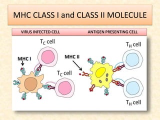
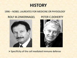
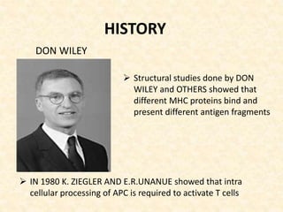
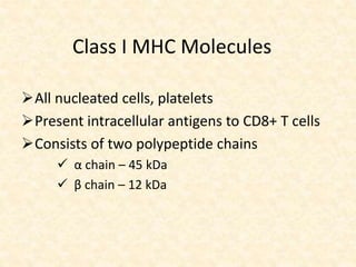
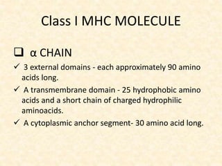
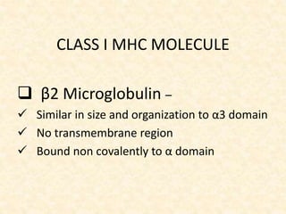
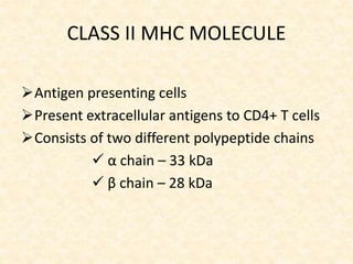
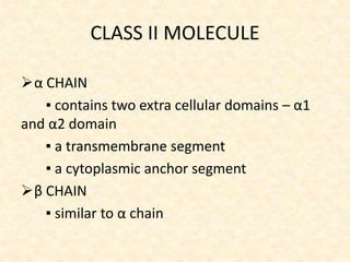
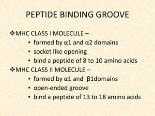
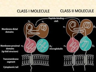
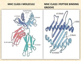
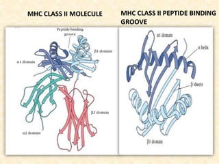
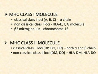
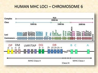
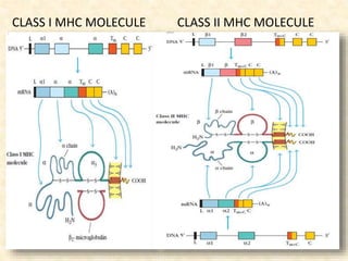
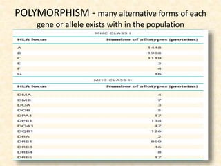
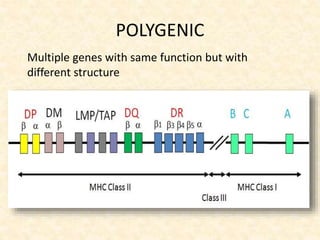
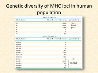
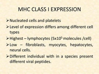
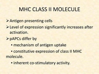
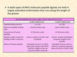
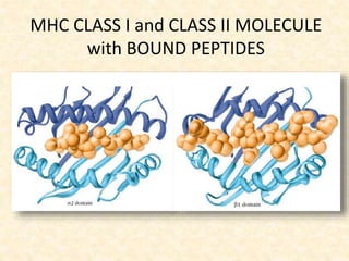
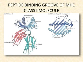
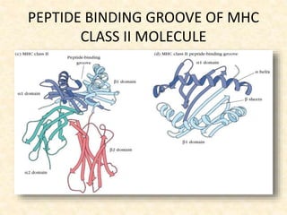
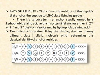
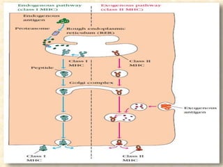
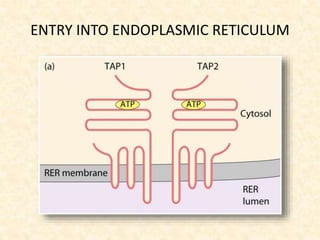
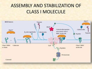
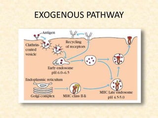
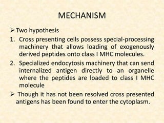
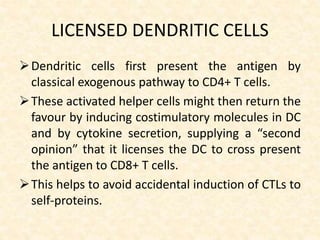
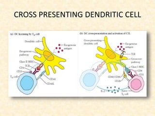
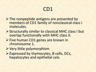
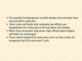
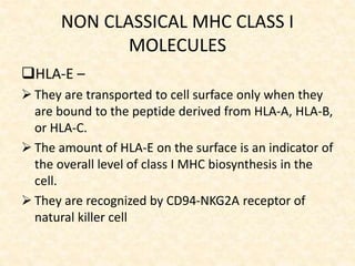
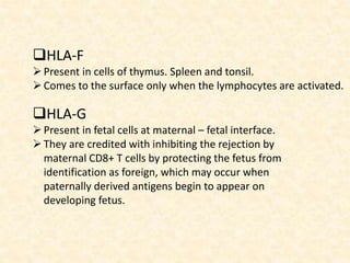
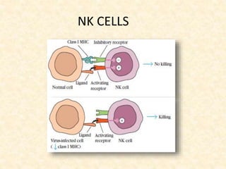
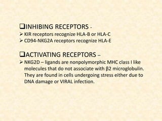
IMMUNOLOGY
MHC CLASS I and CLASS II MOLECULE
VIRUS INFECTED CELL ANTIGEN PRESENTING CELL
ABBREVIATIONS
 MHC - Major Histocompatibility Complex
 HLA - Human Leucocyte Antigen
 APC - Antigen Presenting Cell
 NK - Natural Killer Cell
 CD - Cluster Differentiation
 TNF - Tumor Necrosis Factor
 IFN - Interferon
 KIR - Killer cell Immunoglobulin like Receptor
 TAP - Transporter associated with Antigen Processing
 LMP - Latent Membrane Protein
 ERAP - Endoplasmic Reticulum Amino Peptidase
 MIIC - MHC class II containing Compartment
 CLIP - CLass II associated Invariant chain Peptide
 RAGE - Receptor for Advanced Glycation End Products
 NKG2 - Natural Killer Group 2
HISTORY
 Genetically determined structures on the cell surface that
regulate immunological response.
• 1980 – Nobel Laureates For Medicine Or Physiology
BARUJ BENACERRAFJEAN DAUSSETGEORGE D. SNELL
HISTORY
ROLF M.ZINKERNAGEL PETER C.DOHERTY
1996 – NOBEL LAUREATES FOR MEDICINE OR PHYSIOLOGY
Specificity of the cell mediated immune defense
DON WILEY
 Structural studies done by DON
WILEY and OTHERS showed that
different MHC proteins bind and
present different antigen fragments
HISTORY
 IN 1980 K. ZIEGLER AND E.R.UNANUE showed that intra
cellular processing of APC is required to activate T cells
Structure and
function of MHC
Molecules
CLASSIFICATION
Class I & II MHC Molecules –
 membrane bound glycoproteins
 closely related in both structure and function
Class III MHC Molecule-
 Groups of unrelated proteins
 Do not share any similarity to class I and II
molecules.
 Participates in other aspects of immune response
Class I MHC Molecules
All nucleated cells, platelets
Present intracellular antigens to CD8+ T cells
Consists of two polypeptide chains
 α chain – 45 kDa
 β chain – 12 kDa
Class I MHC MOLECULE
 α CHAIN
 3 external domains - each approximately 90 amino
acids long.
 A transmembrane domain - 25 hydrophobic amino
acids and a short chain of charged hydrophilic
aminoacids.
 A cytoplasmic anchor segment- 30 amino acid long.
CLASS I MHC MOLECULE
 β2 Microglobulin –
 Similar in size and organization to α3 domain
 No transmembrane region
 Bound non covalently to α domain
CLASS II MHC MOLECULE
Antigen presenting cells
Present extracellular antigens to CD4+ T cells
Consists of two different polypeptide chains
 α chain – 33 kDa
 β chain – 28 kDa
CLASS II MOLECULE
α CHAIN
▪ contains two extra cellular domains – α1
and α2 domain
▪ a transmembrane segment
▪ a cytoplasmic anchor segment
β CHAIN
▪ similar to α chain
PEPTIDE BINDING GROOVE
MHC CLASS I MOLECULE –
▪ formed by α1 and α2 domains
▪ socket like opening
▪ bind a peptide of 8 to 10 amino acids
MHC CLASS II MOLECULE –
▪ formed by α1 and β1domains
▪ open-ended groove
▪ bind a peptide of 13 to 18 amino acids
MHC CLASS I MOLECULE MHC CLASS I PEPTIDE BINDING
GROOVE
MHC CLASS II MOLECULE MHC CLASS II PEPTIDE BINDING
GROOVE
GENERAL
ORGANIZATION AND
INHERITANCE OF MHC
MAJOR HISTOCOMPATIBILITY
COMPLEX
MHC is a collection of genes arrayed within a
long continuous stretch of DNA on
▪ chromosome 6 – humans
▪ chromosome 17 – mice
MHC is referred to as
▪ HLA complex – humans
▪ H-2 complex – mice
ORGANIZATION OF MHC COMPLEX
in human chromosome 6
 MHC CLASS I MOLECULE
▪ classical class I loci (A, B, C) - α chain
▪ non classical class I loci - HLA-E, F, G molecule
▪ β2 microglobulin - chromosome 15
 MHC CLASS II MOLECULE
▪ classical class II loci (DP, DQ, DR) – both α and β chain
▪ non classical class II loci (DM, DO) – HLA-DM, HLA-DO
HUMAN MHC LOCI – CHROMOSOME 6
CLASS I MHC MOLECULE CLASS II MHC MOLECULE
POLYMORPHISM - many alternative forms of each
gene or allele exists with in the population
• Many alternative forms of each allele exits in
the population.
POLYGENIC
Multiple genes with same function but with
different structure
LINKAGE DISEQUILIBRIUM
Non random association of alleles of two or more
different genes, at two different loci with a
frequency greater than would be expected by
chance.
Usually these alleles are located close to one
another in the same chromosome
Depends on,
▪ natural selection
▪ population size
▪ mutation rate
GENETIC RECOMBINATION
MHC Molecule expressed on the APC
of a heterozygote offspring
centromere
centromere
Class II LOCI
CLASS I LOCI
Mating of two inbred mice with
different MHC haplotypes
SKIN TRANSPLANTATION
The MHC molecules expressed by an individual are fixed.
However the promiscuity of antigen binding ensures the
enormous flexibility of MHC molecules to bind to various
peptides.
It is rare for any two unrelated individuals to have identical
sets of HLA genes.
Not all alleles encode proteins.
Human class II genes are highly polymorphic and in some
cases an individual can inherit different number of genes.
The theoretical number of combinations possible are 1.7
billion class I haplotypes, 1015 class II haplotypes. Thus
bringing a total of more than 1.7 x1027 combinations.
High level of MHC polymorphism provide better survival
advantage.
FUNCTIONAL RELEVANCE FOR MHC
POLYMORPHISM
• Polymorphism in the MHC is clustered largely
with in the membrane distal α1 and α2
domains of class I, α1 and β1 domains of class
II molecules.
• Allelic differences contribute to the observed
differences in the ability of MHC molecules to
interact with a given peptide ligand
Genetic diversity of MHC loci in human
population
(2013)
(2605)
(1551)
(1200)
(7)
MHC CLASS I EXPRESSION
Nucleated cells and platelets
Level of expression differs among different cell
types
Highest – lymphocytes (5x10⁵ molecules /cell)
Low – fibroblasts, myocytes, hepatocytes,
neural cells.
Different individual with in a species present
different viral peptides.
MHC CLASS II MOLECULE
Antigen presenting cells
Level of expression significantly increases after
activation.
pAPCs differ by
▪ mechanism of antigen uptake
▪ constitutive expression of class II MHC
molecule.
▪ inherent co-stimulatory activity.
 PROFESSIONAL APC
 DENDRITIC CELLS –
▪ most effective
▪ constitutively express high levels of MHC class II molecule
▪ inherent co-stimulatory activity
▪ activate naïve TH cells.
 MACROPHAGES –
to be activated before they express sufficient level of MHC
class II molecule and co-stimulatory activity
 B CELLS –
▪ constitutively express MHC class II molecule
▪ to be activated for co-stimulatory activity.
 NON PROFESSIONAL APC
1.Fibroblasts
2.Glial cells
3.Pancreatic β cells
4.Thymic epithelial cells
5.Thyroid epithelial cells
6.vascular endothelial cells
VARIOUS REGULATORS
 GENETIC REGULATORY COMPONENTS –
CIITA, RFX – class II MHC transactivators. If defective causes
bare lymphocyte syndrome.
 VIRAL INTERFERENCE –
Viral infection interfere with MHC class I expression by,
▪ decreased level of components needed for peptide
transport,
▪ or MHC class I assembly
▪ decreased transcription
 CYTOKINE MEDIATED SIGNALLING –
▪ early stages of infection – IFN α and TNF α
▪ later stages of infection – IFN γ
 The alleles in MHC haplotype
determine which fragment of
peptide to be presented
 This variability in immune response
to an antigen among different
individuals is explained by two
models viz.,
▪ determinant-selection model
▪ holes-in-the-repertoire model
 Determinant-selection model –
 Different class II MHC molecules differ in their
ability to bind particular processed antigens.
 And some peptides may be more crucial to
eliminate the pathogen than others.
 Holes-in-the-repertoire model –
T cells bearing receptors that recognize certain
foreign antigens which happen to closely
resemble self antigens may be eliminated during
T cell development, leaving the organism without
the cells/receptors for future responses to foreign
molecules.
PEPTIDE BINDING
Each individual has 6 different class I molecule
and 12 or more different class II molecule.
Peptide binding by class I and II molecules
does not exhibit fine specificity.
A given MHC molecule can bind to several
different peptides and some peptides can bind
to several different MHC molecules.
So the binding between peptide and MHC is
often referred to as promiscuous.
 In both types of MHC molecules peptide ligands are held in
largely extended conformation that runs along the length of
the groove.
MHC CLASS I and CLASS II MOLECULE
with BOUND PEPTIDES
PEPTIDE BINDING GROOVE OF MHC
CLASS I MOLECULE
PEPTIDE BINDING GROOVE OF MHC
CLASS II MOLECULE
 ANCHOR RESIDUES – The amino acid residues of the peptide
that anchor the peptide to MHC class I binding groove.
▪ There is a carboxy terminal anchor usually formed by a
hydrophobic amino acid and amino terminal anchor either in 2nd
or 2nd and 3rd position also formed by hydrophobic amino acid.
 The amino acid residues lining the binding site vary among
different class I allelic molecule which determines the
classical identity of anchor residues.
PROCESSING OF ANTIGEN
K. ZIEGLER , E.R. UNANUE
1980
ENDOGENOUS PATHWAY
 STANDARD PROTEASOME – 14 subunits.
 IMMUNOPROTEASOME –
▪ present in pAPCs and infected cells
▪ in response to the cytokines IFN γ and TNFα the
genes LMP2 and LMP7 encode replacement catalytic
protein subunits that convert standard proteasomes to
immunoproteasomes.
▪ increased level of protein degradation and turn
over more rapidly.
 The proteasome complex cleaves peptide bonds in an
ATP-dependent manner.
ENDOGENOUS PATHWAY
CYTOSOLIC PROTEOLYTIC SYSTEM
TAP PROTEIN
 Membrane spanning heterodimer.
 Consists of two proteins : TAP1 and TAP2
 Has multiple transmembrane segments, a domain
projecting into the lumen of RER and an ATP-binding
domain that projects into the cytosol.
 Encoded by TAP1 and TAP2 genes within class II MHC region
and different allelic forms of these regions exists.
 Both TAP1 and TAP2 belong to the family of ATP-binding
cassette proteins.
 These proteins mediate ATP dependent transport of
aminoacids, sugars, ions, and peptides.
 TAP has affinity for peptides containing 8 to16 aminoacids.
ENTRY INTO ENDOPLASMIC RETICULUM
ASSEMBLY AND STABILIZATION OF
CLASS I MOLECULE
EXOGENOUS PATHWAY
INVARIANT CHAIN
CD 74
Non MHC encoded protein
Interacts with class II molecule preventing any
endogenously derived peptide from binding
Also involved in the folding of class II α andβ
chains, their exit from RER, and subsequent
routing.
ASSEMBLY OF CLASS II MHC
MOLECULE
CROSS PRESENTATION
First reported by Michael Bevan and later
described in detail by Peter Cresswell and
colleagues.
Internalized antigens that would normally be
handled by the exogenous pathway leading to
class II MHC presentation is redirected to class I
peptide loading pathway.
When this form of antigen presentation leads to
the activation of CTL responses, it is referred to as
cross-priming.
Dendritic cells are most efficient cross-presenters.
MECHANISM
Two hypothesis
1. Cross presenting cells possess special-processing
machinery that allows loading of exogenously
derived peptides onto class I MHC molecules.
2. Specialized endocytosis machinery that can send
internalized antigen directly to an organelle
where the peptides are loaded to class I MHC
molecule
 Though it has not been resolved cross presented
antigens has been found to enter the cytoplasm.
LICENSED DENDRITIC CELLS
Dendritic cells first present the antigen by
classical exogenous pathway to CD4+ T cells.
These activated helper cells might then return the
favour by inducing costimulatory molecules in DC
and by cytokine secretion, supplying a “second
opinion” that it licenses the DC to cross present
the antigen to CD8+ T cells.
This helps to avoid accidental induction of CTLs to
self-proteins.
CROSS PRESENTING DENDRITIC CELL
CD1
The nonpeptide antigens are presented by
members of CD1 family of nonclassical class I
molecules.
Structurally similar to classical MHC class I but
overlap functionally with MHC class II.
Five human CD1 genes are known in
chromosome 1.
Very little polymorphism
Expressed by thymocytes, B cells, DCs,
hepatocytes and epithelial cels.
The peptide binding groove as both deeper and narrower than
classical MHC molecules
 Short chain self lipids with relatively low affinity are
loadedonto CD1 molecules in ER and allow CD1 folding.
When they encounter long chain, high affinity lipid antigens
self lipids are exchanged.
These newly loaded CD1 molecules return to the surface for
recognition by CD1-restricted T cells.
NON CLASSICAL MHC CLASS I
MOLECULES
HLA-E –
 They are transported to cell surface only when they
are bound to the peptide derived from HLA-A, HLA-B,
or HLA-C.
 The amount of HLA-E on the surface is an indicator of
the overall level of class I MHC biosynthesis in the
cell.
 They are recognized by CD94-NKG2A receptor of
natural killer cell
HLA-F
Present in cells of thymus. Spleen and tonsil.
Comes to the surface only when the lymphocytes are activated.
HLA-G
Present in fetal cells at maternal – fetal interface.
They are credited with inhibiting the rejection by
maternal CD8+ T cells by protecting the fetus from
identification as foreign, which may occur when
paternally derived antigens begin to appear on
developing fetus.
HLA DM
Non classical MHC class II molecule
Catalyze the exchange of CLIP with antigenic
peptides.
It is a heterodimer and relatively non
polymorphic
Found predominantly in the endosomal
compartment
HLA DO
Observed in B cells, dendritic cells
Relatively non polymorphic class II molecule
Expression is not induced by IFNγ.
Negative regulator of antigen binding by
modulating the function of HLA-DM.
Following cell activation DO expression is
down regulated.
CLASS III MHC MOLECULE
 Extremely gene dense
 Contain about 62 genes
 The genes C4A and C4B are highly polymorphic.
 Some important products include
I. C4, C2 and factor B
II. TNF α and β
III. NOTCH-4
IV. PBX2 gene product – regulating the expansion of
hematopoietic precursor
V. RAGE – upregulated in vasculopathies, atherosclerosis,
and vasculitis.
NK CELLS
INHIBING RECEPTORS -
KIR receptors recognize HLA-B or HLA-C
CD94-NKG2A receptors recognize HLA-E
ACTIVATING RECEPTORS –
NKG2D – ligands are nonpolymorphic MHC class I like
molecules that do not associate with β2 microglobulin.
They are found in cells undergoing stress either due to
DNA damage or VIRAL infection.
MHC

MHC

  • 1.
  • 2.
    MHC CLASS Iand CLASS II MOLECULE VIRUS INFECTED CELL ANTIGEN PRESENTING CELL
  • 3.
    ABBREVIATIONS  MHC -Major Histocompatibility Complex  HLA - Human Leucocyte Antigen  APC - Antigen Presenting Cell  NK - Natural Killer Cell  CD - Cluster Differentiation  TNF - Tumor Necrosis Factor  IFN - Interferon  KIR - Killer cell Immunoglobulin like Receptor  TAP - Transporter associated with Antigen Processing  LMP - Latent Membrane Protein  ERAP - Endoplasmic Reticulum Amino Peptidase  MIIC - MHC class II containing Compartment  CLIP - CLass II associated Invariant chain Peptide  RAGE - Receptor for Advanced Glycation End Products  NKG2 - Natural Killer Group 2
  • 4.
    HISTORY  Genetically determinedstructures on the cell surface that regulate immunological response. • 1980 – Nobel Laureates For Medicine Or Physiology BARUJ BENACERRAFJEAN DAUSSETGEORGE D. SNELL
  • 5.
    HISTORY ROLF M.ZINKERNAGEL PETERC.DOHERTY 1996 – NOBEL LAUREATES FOR MEDICINE OR PHYSIOLOGY Specificity of the cell mediated immune defense
  • 6.
    DON WILEY  Structuralstudies done by DON WILEY and OTHERS showed that different MHC proteins bind and present different antigen fragments HISTORY  IN 1980 K. ZIEGLER AND E.R.UNANUE showed that intra cellular processing of APC is required to activate T cells
  • 7.
  • 8.
    CLASSIFICATION Class I &II MHC Molecules –  membrane bound glycoproteins  closely related in both structure and function Class III MHC Molecule-  Groups of unrelated proteins  Do not share any similarity to class I and II molecules.  Participates in other aspects of immune response
  • 9.
    Class I MHCMolecules All nucleated cells, platelets Present intracellular antigens to CD8+ T cells Consists of two polypeptide chains  α chain – 45 kDa  β chain – 12 kDa
  • 10.
    Class I MHCMOLECULE  α CHAIN  3 external domains - each approximately 90 amino acids long.  A transmembrane domain - 25 hydrophobic amino acids and a short chain of charged hydrophilic aminoacids.  A cytoplasmic anchor segment- 30 amino acid long.
  • 11.
    CLASS I MHCMOLECULE  β2 Microglobulin –  Similar in size and organization to α3 domain  No transmembrane region  Bound non covalently to α domain
  • 12.
    CLASS II MHCMOLECULE Antigen presenting cells Present extracellular antigens to CD4+ T cells Consists of two different polypeptide chains  α chain – 33 kDa  β chain – 28 kDa
  • 13.
    CLASS II MOLECULE αCHAIN ▪ contains two extra cellular domains – α1 and α2 domain ▪ a transmembrane segment ▪ a cytoplasmic anchor segment β CHAIN ▪ similar to α chain
  • 14.
    PEPTIDE BINDING GROOVE MHCCLASS I MOLECULE – ▪ formed by α1 and α2 domains ▪ socket like opening ▪ bind a peptide of 8 to 10 amino acids MHC CLASS II MOLECULE – ▪ formed by α1 and β1domains ▪ open-ended groove ▪ bind a peptide of 13 to 18 amino acids
  • 16.
    MHC CLASS IMOLECULE MHC CLASS I PEPTIDE BINDING GROOVE
  • 17.
    MHC CLASS IIMOLECULE MHC CLASS II PEPTIDE BINDING GROOVE
  • 18.
  • 19.
    MAJOR HISTOCOMPATIBILITY COMPLEX MHC isa collection of genes arrayed within a long continuous stretch of DNA on ▪ chromosome 6 – humans ▪ chromosome 17 – mice MHC is referred to as ▪ HLA complex – humans ▪ H-2 complex – mice
  • 20.
    ORGANIZATION OF MHCCOMPLEX in human chromosome 6
  • 21.
     MHC CLASSI MOLECULE ▪ classical class I loci (A, B, C) - α chain ▪ non classical class I loci - HLA-E, F, G molecule ▪ β2 microglobulin - chromosome 15  MHC CLASS II MOLECULE ▪ classical class II loci (DP, DQ, DR) – both α and β chain ▪ non classical class II loci (DM, DO) – HLA-DM, HLA-DO
  • 22.
    HUMAN MHC LOCI– CHROMOSOME 6
  • 23.
    CLASS I MHCMOLECULE CLASS II MHC MOLECULE
  • 24.
    POLYMORPHISM - manyalternative forms of each gene or allele exists with in the population • Many alternative forms of each allele exits in the population.
  • 25.
    POLYGENIC Multiple genes withsame function but with different structure
  • 26.
    LINKAGE DISEQUILIBRIUM Non randomassociation of alleles of two or more different genes, at two different loci with a frequency greater than would be expected by chance. Usually these alleles are located close to one another in the same chromosome Depends on, ▪ natural selection ▪ population size ▪ mutation rate
  • 27.
  • 28.
    MHC Molecule expressedon the APC of a heterozygote offspring centromere centromere Class II LOCI CLASS I LOCI
  • 29.
    Mating of twoinbred mice with different MHC haplotypes
  • 30.
  • 31.
    The MHC moleculesexpressed by an individual are fixed. However the promiscuity of antigen binding ensures the enormous flexibility of MHC molecules to bind to various peptides. It is rare for any two unrelated individuals to have identical sets of HLA genes. Not all alleles encode proteins. Human class II genes are highly polymorphic and in some cases an individual can inherit different number of genes. The theoretical number of combinations possible are 1.7 billion class I haplotypes, 1015 class II haplotypes. Thus bringing a total of more than 1.7 x1027 combinations. High level of MHC polymorphism provide better survival advantage.
  • 32.
    FUNCTIONAL RELEVANCE FORMHC POLYMORPHISM • Polymorphism in the MHC is clustered largely with in the membrane distal α1 and α2 domains of class I, α1 and β1 domains of class II molecules. • Allelic differences contribute to the observed differences in the ability of MHC molecules to interact with a given peptide ligand
  • 33.
    Genetic diversity ofMHC loci in human population (2013) (2605) (1551) (1200) (7)
  • 35.
    MHC CLASS IEXPRESSION Nucleated cells and platelets Level of expression differs among different cell types Highest – lymphocytes (5x10⁵ molecules /cell) Low – fibroblasts, myocytes, hepatocytes, neural cells. Different individual with in a species present different viral peptides.
  • 36.
    MHC CLASS IIMOLECULE Antigen presenting cells Level of expression significantly increases after activation. pAPCs differ by ▪ mechanism of antigen uptake ▪ constitutive expression of class II MHC molecule. ▪ inherent co-stimulatory activity.
  • 37.
     PROFESSIONAL APC DENDRITIC CELLS – ▪ most effective ▪ constitutively express high levels of MHC class II molecule ▪ inherent co-stimulatory activity ▪ activate naïve TH cells.  MACROPHAGES – to be activated before they express sufficient level of MHC class II molecule and co-stimulatory activity  B CELLS – ▪ constitutively express MHC class II molecule ▪ to be activated for co-stimulatory activity.  NON PROFESSIONAL APC 1.Fibroblasts 2.Glial cells 3.Pancreatic β cells 4.Thymic epithelial cells 5.Thyroid epithelial cells 6.vascular endothelial cells
  • 38.
    VARIOUS REGULATORS  GENETICREGULATORY COMPONENTS – CIITA, RFX – class II MHC transactivators. If defective causes bare lymphocyte syndrome.  VIRAL INTERFERENCE – Viral infection interfere with MHC class I expression by, ▪ decreased level of components needed for peptide transport, ▪ or MHC class I assembly ▪ decreased transcription  CYTOKINE MEDIATED SIGNALLING – ▪ early stages of infection – IFN α and TNF α ▪ later stages of infection – IFN γ
  • 39.
     The allelesin MHC haplotype determine which fragment of peptide to be presented  This variability in immune response to an antigen among different individuals is explained by two models viz., ▪ determinant-selection model ▪ holes-in-the-repertoire model
  • 40.
     Determinant-selection model–  Different class II MHC molecules differ in their ability to bind particular processed antigens.  And some peptides may be more crucial to eliminate the pathogen than others.  Holes-in-the-repertoire model – T cells bearing receptors that recognize certain foreign antigens which happen to closely resemble self antigens may be eliminated during T cell development, leaving the organism without the cells/receptors for future responses to foreign molecules.
  • 41.
    PEPTIDE BINDING Each individualhas 6 different class I molecule and 12 or more different class II molecule. Peptide binding by class I and II molecules does not exhibit fine specificity. A given MHC molecule can bind to several different peptides and some peptides can bind to several different MHC molecules. So the binding between peptide and MHC is often referred to as promiscuous.
  • 42.
     In bothtypes of MHC molecules peptide ligands are held in largely extended conformation that runs along the length of the groove.
  • 43.
    MHC CLASS Iand CLASS II MOLECULE with BOUND PEPTIDES
  • 44.
    PEPTIDE BINDING GROOVEOF MHC CLASS I MOLECULE
  • 45.
    PEPTIDE BINDING GROOVEOF MHC CLASS II MOLECULE
  • 46.
     ANCHOR RESIDUES– The amino acid residues of the peptide that anchor the peptide to MHC class I binding groove. ▪ There is a carboxy terminal anchor usually formed by a hydrophobic amino acid and amino terminal anchor either in 2nd or 2nd and 3rd position also formed by hydrophobic amino acid.  The amino acid residues lining the binding site vary among different class I allelic molecule which determines the classical identity of anchor residues.
  • 48.
    PROCESSING OF ANTIGEN K.ZIEGLER , E.R. UNANUE 1980
  • 50.
    ENDOGENOUS PATHWAY  STANDARDPROTEASOME – 14 subunits.  IMMUNOPROTEASOME – ▪ present in pAPCs and infected cells ▪ in response to the cytokines IFN γ and TNFα the genes LMP2 and LMP7 encode replacement catalytic protein subunits that convert standard proteasomes to immunoproteasomes. ▪ increased level of protein degradation and turn over more rapidly.  The proteasome complex cleaves peptide bonds in an ATP-dependent manner.
  • 51.
  • 52.
    TAP PROTEIN  Membranespanning heterodimer.  Consists of two proteins : TAP1 and TAP2  Has multiple transmembrane segments, a domain projecting into the lumen of RER and an ATP-binding domain that projects into the cytosol.  Encoded by TAP1 and TAP2 genes within class II MHC region and different allelic forms of these regions exists.  Both TAP1 and TAP2 belong to the family of ATP-binding cassette proteins.  These proteins mediate ATP dependent transport of aminoacids, sugars, ions, and peptides.  TAP has affinity for peptides containing 8 to16 aminoacids.
  • 53.
  • 54.
    ASSEMBLY AND STABILIZATIONOF CLASS I MOLECULE
  • 55.
  • 56.
    INVARIANT CHAIN CD 74 NonMHC encoded protein Interacts with class II molecule preventing any endogenously derived peptide from binding Also involved in the folding of class II α andβ chains, their exit from RER, and subsequent routing.
  • 57.
    ASSEMBLY OF CLASSII MHC MOLECULE
  • 60.
    CROSS PRESENTATION First reportedby Michael Bevan and later described in detail by Peter Cresswell and colleagues. Internalized antigens that would normally be handled by the exogenous pathway leading to class II MHC presentation is redirected to class I peptide loading pathway. When this form of antigen presentation leads to the activation of CTL responses, it is referred to as cross-priming. Dendritic cells are most efficient cross-presenters.
  • 61.
    MECHANISM Two hypothesis 1. Crosspresenting cells possess special-processing machinery that allows loading of exogenously derived peptides onto class I MHC molecules. 2. Specialized endocytosis machinery that can send internalized antigen directly to an organelle where the peptides are loaded to class I MHC molecule  Though it has not been resolved cross presented antigens has been found to enter the cytoplasm.
  • 62.
    LICENSED DENDRITIC CELLS Dendriticcells first present the antigen by classical exogenous pathway to CD4+ T cells. These activated helper cells might then return the favour by inducing costimulatory molecules in DC and by cytokine secretion, supplying a “second opinion” that it licenses the DC to cross present the antigen to CD8+ T cells. This helps to avoid accidental induction of CTLs to self-proteins.
  • 63.
  • 65.
    CD1 The nonpeptide antigensare presented by members of CD1 family of nonclassical class I molecules. Structurally similar to classical MHC class I but overlap functionally with MHC class II. Five human CD1 genes are known in chromosome 1. Very little polymorphism Expressed by thymocytes, B cells, DCs, hepatocytes and epithelial cels.
  • 66.
    The peptide bindinggroove as both deeper and narrower than classical MHC molecules  Short chain self lipids with relatively low affinity are loadedonto CD1 molecules in ER and allow CD1 folding. When they encounter long chain, high affinity lipid antigens self lipids are exchanged. These newly loaded CD1 molecules return to the surface for recognition by CD1-restricted T cells.
  • 67.
    NON CLASSICAL MHCCLASS I MOLECULES HLA-E –  They are transported to cell surface only when they are bound to the peptide derived from HLA-A, HLA-B, or HLA-C.  The amount of HLA-E on the surface is an indicator of the overall level of class I MHC biosynthesis in the cell.  They are recognized by CD94-NKG2A receptor of natural killer cell
  • 68.
    HLA-F Present in cellsof thymus. Spleen and tonsil. Comes to the surface only when the lymphocytes are activated. HLA-G Present in fetal cells at maternal – fetal interface. They are credited with inhibiting the rejection by maternal CD8+ T cells by protecting the fetus from identification as foreign, which may occur when paternally derived antigens begin to appear on developing fetus.
  • 69.
    HLA DM Non classicalMHC class II molecule Catalyze the exchange of CLIP with antigenic peptides. It is a heterodimer and relatively non polymorphic Found predominantly in the endosomal compartment
  • 70.
    HLA DO Observed inB cells, dendritic cells Relatively non polymorphic class II molecule Expression is not induced by IFNγ. Negative regulator of antigen binding by modulating the function of HLA-DM. Following cell activation DO expression is down regulated.
  • 71.
    CLASS III MHCMOLECULE  Extremely gene dense  Contain about 62 genes  The genes C4A and C4B are highly polymorphic.  Some important products include I. C4, C2 and factor B II. TNF α and β III. NOTCH-4 IV. PBX2 gene product – regulating the expansion of hematopoietic precursor V. RAGE – upregulated in vasculopathies, atherosclerosis, and vasculitis.
  • 72.
  • 73.
    INHIBING RECEPTORS - KIRreceptors recognize HLA-B or HLA-C CD94-NKG2A receptors recognize HLA-E ACTIVATING RECEPTORS – NKG2D – ligands are nonpolymorphic MHC class I like molecules that do not associate with β2 microglobulin. They are found in cells undergoing stress either due to DNA damage or VIRAL infection.

Editor's Notes

  • #24 CIITA, RFX – class II MHC trans activators. Defects in these ta causes BARE LYMPHOCYTE SYNDROME
  • #40 Eg – 1. in CMV viral proteins bind to β2M preventing assembly and transport of MHC class I molecule. 2. ADENO virus – reduced transcription of TAP gene.