More Related Content
Similar to Amyloid and alzheimer’s disease
Similar to Amyloid and alzheimer’s disease (20)
Amyloid and alzheimer’s disease
- 1. PERSPECTIVE
Amyloid and Alzheimer’s disease
Hongxing Lei ✉
Beijing Institute of Genomics, Chinese Academy of Sciences, Beijing 100029, China
✉ Correspondence: leihx@big.ac.cn
A CLASSICAL HISTOPATHOLOGICAL HALLMARK
Alzheimer’s disease (AD) is the most prevalent neurodegen-
erative disease afflicting over 30 million patients worldwide.
The typical symptoms of AD include memory loss and
impairment of cognitive function, and currently, there is no
available approach to cure the disease. The projected fast
increase of the senior population is a growing burden for the
international society in terms of both medical cost and patient
care. Since the first case examination in1907, amyloid has
been associated with the disease named after its pioneer
Dr. Alois Alzheimer. A classical histopathological hallmark for
AD is the extracellular deposition of amyloid plaques found in
the postmortem brain of AD patients, along with the
intracellular neurofibrillary tangles (NFT). It is widely believed
that amyloid is the cause of all the symptoms and the eventual
death of AD patients. This so called “amyloid hypothesis” is
dominant in the field of AD research, and a good portion of the
work in this field has been devoted to the mechanism and
pathological effect of amyloid formation.
A CHALLENGING PROTEIN FOLDING PROBLEM
Structure characterization has revealed that amyloid is a fibril
structure consisting of several protofibrils, each of which is
formed by the stacking of two or more prolonged β-sheets.
The hydrogen bonding within a β-sheet is along the fibril axis
while each β strand is perpendicular to the fibril axis, forming
the so called “cross-β” architecture. The building block of this
esthetically pleasing architecture is the Aβ protein. Unlike
most proteins featuring a stable globular structure, Aβ protein
is unstructured in the cytosol under physiologic condition. The
exact mechanism by which this unstructured entity forms fibril
has been pursued by experimentalists and theoreticians for
many years.
From structural studies by solid-state NMR (nuclear
magnetic resonance) (Petkova et al., 2002), site-directed
spin-labeling EPR (electron paramagnetic resonance) ( Török
et al., 2002), and hydrogen/deuterium-exchange (HX) ( Lührs
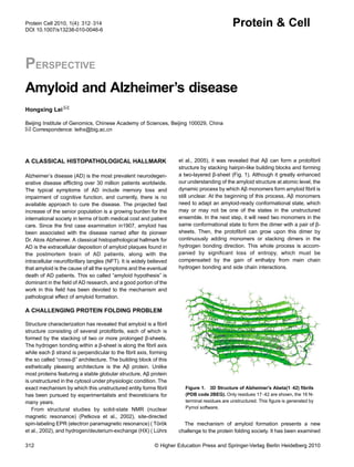
et al., 2005), it was revealed that Aβ can form a protofibril
structure by stacking hairpin-like building blocks and forming
a two-layered β-sheet (Fig. 1). Although it greatly enhanced
our understanding of the amyloid structure at atomic level, the
dynamic process by which Aβ monomers form amyloid fibril is
still unclear. At the beginning of this process, Aβ monomers
need to adapt an amyloid-ready conformational state, which
may or may not be one of the states in the unstructured
ensemble. In the next step, it will need two monomers in the
same conformational state to form the dimer with a pair of β-
sheets. Then, the protofibril can grow upon this dimer by
continuously adding monomers or stacking dimers in the
hydrogen bonding direction. This whole process is accom-
panied by significant loss of entropy, which must be
compensated by the gain of enthalpy from main chain
hydrogen bonding and side chain interactions.
The mechanism of amyloid formation presents a new
challenge to the protein folding society. It has been examined
Figure 1. 3D Structure of Alzheimer's Abeta(1–42) fibrils
(PDB code 2BEG). Only residues 17–42 are shown, the 16 N-
terminal residues are unstructured. This figure is generated by
Pymol software.
312 © Higher Education Press and Springer-Verlag Berlin Heidelberg 2010
Protein Cell 2010, 1(4): 312–314
DOI 10.1007/s13238-010-0046-6
Protein & Cell
- 2. by almost every imaginable techniques for structural char-
acterization (Langkilde and Vestergaard, 2009; Tompa,
2009), including circular dichroism (CD), fluorescence, Four-
ier-transform infrared spectroscopy (FTIR), X-ray crystal-
lography, small angle X-ray scattering (SAXS), NMR, solid-
state NMR, cryo-EM (electron microscopy), STEM (scanning
tunneling EM) and AFM (atomic force microscopy). Never-
theless, due to the dynamic nature of the oligomerization
process, information gathered from experiments regarding
the initial stage of oligomerization has been scarce to date. In
the mean time, molecular modeling and simulation has
provided some insight about the oligomerization process at
atomic level (Lei et al., 2006). However, the inaccuracy in the
modeling and simulation tools has severely hampered the
understanding of the kinetics and thermodynamics and
further dissection of energetic and entropic contributions.
To make things even more complicated, it has been
recently discovered that amyloid has structural polymorphism
(Fändrich et al., 2009). Many fibril species may coexist for the
same amyloidogenic protein/peptide and different physico-
chemical environment can result in the shift of the equilibrium.
These fibril species differ in the number of protofibrils,
arrangement of protofibrils in amyloid fibril, and the polypep-
tide conformation within protofibrils. This phenomenon adds
another level of complexity that has yet to be understood
quantitatively. Nevertheless, the strong interest from diverse
fields such as basic science, drug development and materials
design will continue to drive the research forward.
A PLETHORA OF INVOLVED CELLULAR
PATHWAYS
Adding to the complexity at the structural level is the
existence of a plethora of cellular pathways that amyloid is
involved in. Aβ is generated from amyloid precursor protein
(APP) by subsequent cleavage by β and g secretases, while
α secretase cuts in the middle of Aβ thereby preventing Aβ
aggregation. Aβ mainly exist in two forms: Aβ42 and Aβ40.
From genetic association studies, it has been found that all
familiar form of AD are associated with mutations in either
APP or two of the subunits in g secretase (PS1 and PS2),
while sporadic AD is mainly associated with apolipoprotein E
(ApoE), a protein involved in the transport of cholesterol,
lipoproteins and fat-soluble vitamins.
The processing and metabolism of APP can be regulated
by extracellular stimuli or the binding of adaptor proteins to the
YENPTY motif of its intracellular domain (Jacobsen and
Iverfeldt, 2009). APP intracellular domain (AICD) possesses
transcriptional regulatory activity and alters signaling path-
ways (Pimplikar, 2009). Aβ has been implicated in the
regulation of lipid metabolism, demonstrating inhibition
against hydroxymethylglutaryl-CoA reductase (HMGR) and
activation of sphingomyelinases (SMases) (Normando et al.,
2009). Aβ can disrupt calcium homeostasis by forming
channel with oligomers in the membrane (Kawahara et al.,
2009), it can also disrupt iron homeostasis through MAPK
(mitogen activated protein kinase) cascade (Cahill et al.,
2009). Aβ can increase the production of ROS (reactive
oxygen species) in mitochondrion and nucleus which lead to
apoptosis of neurons (Kaminsky et al., 2010). The induced
neural cell death by Aβ can also be achieved by the activation
of nicotinic acetylcholine receptors with the involvement of
ERK/MAPK pathway, JNK pathway, PI3K/AKT pathway and
JAK-2/STAT-3 pathway (Buckingham et al., 2009).
In a proposed positive feedback loop, Aβ can have
signaling effect on the transcription of BACE1, a candidate
β secretase (Tabaton et al., 2010). This can be achieved by
the activation of G-protein coupled receptors (GPCR) or
calcium ion channels, or the inhibition of insulin receptors. It
can also be achieved by the interaction with ER (endoplasmic
reticulum). This signaling may involve the JNK and ERK/Akt
pathways and eventually lead to the transcriptional activation
of BACE1 by transcription factor AP1, which result in more
production of Aβ and complete the positive feed back loop.
The binding of Aβ to RAGE (receptor for advanced glycation
end products) can also activate the MAPK signaling pathway
and activation of transcription factor NF-κB through Ras-
ERK1/2 pathway, Cdc42/Rac pathway, p38 and JNK path-
ways (Origlia et al., 2009).
AN ATTRACTIVE THERAPEUTIC TARGET
Due to the proposed central role of amyloid in AD develop-
ment, it has enjoyed great attention from the pharmaceutical
industry as well as the academic society (Amijee and Scopes,
2009). Currently, FDA and EMEA approved drugs for AD are
all symptomatic, including four acetylcholinesterase inhibitors
and one NMDA-antagonist. These drugs can only alleviate
the symptoms and cannot cure the disease. To find disease-
modifying treatments, many people have turned to the
amyloid formation process. The basic idea is to develop
drugs that reduce the load of amyloid plaques by shifting the
equilibrium toward the non-toxic Aβ monomer. This can be
accomplished by binding preferentially to the Aβ monomer or
oligomers therefore inhibiting the fibrilization process. It can
also be achieved by disrupting the fibrils, protofibrils and
oligomers. The involvement of zinc and copper ions in the
fibrilization process has also been investigated. Based on this
idea, many chemical compounds and peptide analogs have
been discovered and/or developed, some of which target
specific region of Aβ such as HHQK(13–16) and KLVFF
(16–20). Encouraging results has been observed in mouse
models and clinical trials, including the reduction of amyloid
plaques and improvement of the symptoms. Unfortunately,
most of them have been withdrawn at one stage or another
due to various concerns and none has reached the market.
Another strategy is the reduction of Aβ production by
modifying the proteolytic activity or expression level of the
© Higher Education Press and Springer-Verlag Berlin Heidelberg 2010 313
Amyloid and Alzheimer’s disease Protein & Cell
- 3. secretases involved in the cleavage of APP and production of
Aβ. However, these secretases also participate in many other
cellular pathways, some known and some unknown, raising
serious concern and driving many people away from further
pursuing in this direction. Yet another strategy is the facilitated
clearance of amyloid from the central nervous system (CNS).
A hot topic along this line is the development of antibodies to
stimulate the immune system to accomplish this mission
(Pahnke et al., 2009). One caveat of this strategy is that most
antibodies can only be developed against the mature amyloid
fibril with well-formed structure while the toxic oligomers are
difficult to be targeted. Another hurdle is the blood brain
barrier (BBB) which may limit the transport of the amyloid and
liberated toxic oligomers away from the CNS and lead to
increase in severity of cerebral amyloid angiopathy (CAA).
A LONG-STANDING CONTROVERSY
Ever since the inception of the “amyloid hypothesis”, the
controversy has always been around it (Pimplikar, 2009).
Many evidences have been presented to support this
hypothesis, but on the other hand, many evidences against
this hypothesis also exist. Another histopathological hallmark
of AD is the taupathy caused by NFT, which originated from
the aggregation of hyperphosphorylated tau, another natively
unstructured protein. It has been observed that the severity of
AD symptoms has better correlation with the load of NFT than
that of amyloid plaques, and amyloid plaques have also been
found in cognitively normal people. In addition, the Aβ42/
Aβ40 ratio has been found to have good correlation with the
severity of the disease. It has also been found that some
oligomer species, including dimer, trimer and dodecamer are
much more toxic than amyloid fibrils. In summary, some
doubts have been raised regarding the central role of amyloid
from pathology, cell biology, animal models and genetics
studies. The original theme of amyloid fibril standing alone at
the top of the hierarchy has been modified to include other Aβ
species during amyloid formation. Furthermore, increasing
evidences have suggested that the disruption of other cellular
pathways independent of amyloid formation may also act as
the source of AD development. After more than 100 years
since the discovery of AD, enormous hurdles are still ahead of
us before we can reach a clear understanding and eventual
cure of the disease.
ACKNOWLEDGMENTS
This work was supported by research grants from National Natural
Science Foundation of China (Grant No. 30870474) and SRF for
ROCS, SEM.
REFERENCES
Amijee, H., and Scopes, D.I.C. (2009). The quest for small molecules
as amyloid inhibiting therapies for Alzheimer’s disease. J
Alzheimers Dis 17, 33–47.
Buckingham, S.D., Jones, A.K., Brown, L.A., and Sattelle, D.B.
(2009). Nicotinic acetylcholine receptor signalling: roles in Alzhei-
mer’s disease and amyloid neuroprotection. Pharmacol Rev 61,
39–61.
Cahill, C.M., Lahiri, D.K., Huang, X.D., and Rogers, J.T. (2009).
Amyloid precursor protein and alpha synuclein translation,
implications for iron and inflammation in neurodegenerative
diseases. Biochimica Et Biophysica Acta-General Subjects 1790,
615–628.
Fändrich, M., Meinhardt, J., and Grigorieff, N. (2009). Structural
polymorphism of Alzheimer Abeta and other amyloid fibrils. Prion
3, 89–93.
Jacobsen, K.T., and Iverfeldt, K. (2009). Amyloid precursor protein
and its homologues: a family of proteolysis-dependent receptors.
Cell Mol Life Sci 66, 2299–2318.
Kaminsky, Y.G., Marlatt, M.W., Smith, M.A., and Kosenko, E.A.
(2010). Subcellular and metabolic examination of amyloid-beta
peptides in Alzheimer disease pathogenesis: evidence for Abeta
(25–35). Exp Neurol 221, 26–37.
Kawahara, M., Negishi-Kato, M., and Sadakane, Y. (2009). Calcium
dyshomeostasis and neurotoxicity of Alzheimer’s beta-amyloid
protein. Expert Rev Neurother 9, 681–693.
Langkilde, A.E., and Vestergaard, B. (2009). Methods for structural
characterization of prefibrillar intermediates and amyloid fibrils.
FEBS Lett 583, 2600–2609.
Lei, H., Wu, C., Wang, Z., and Duan, Y. (2006). Molecular dynamics
simulations and free energy analyses on the dimer formation of
an amyloidogenic heptapeptide from human beta2-microglobulin:
implication for the protofibril structure. J Mol Biol 356, 1049–1063.
Lührs, T., Ritter, C., Adrian, M., Riek-Loher, D., Bohrmann, B., Döbeli,
H., Schubert, D., and Riek, R. (2005). 3D structure of Alzheimer’s
amyloid-beta(1–42) fibrils. Proc Natl Acad Sci U S A 102,
17342–17347.
Normando, E.M., Coxon, K.M., Guo, L., and Cordeiro, M.F. (2009).
Focus on: amyloid beta. Exp Eye Res 89, 446–447.
Origlia, N., Arancio, O., Domenici, L., and Yan, S.S. (2009). MAPK,
beta-amyloid and synaptic dysfunction: the role of RAGE. Expert
Rev Neurother 9, 1635–1645.
Pahnke, J., Walker, L.C., Scheffler, K., and Krohn, M. (2009).
Alzheimer’s disease and blood-brain barrier function-Why have
anti-beta-amyloid therapies failed to prevent dementia progres-
sion? Neurosci Biobehav Rev 33, 1099–1108.
Petkova, A.T., Ishii, Y., Balbach, J.J., Antzutkin, O.N., Leapman, R.D.,
Delaglio, F., and Tycko, R. (2002). A structural model for
Alzheimer’s beta-amyloid fibrils based on experimental constraints
from solid state NMR. Proc Natl Acad Sci USA 99, 16742–16747.
Pimplikar, S.W. (2009). Reassessing the amyloid cascade hypothesis
of Alzheimer’s disease. Int J Biochem Cell Biol 41, 1261–1268.
Tabaton, M., Zhu, X.W., Perry, G., Smith, M.A., and Giliberto, L.
(2010). Signaling effect of amyloid-beta(42) on the processing of
AbetaPP. Exp Neurol 221, 18–25.
Tompa, P. (2009). Structural disorder in amyloid fibrils: its implication
in dynamic interactions of proteins. FEBS J 276, 5406–5415.
Török, M., Milton, S., Kayed, R., Wu, P., McIntire, T., Glabe, C.G., and
Langen, R. (2002). Structural and dynamic features of Alzheimer’s
Abeta peptide in amyloid fibrils studied by site-directed spin
labeling. J Biol Chem 277, 40810–40815.
Protein & Cell
314 © Higher Education Press and Springer-Verlag Berlin Heidelberg 2010
Hongxing Lei