2. 3262
functions manifested by ABPs that can
adequately explain many aspects of
cortical actin remodeling, each step
being responsive to signaling cascades.
Here, we briefly summarize the key
features of this cycle and their regulation.
Notice that the outline of actin
remodeling provided is based on
information obtained from studies with
different cell types but predominantly
mammalian platelets, leukocytes,
fibroblasts, epithelial cells, neuronal
cells and tumor cells.
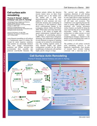
Initiation
Initiation (see poster, step 1) defines
where and when actin filament
elongation occurs at the cell surface.
Cells have several strategies for initiating
new actin polymerization, including de
novo nucleation by the Arp2/3 complex,
formins and Spir (Nicholson-Dykstra et
al., 2005; Rafelski and Theriot, 2004).
Polymerization of actin from newly
exposed actin filament barbed ends is
also a compelling mechanism for
initiating new filament assembly. Free
barbed ends elicit diffusion-limited
polymerization of actin subunits bound
to actin-monomer-sequestering proteins,
the thymosins and profilins (step 9)
(Yarmola and Bubb, 2004). Barbed-end
exposure can result from uncapping – the
removal of numerous barbed-end-
capping proteins (gelsolin family, CapZ,
Hsp70, ankyrins, Eps8) (step 1a) (Allen,
2003; Barkalow et al., 2003; Disanza et
al., 2004) – or from the action of ABPs
that sever actin filaments without
capping them (step 7b) (DesMarais et al.,
2005). One thing that makes these
mechanisms attractive is the fact that half
of the nearly millimolar actin in most
eukaryotic non-muscle cells exists as
short filaments, which provide ample
barbed ends for rapid elongatation when
uncapped and exposed to the large
reservoir of sequestered actin monomers
that cannot spontaneously nucleate. This
mechanism is linked to signaling
cascades that regulate ABPs to promote
new polymerization from pre-existing
barbed ends. Key participants in these
are polyphosphoinositides, which
remove all known capping proteins from
barbed ends. Agonists that promote
cortical actin assembly generally do so
through activation of the small GTPases
Rac, Rho and Cdc42 (Jaffe and Hall,
2005), that in turn stimulate enzymes
leading to focal polyphosphoinositide
accumulation and actin filament
assembly and rearrangement (Niggli,
2005; Yin and Janmey, 2003).
Elongation
Actin filament barbed-end capping by
factors that promote or inhibit capping
(see below) regulates the extent of actin
filament elongation (see poster, step 2).
Termination
Degradation of polyphosphoinositides,
activation of the barbed-end-capping
activity of CapG by Ca2+
and activation of
Hsp70 or CapZ-interacting protein by
phosphorylation (During et al., 2005;
Eyers et al., 2005) terminate actin
filament elongation (see poster, step 3a).
By contrast, the capping protein inhibitors
ENA, VASP, profilin, formins and
CARMIL promote elongation even in the
presence of active capping proteins (step
3b) (Barzik et al., 2005; Bubb et al., 2003;
Higgs, 2005; Kovar, 2006; Yang et al.,
2005). Actin-filament-stabilizing proteins
such as tropomyosins, caldesmons,
calponins, and tropomodulin (which also
caps pointed ends in the presence of
tropomyosin) (Fischer and Fowler, 2003)
also promote elongation by retarding
subunit depolymerization and inhibiting
actin-depolymerizing ABPs, as described
below (step 3c) (Bakin et al., 2004; Eyers
et al., 2005; Mirzapoiazova et al., 2005).
Branching
Nucleation and transient 70° branching
(see poster, step 4) of actin filaments
mediated by the Arp2/3 complex
(Pollard and Borisy, 2003; Stradal and
Scita, 2006; Vicente-Manzanares et al.,
2005) is essential for the intracellular
movements of certain pathogens
(Listeria monocytogenes, Shigella,
Salmonella and Rickettsia species and
poxviruses) (Gouin et al., 2005), some
vesicles, and for the normal dynamics of
adhesive podosomes and related
structures (Linder and Kopp, 2005). A
role for Arp2/3-mediated actin filament
branching in leading-edge actin
elongation is less certain, given high-
resolution images preserving three-
dimensionality (Medalia et al., 2002;
Small et al., 2002), the absence of
Arp2/3 from the leading edges of certain
cells (Gupton et al., 2005; Strasser et al.,
2004), the lack of an effect of Arp2/3 on
actin filament network rigidity required
for lamellar extension (Nakamura et al.,
2002) and the results of experiments
employing RNAi in fibroblasts (Di
Nardo et al., 2005).
Actin filament crosslinking
Bivalent actin-filament-crosslinking
proteins either abet or repel the inherent
parallel alignment of actin filaments
promoted by thermodynamic and ionic
factors (see poster, step 5). Relatively
small globular or rod-like ABPs, such as
fimbrin, scruin, ␣-actinins and espins,
stabilize actin bundles, whereas larger
ABPs that have inherent spring-like
properties, such as the filamins, instead
promote high-angle (orthogonal) filament
organization (Gardel et al., 2006; Gardel
et al., 2004). ␣-Actinins, filamins and
spectrins, a family of membrane-
associated crosslinking proteins,
also function as scaffolds for signaling
intermediates that stimulate actin
elongation; so they are well positioned to
direct the orientation of elongating actin
filaments (Broderick and Winder, 2005;
Feng and Walsh, 2004).
Actin filament contraction, cargo
motoring, and membrane binding
Parallel bundles and orthogonal networks
represent extremes of the highly complex
actin filament arrangements observable at
the cell surface by electron microscopy
and other high-resolution techniques.
Actin filament configurations are
susceptible to deformation by contractile
forces generated by bipolar myosin
filaments (predominantly myosin II)
(Landsverk and Epstein, 2005), which act
especially on actin networks attached to
membranes. Unconventional myosins
primarily move vesicles and other cargoes
along actin filaments (‘motoring’) (see
poster, step 6). Signals contributing to
actin elongation, such as
polyphosphoinositides, also increase
binding of actin filaments and
intermediary ABPs, including talin,
vinculin, filamins, catenins, ␣-actinins,
and zyxin to certain receptors, including
integrins (Ginsberg et al., 2005),
cadherins (Drees et al., 2005), and
proteins of the FERM family (Cho and
Stahelin, 2005) (step 7). This brings actin
filament barbed ends close to the same
signals that promote their elongation,
Journal of Cell Science 119 (16)
JournalofCellScience
3. 3263
potentially amplifying the mass of
elongating actin at the surface.
The linkage between actin filaments and
membranes is important for mechanical
traction against substrates and retraction
of membranes for shape changes and
locomotion in response to contractile
forces. This linkage is also essential for
localizing signaling factors to initiate the
formation of cell-substratum and cell-
cell adhesions, as well as other cellular
processes.
Actin filament disassembly
The most efficient way to break down a
network dominated by thread-like
elements is to cut the threads. This
approach disperses lattices of long actin
filaments immobilized by interpenetration
of filaments and shorter filaments cross-
linked by ABPs. Two ABP families
accomplish this task. The most efficient
are proteins of the gelsolin family, which
disrupt the interactions between actin
subunits in filaments in response to Ca2+
or phosphorylation by Src kinase and then
tightly cap the barbed ends of the severed
filaments (Kumar et al., 2004) (see poster,
step 8a). Ca2+
can also interfere with
binding of crosslinking ABPs and thus
destabilize actin networks. Ca2+
works
with calmodulin to inhibit binding of
filamin to actin (Nakamura et al., 2005)
and directly inhibits the binding of some
␣-actinins (Broderick and Winder, 2005).
The second major actin-filament-severing
ABPs are proteins of the actin-
depolymerizing factor (ADF)/cofilin
family, which weakly sever but do not cap
the barbed ends of actin filaments (step
8b) (Fass et al., 2004). Barbed ends
generated by cofilin either serve as
initiation sites for new elongation or
become capped (step 3a), depending upon
the signals present. A cofilin-binding
protein, Aip1, enhances cofilin activity
(Okada et al., 2002), as do two families of
phosphatases, the slingshots and
cronophin. The adaptor protein 14-3-3
antagonizes this effect. Phosphorylation
of cofilin by LIM kinase, downstream of
Rac activation, inactivates cofilin (Huang
et al., 2005; Nishita et al., 2005). Actin-
filament-stabilizing proteins, particularly
tropomyosins, also inhibit severing of
actin filaments by cofilin but are less
effective against gelsolin family
members, and different tropomyosin
isoforms generated by alternative mRNA
splicing confer subtlety on this
inhibition (Gunning et al., 2005).
Polyphosphoinositides strongly inhibit
actin filament severing by both protein
families, which is consistent with their
general propensity to promote actin
filament assembly.
Although efficient at breaking down
actin networks and providing more
filament ends, actin filament severing
does not directly contribute to
maintenance of an actin monomer pool
required for new filament assembly.
However, the cofilin proteins, by
accelerating subunit dissociation from
pointed ends (‘nibbling’) (step 8c) are
the major drivers for this (Carlier et al.,
1999).
Monomer sequestration
Profilins and, in mammalian cells,
thymosins bind to actin monomers (see
poster, step 9), preventing them from
spontaneous nucleation.
References
Allen, P. (2003). Actin filament uncapping localizes to
ruffling lamellae and rocketing vesicles. Nat. Cell Biol. 5,
972-979.
Bakin, A., Safina, A., Rinehart, C., Daroqui, C.,
Darbary, H. and Helfman, D. (2004). A critical role of
tropomyosins in TGF-beta regulation of the actin
cytoskeleton and cell motility in epithelial cells. Mol. Biol.
Cell 15, 4682-4694.
Barkalow, K., Italiano, J., Jr, Chou, D., Matsuoka, Y.,
Bennett, V. and Hartwig, J. (2003). a-Adducin
dissociates from F-actin and spectrin during platelet
activation. J. Cell Biol. 161, 557-570.
Barzik, M., Kotova, T., Higgs, H., Hazelwood, L.,
Hanein, D., Gertler, F. and Shafer, D. (2005). Ena/VASP
proteins enhance actin polymerization in the presence of
barbed end capping proteins. J. Biol. Chem. 280, 28653-
28662.
Broderick, M. and Winder, S. (2005). Spectrin, alpha-
actinin, and dystrophin. Adv. Protein Chem. 70, 203-246.
Bubb, M., Yarmola, E., Gibson, B. and Southwick, F.
(2003). Depolymerization of actin filaments by profilin.
Effects of profilin on capping protein function. J. Biol.
Chem. 278, 24629-24635.
Carlier, M.-F., Ressad, F. and Pantaloni, D. (1999).
Control of actin dynamics in cell motility. Role of
ADF/Cofilin. J. Biol. Chem. 274, 33827-33830.
Cho, W. and Stahelin, R. (2005). Membrane-protein
interactions in cell signaling and membrane trafficking.
Annu. Rev. Biophys. Biomol. Struct. 34, 119-151.
DesMarais, V., Ghosh, M., Eddy, R. and Condeelis, J.
(2005). Cofilin takes the lead. J. Cell Sci. 118, 19-26.
Di Nardo, A., Cicchetti, G., Falet, H., Hartwig, J.,
Stossel, T. and Kwiatkowski, D. (2005). Arp2/3
complex-deficient mouse fibroblasts are viable and have
normal leading-edge structure and function. Proc. Natl.
Acad. Sci. USA 102, 16263-16268.
Disanza, A., Carlier, M.-F., Stradal, T., Didry, D.,
Frittoli, E., Confalonieri, S., Croce, A., Wehland, J.,
DiFiore, P. and Scita, G. (2004). Eps8 controls actin-
based motility by capping the barbed ends of actin
filaments. Nat. Cell Biol. 6, 1180-1188.
Drees, F., Pokutta, S., Yamada, S., Nelson, S. and Weis,
W. (2005). a-Catenin is a molecular switch that binds E-
cadherin--catenin and regulates actin-filament assembly.
Cell 123, 903-915.
During, R., Li, W., Hao, B., Koenig, J., Stephens, D.,
Quinn, C. and Southwick, F. (2005). Anthrax lethal toxin
paralyzes neutrophil actin-based motility. J. Infect. Dis.
192, 837-845.
Eyers, C., McNeill, H., Knebel, A., Morrice, N., Arthur,
S., Cuenda, A. and Cohen, P. (2005). The
phosphorylation of CapZ-interacting protein (CapZIP) by
stress-activated protein kinases triggers its dissociation
from CapZ. Biochem. J. 389, 127-135.
Fass, J., Gehler, S., Sarmiere, P., Letourneau, P. and
Bamburg, J. (2004). Regulating filopodial dynamics
through actin-depolymerizing factor/cofilin. Anat. Sci. Int.
79, 173-183.
Feng, Y. and Walsh, C. (2004). The many faces of
filamin: a versatile molecular scaffold for cell motility and
signalling. Nat. Cell Biol. 6, 1034-1038.
Fischer, R. and Fowler, V. (2003). Tropomodulins: life at
the slow end. Trends Cell Biol. 13, 593-601.
Gardel, M., Shin, J., MacKintosh, F., Madhadevan, L.,
Matsudaira, P. and Weitz, D. (2004). Elastic behavior of
cross-linked and bundled actin networks. Science 304,
1301-1305.
Gardel, M., Nakamura, F., Hartwig, J., Crocker, J.,
Stossel, T. and Weitz, D. (2006). Pre-stressed F-actin
networks cross-linked by hinged filamins replicate
mechanical properties of cells. Proc. Natl. Acad. Sci. USA
103, 1762-1767.
Ginsberg, M., Partridge, A. and Shattil, S. (2005).
Integrin regulation. Curr. Opin. Cell Biol. 17, 509-516.
Gouin, E., Welch, M. and Cossart, P. (2005). Actin-
based motility of intracellular pathogens. Curr. Opin.
Microbiol. 8, 35-45.
Gunning, P., Schevzov, G., Kee, A. and Hardeman, E.
(2005). Tropomyosin isoforms: divining rods for actin
cytoskeleton function. Trends Cell Biol. 15, 333-341.
Gupton, S., Anderson, K., Kole, T., Fischer, R., Ponti,
A., Hitchcock-DeGregori, S., Danuser, G., Fowler, V.,
Wirtz, D., Hanein, D. et al. (2005). Cell migration
without a lamellipodium: translation of actin dynamics
into cell movement mediated by tropomyosin. J. Cell Biol.
168, 619-631.
Higgs, H. (2005). Formin proteins: a domain-based
approach. Trends Biochem. Sci. 30, 342-352.
Huang, T., Dermardirossian, C. and Bokoch, G. (2005).
Cofilin phosphatases and regulation of actin dynamics.
Curr. Opin. Cell Biol. 18, 26-31.
Jaffe, A. and Hall, A. (2005). Rho GTPases: biochemistry
and biology. Annu. Rev. Cell Dev. Biol. 21, 247-269.
Kovar, D. (2006). Molecular details of formin-mediated
actin assembly. Curr. Opin. Cell Biol. 18, 11-17.
Kuhn, J. and Pollard, T. (2005). Real-time measurements
of actin filament polymerization by total internal reflection
fluorescence microscopy. Biophys. J. 88, 1387-1402.
Kumar, N., Tomar, A., Parrill, A. and Khurana, S.
(2004). Functional dissection and molecular
characterization of calcium-sensitive actin-capping and
actin-depolymerizing sites in villin. J. Biol. Chem. 279,
45036-45046.
Landsverk, M. and Epstein, H. (2005). Genetic analysis
of myosin II assembly and organization in model
organisms. Cell. Mol. Life Sci. 62, 2270-2282.
Linder, S. and Kopp, P. (2005). Podosomes at a glance.
J. Cell Sci. 118, 2079-2082.
Medalia, O., Weber, I., Frangakis, A., Nicastro, D.,
Gerisch, G. and Baumeister, W. (2002). Macromolecular
architecture in eukaryotic cells visualized by cryoelectron
tomography. Science 298, 1209-1213.
Mirzapoiazova, T., Kolosova, I., Romer, L., Garcia, J.
and Verin, A. (2005). The role of caldesmon in the
regulation of endothelial cytoskeleton and migration. J.
Cell Physiol. 203, 520-528.
Nakamura, F., Osborn, E., Janmey, P. and Stossel, T.
(2002). Comparison of filamin A-induced cross-linking
and Arp2/3 complex-mediated branching on the mechanics
of actin filaments. J. Biol. Chem. 277, 9148-9154.
Nakamura, F., Hartwig, J., Stossel, T. and Syzmanski,
P. (2005). Ca2+
and calmodulin regulate binding of filamin
A to actin filaments. J. Biol. Chem. 280, 32426-32433.
Nicholson-Dykstra, S., Higgs, H. N. and Harris, E. S.
Journal of Cell Science 119 (16)
JournalofCellScience
4. 3264
(2005). Actin dynamics: growth from dendritic branches.
Curr. Biol. 15, R346-R357.
Niggli, V. (2005). Regulation of protein activities by
phosphoprotein phosphates. Annu. Rev. Cell Dev. Biol. 21,
57-80.
Nishita, M., Tomizawa, C., Yamamoto, M., Horita, Y.,
Ohashi, K. and Mizuno, K. (2005). Spatial and temporal
regulation of cofilin activity by LIM kinase and Slingshot
is critical for directional cell migration. J. Cell Biol. 171,
349-359.
Okada, K., Blanchoin, L., Abe, H., Chen, H., Pollard,
T. and Bamburg, J. (2002). Xenopus actin-interacting
protein 1 (XAip1) enhances cofilin fragmentation of
filaments by capping filament ends. J. Biol. Chem. 277,
43011-43016.
Pollard, T. and Borisy, G. (2003). Cellular motility driven
by assembly and disassembly of actin filaments. Cell 112,
453-465.
Rafelski, S. and Theriot, J. (2004). Crawling toward a
unified model of cell motiility: spatial and temporal
regulation of actin dynamics. Annu. Rev. Biochem. 73,
209-239.
Small, J., Stradal, T., Vignal, E. and Rottner, K. (2002).
The lamellipodium: where motility begins. Trends Cell
Biol. 12, 112-120.
Stradal, T. and Scita, G. (2006). Protein complexes
regulating Arp2/3-mediated actin assembly. Curr. Opin.
Cell Biol. 18, 4-10.
Strasser, G., Rahim, N., VanderWaal, K., Gertler, F.
and Lanier, L. (2004). Arp2/3 is a negative regulator of
growth cone translocation. Neuron 43, 81-94.
Vicente-Manzanares, M., Webb, D. and Horwitz, A.
(2005). Cell migration at a glance. J. Cell Sci. 118, 4917-
4919.
Yang, C., Pring, M., Wear, M., Huang, M., Cooper, J.,
Svitkina, T. and Zigmond, S. (2005). Mammalian
CARMIL inhibits actin filament capping by capping
protein. Dev. Cell 9, 209-221.
Yarmola, E. and Bubb, M. (2004). Effects of profilin and
thymosin 4 on the critical concentration of actin
demonstrated in vitro and in cell extracts with a novel
direct assay. J. Biol. Chem. 279, 33519-33527.
Yin, H. and Janmey, P. (2003). Phosphoinositide
regulation of the actin cytoskeleton. Annu. Rev. Physiol.
65, 761-789.
Journal of Cell Science 119 (16)
Cell Science at a Glance on the Web
Electronic copies of the poster insert are
available in the online version of this article
at jcs.biologists.org. The JPEG images can
be downloaded for printing or used as
slides.
Commentaries
JCS Commentaries highlight and critically discuss recent exciting work that will interest those working
in cell biology, molecular biology, genetics and related disciplines. These short reviews are
commissioned from leading figures in the field and are subject to rigorous peer-review and in-house
editorial appraisal. Each issue of the journal usually contains at least two Commentaries. JCS thus
provides readers with more than 50 Commentaries over the year, which cover the complete spectrum
of cell science. The following are just some of the Commentaries appearing in JCS over the coming
months.
Roles of the centrosome Michel Bornens Spir proteins R. Dyche Mullins
Non-apoptotic functions of caspases Bruce Hay Golgi fragmentation Jennifer Lippincott-Schwartz
Mechanotransduction Chris Chen Nuclear actin Pavel Hozak
Dorsal closure Daniel Kiehart p120 catenin Albert Reynolds
Cargo-selective adaptors Linton Traub Non-centrosomal MT networks Greg Gundersen
Filopodia Richard Cheney p53 outputs Karen Vousden
Cancer stem cells Max Wicha Endomembrane evolution Joel Dacks
Although we discourage submission of unsolicited Commentaries to the journal, ideas for future articles
– in the form of a short proposal and some key references – are welcome and should be sent to the
Executive Editor at the address below.
Journal of Cell Science, Bidder Building, 140 Cowley Rd, Cambridge, CB4 0DL, UK
E-mail: jcs@biologists.com; http://jcs.biologists.org
JournalofCellScience