SlideShare a Scribd company logo
1 of 19
Download to read offline
1
© 2012 Jones & Bartlett Learning, LLC (www.jblearning.com)
Knowledge Objectives
1.	 Discuss the indications, contraindications, technique, and
complications of endotracheal intubation. (p 2)
2.	 Explain how to perform direct laryngoscopy, including how
to find the visual landmarks. (pp 4–9)
3.	 Discuss how to confirm correct placement of an endotra-
cheal tube, including the use of capnography. (pp 9, 12)
4.	 Describe how to secure an endotracheal tube with tape.
(p 14)
5.	 Describe how to secure an endotracheal tube with a com-
mercial device. (p 14)
6.	 Discuss the indications, contraindications, technique, and
complications of extubation. (p 17)
Skills Objectives
1.	 Demonstrate endotracheal tube insertion. (pp 9–11, Skill
Drill 1)
2.	 Demonstrate how to confirm correct placement of an endo-
tracheal tube. (pp 9–12)
3.	 Demonstrate end-tidal carbon dioxide detection. (p 9)
4.	 Demonstrate how to perform capnography. (pp 12–13, Skill
Drill 2)
5.	 Demonstrate how to secure an endotracheal tube with
tape. (pp 14, 15, Skill Drill 3)
6.	 Demonstrate how to secure an endotracheal tube with a
commercial device. (pp 14, 16, Skill Drill 4)
7.	 Demonstrate how to perform extubation. (pp 17–18, Skill
Drill 5)
Contents
■
■ Intubation of the Trachea Using Direct
Laryngoscopy Skill Drill 1
Skill Drill 1
■
■ Using Colorimetric Capnography for Carbon
Dioxide Detection Skill Drill 2
Skill Drill 2
■
■ Securing an Endotracheal Tube With Tape
Skill Drill 3
Skill Drill 3
■
■ Securing an Endotracheal Tube With a
Commercial Device Skill Drill 4
Skill Drill 4
■
■ Performing Extubation Skill Drill 5
Skill Drill 5
ADDITIONAL SKILLS
Endotracheal
Intubation
© Jones & Bartlett Learning, LLC
NOT FOR SALE OR DISTRIBUTION
© Jones & Bartlett Learning, LLC
NOT FOR SALE OR DISTRIBUTION
© Jones & Bartlett Learning, LLC
NOT FOR SALE OR DISTRIBUTION
© Jones & Bartlett Learning, LL
NOT FOR SALE OR DISTRIBUT
© Jones & Bartlett Learning, LLC
NOT FOR SALE OR DISTRIBUTION
© Jones & Bartlett Learning, LLC
NOT FOR SALE OR DISTRIBUTION
© Jones & Bartlett Learning, LLC
NOT FOR SALE OR DISTRIBUTION
© Jones & Bartlett Learning, LLC
NOT FOR SALE OR DISTRIBUTION
© Jones & Bartlett Learning, LLC
NOT FOR SALE OR DISTRIBUTION
© Jones & Bartlett Learning, LL
NOT FOR SALE OR DISTRIBUT
© Jones & Bartlett Learning, LLC
NOT FOR SALE OR DISTRIBUTION
© Jones & Bartlett Learning, LLC
NOT FOR SALE OR DISTRIBUTION
© Jones & Bartlett Learning, LLC
NOT FOR SALE OR DISTRIBUTION
© Jones & Bartlett Learning, LLC
NOT FOR SALE OR DISTRIBUTION
2	 Endotracheal Intubation
© 2012 Jones & Bartlett Learning, LLC (www.jblearning.com)
6	
Introduction
For patients unable to maintain their own airway, endotra-
cheal intubation provides not only airway management, but
also a medication route for several prehospital drugs. The
endotracheal (ET) tube can also be used for deep tracheal
suctioning when needed.
  Endotracheal intubation.
  Endotracheal intubation.
Table 1	 Endotracheal Intubation
Indications ■
■ Present or impending respiratory
failure
■
■ Apnea
■
■ Hypoxia
■
■ Poor respiratory effort
■
■ Suppression or absence of gag
reflex
■
■ Inability of patient to protect own
airway
Contraindications ■
■ None in emergency situations
■
■ Caution in unsuppressed gag
reflex
Advantages ■
■ Provides a secure airway
■
■ Protects against aspiration
■
■ Provides a route for certain
medications (as a last resort;
absorption of medications is
unpredictable at best)
■
■ Direct visualization of anatomy
and tube placement
■
■ Ideal method for confirming
placement
■
■ May be performed in breathing or
apneic patients
Disadvantages ■
■ Requires special equipment and
training
■
■ Bypasses physiologic function of
upper airway
■
■ Warming
■
■ Filtering
■
■ Humidifying
Complications ■
■ Bleeding
■
■ Hypoxia
■
■ Laryngeal swelling and trauma
■
■ Laryngospasm
■
■ Vocal cord damage
■
■ Dental trauma
■
■ Mucosal necrosis
■
■ Barotrauma
■
■ Misplacement
■
■ Right mainstem bronchus
■
■ Esophagus
Endotracheal intubation involves placing a tube through
the glottic opening and sealing the tube with a cuff inflated
against the wall of the trachea. Endotracheal intubation pro-
vides an airtight seal between the patient’s lungs and the ven-
tilation device. A sealed cuff placed below the level of the
vocal cords is the only form of definitive airway management.
A solid understanding of the basics of endotracheal intuba-
tion is needed when making urgent decisions about when
and how to intubate a patient. Table 1
Table 1 lists indications,
contraindications, advantages, disadvantages, and complica-
tions of endotracheal intubation.
By definition, endotracheal intubation means simply
placing a tube into the trachea Figure 1
Figure 1 . In common usage,
however, this term is used in a variety of contexts. For the
purposes of this chapter, the phrase endotracheal intubation
means the process of placing a tube into the trachea. It is a
common error to consider the term endotracheal intubation
as synonymous with direct laryngoscopy, the most common
method of putting a tube in the trachea. Direct laryngoscopy
is only one of many techniques used to place a tube into
the trachea. The tube is passed into the trachea to provide
externally controlled breathing through a bag-mask device
or other ventilation device.
6	
Equipment
Endotracheal (ET) tubes range in size from 2.5 to 9.0 mm
for the inside diameter, and the length ranges from 12 to 32
cm Figure 2
Figure 2 . Sizes ranging from 5.0 to 9.0 mm are cuffed
to make an airtight seal with the tracheal wall. They have a
proximal end 15/22-mm adapter that allows for ventilation
with a standard device. There is also an inflation port with a
pilot balloon at the proximal end. The distal cuff is inflated
© Jones & Bartlett Learning, LLC
NOT FOR SALE OR DISTRIBUTION
© Jones & Bartlett Learning, LLC
NOT FOR SALE OR DISTRIBUTION
© Jones & Bartlett Learning, LLC
NOT FOR SALE OR DISTRIBUTION
© Jones & Bartlett Learning, LL
NOT FOR SALE OR DISTRIBUT
© Jones & Bartlett Learning, LLC
NOT FOR SALE OR DISTRIBUTION
© Jones & Bartlett Learning, LLC
NOT FOR SALE OR DISTRIBUTION
© Jones & Bartlett Learning, LLC
NOT FOR SALE OR DISTRIBUTION
© Jones & Bartlett Learning, LLC
NOT FOR SALE OR DISTRIBUTION
© Jones & Bartlett Learning, LLC
NOT FOR SALE OR DISTRIBUTION
© Jones & Bartlett Learning, LL
NOT FOR SALE OR DISTRIBUT
© Jones & Bartlett Learning, LLC
NOT FOR SALE OR DISTRIBUTION
© Jones & Bartlett Learning, LLC
NOT FOR SALE OR DISTRIBUTION
© Jones & Bartlett Learning, LLC
NOT FOR SALE OR DISTRIBUTION
© Jones & Bartlett Learning, LLC
NOT FOR SALE OR DISTRIBUTION
Endotracheal Intubation	 3	
© 2012 Jones & Bartlett Learning, LLC (www.jblearning.com)
with a syringe attached to a one-way valve. The pilot bal-
loon indicates whether the cuff is inflated or deflated once
the distal end of the tube is inserted into the patient’s airway.
Centimeter markings along the length of the tube provide a
measurement of the depth of the tube. The distal end has a
beveled tip to facilitate insertion.
Tubes ranging from 2.5 to 5.0 mm are used in pediatric
patients and are generally uncuffed. The narrowest portion
of the pediatric airway is the cricoid ring. In children the
cricoid ring is funnel-shaped and forms an anatomic seal
with the ET tube, eliminating the need for a cuff. The proxi-
mal end still has a 15/22-mm adapter for use with standard
ventilating devices. The distal end has a beveled tip with
distal end depth markings. Because there is no balloon cuff,
there is no pilot balloon.
Selecting the proper tube size is important. When the
tube selected is too small for the patient, it increases the
resistance to flow and, therefore, difficulty in ventilating.
When the tube selected is too large, it can be difficult to
insert and can cause trauma. The average-sized adult female
uses a 7.0- to 8.0-mm tube. An average-sized adult male uses
a 7.5- to 8.5-mm tube.
A number of anatomic clues can help determine the
proper tube size for adults and children. First, the internal
diameter of the nostril is a good approximation of the diam-
eter of the glottic opening. The diameter of the little finger
or the size of the thumbnail is also a good approximation
of airway size.
There are many types of laryngoscope blades, each with
its own advantages and disadvantages, depending on per-
sonal preferences. The two most common blades are the
straight (Miller) and the curved (Macintosh) blades Figure 3
Figure 3 .
Many other blade designs are manufactured for specialty
purposes, with the Wisconsin blade having gained much
popularity, especially for use in intubating children. Various
  Endotracheal tube.
  Endotracheal tube.
  A. A straight (Miller)
blade. B. A laryngoscope and an
assortment of curved (Macintosh)
blades.
  A. A straight (Miller)
blade. B. A laryngoscope and an
assortment of curved (Macintosh)
blades.
blade designs have
been shown to be
effective, although
the curved and
straight blades are
the most commonly
used and readily
available. In most
emergency situa-
tions, it is unlikely
that you will have
immediate access to
other blades.
Blade choice is
mainly a matter of
personal preference
and is more related
to experience than
to functional dif-
ferences. Neverthe-
less, the two blades
are used differently.
The straight blade
is narrow and has a
curved channel. Its
tip is rounded and
is designed to lift the
epiglottis to provide
the laryngoscopic
view. The curved
blade has a broad
flange that is used to
move the tongue out
of the way. The tip
of the curved blade
is flat and broader
and is designed to
fit into the vallecula.
Because of the hypoepiglottic ligament, upward pressure in
the vallecula moves the epiglottis, providing the laryngo-
scopic view.
Blade sizes range from 0 to 4. Infants and children use
sizes 0, 1, and 2, whereas 3 and 4 are considered adult sizes.
In most adults of average size, a size 3 straight or curved
blade provides the best visualization. For pediatric patients,
blade sizes are often recommended based on patient age or
height. Most providers choose the blade for adults based on
experience and the size of the patient (size 3 for average-sized
adults and size 4 for larger persons).
Two other pieces of equipment have specific uses. The
first is the stylet. It is common, especially in emergency situa-
tions, to be unable to obtain a full view of the glottic opening.
The stylet enables you to guide the tip of the tube over the
arytenoid cartilage, even if you cannot see the entire glottic
opening. The second is the Magill forceps. Magill forceps
have two uses in the emergency setting. First, they are used
© Jones  Bartlett Learning, LLC
NOT FOR SALE OR DISTRIBUTION
© Jones  Bartlett Learning, LLC
NOT FOR SALE OR DISTRIBUTION
© Jones  Bartlett Learning, LLC
NOT FOR SALE OR DISTRIBUTION
© Jones  Bartlett Learning, LL
NOT FOR SALE OR DISTRIBUT
© Jones  Bartlett Learning, LLC
NOT FOR SALE OR DISTRIBUTION
© Jones  Bartlett Learning, LLC
NOT FOR SALE OR DISTRIBUTION
© Jones  Bartlett Learning, LLC
NOT FOR SALE OR DISTRIBUTION
© Jones  Bartlett Learning, LLC
NOT FOR SALE OR DISTRIBUTION
© Jones  Bartlett Learning, LLC
NOT FOR SALE OR DISTRIBUTION
© Jones  Bartlett Learning, LL
NOT FOR SALE OR DISTRIBUT
© Jones  Bartlett Learning, LLC
NOT FOR SALE OR DISTRIBUTION
© Jones  Bartlett Learning, LLC
NOT FOR SALE OR DISTRIBUTION
© Jones  Bartlett Learning, LLC
NOT FOR SALE OR DISTRIBUTION
© Jones  Bartlett Learning, LLC
NOT FOR SALE OR DISTRIBUTION
4	 Endotracheal Intubation
© 2012 Jones  Bartlett Learning, LLC (www.jblearning.com)
to remove obstructions from the airway under direct visual-
ization. If you see a solid obstruction in the airway, hold the
Magill forceps in your right hand and remove the obstruction.
The Magill forceps can also be used to guide the tip of the ET
tube through the glottic opening if you are unable to get the
proper angle with simple manipulation of the tube.
6	
Orotracheal Intubation by Direct
6	
Laryngoscopy
Orotracheal intubation by direct laryngoscopy involves
inserting an ET tube through the mouth and into the trachea
while visualizing the glottic opening with a laryngoscope; it is
by far the most common method of performing endotracheal
intubation in the emergency setting.
It is important to always prepare and check the equip-
ment before beginning the intubation attempt Table 2
Table 2 .
However, it is easy to become complacent with this step.
With practice, you will find that most intubations are rela-
tively easy and uncomplicated and you will most likely not
use all of the extra equipment that you assembled for the
intubation attempt. Unfortunately, this usual experience may
lull you into a false sense of security. When you do encounter
Table 2	 Preparing Equipment for Intubation
Equipment What to Check, Prepare, and Assemble
Ventilation equipment Have an assistant ventilate the patient while you are assembling, checking, and preparing
your equipment. Check to make sure that the patient is being ventilated with 100% oxygen
and that the pulse oximeter is reading 96% to 100%.
Endotracheal tube, stylet,
10-mL syringe (or appropriate
size for the type of tube you
are using), water-soluble
lubricant
Select the proper size endotracheal tube (7.0 to 8.0 for adult female; 7.5 to 8.5 for adult
male). Inject 10 mL of air into the cuff, and check that it holds air. Check to make sure that the
15/22-mm adapter is firmly inserted into the tube. Insert the stylet, and be sure that the tip
is proximal to Murphy’s eye. Bend it to be sure that the stylet does not protrude. Bend the
tube/stylet into a hockey stick configuration. Increase the angle of the bend if you anticipate
a difficult intubation.
Laryngoscope handle and
blades
It is best to have an assortment of blades available because some patients are easier to
intubate with one than another. Check the blade that you plan to use. Be sure that it is free
from any nicks in the metal (which could easily cause a laceration). Check the bulb to be sure
that the light is “bright, white, steady, and tight.” The light should be bright enough that it
is uncomfortable to look directly at it. It should be white, not yellow or dim. The light should
not flicker, especially as the blade is moved on the handle. Most important, the bulb must be
tightly screwed into the handle to prevent its loss in the airway.
Magill forceps Have available to guide the tip of the tube into the trachea or to remove foreign body airway
obstructions.
Suction You will not need it in most patients, but when you need it, you need it fast!
Towels Needed to position the patient’s head.
Confirmation devices Stethoscope and an end-tidal carbon dioxide detector or an esophageal detector device.
ET tube securing device Have the appropriate device (eg, tape or a commercial ET tube securing device) ready before
you start.
a difficult airway in a patient, you need to have the necessary
extra equipment immediately available. Remember that you
are preparing for possible difficulty, even though you hope
that you have none. Therefore, it is best to have the extra
equipment assembled because in a small percentage of cases
you will be glad to have it when you need it.
Standard Precautions
Intubation is a procedure that may expose you to body fluids;
therefore, proper precautions should be taken. In addition
to gloves, you should wear a mask and eye shield because
your face will be relatively close to the patient’s mouth and
nose. The mask and eye shield will protect you if the patient
vomits. Masks with eye protection included are inexpensive,
easy to put on, and unobtrusive. They should be considered
mandatory parts of your airway kit.
Preoxygenation
The importance of preoxygenation cannot be overstated.
During an intubation attempt, the patient will undergo a
period of forced apnea. The goal is to prevent hypoxia from
occurring during this period in which the patient is not
breathing or being ventilated. In general, patients in stable
© Jones  Bartlett Learning, LLC
NOT FOR SALE OR DISTRIBUTION
© Jones  Bartlett Learning, LLC
NOT FOR SALE OR DISTRIBUTION
© Jones  Bartlett Learning, LLC
NOT FOR SALE OR DISTRIBUTION
© Jones  Bartlett Learning, LL
NOT FOR SALE OR DISTRIBUT
© Jones  Bartlett Learning, LLC
NOT FOR SALE OR DISTRIBUTION
© Jones  Bartlett Learning, LLC
NOT FOR SALE OR DISTRIBUTION
© Jones  Bartlett Learning, LLC
NOT FOR SALE OR DISTRIBUTION
© Jones  Bartlett Learning, LLC
NOT FOR SALE OR DISTRIBUTION
© Jones  Bartlett Learning, LLC
NOT FOR SALE OR DISTRIBUTION
© Jones  Bartlett Learning, LL
NOT FOR SALE OR DISTRIBUT
© Jones  Bartlett Learning, LLC
NOT FOR SALE OR DISTRIBUTION
© Jones  Bartlett Learning, LLC
NOT FOR SALE OR DISTRIBUTION
© Jones  Bartlett Learning, LLC
NOT FOR SALE OR DISTRIBUTION
© Jones  Bartlett Learning, LLC
NOT FOR SALE OR DISTRIBUTION
Endotracheal Intubation	 5	
© 2012 Jones  Bartlett Learning, LLC (www.jblearning.com)
20° extension of the atlanto-occipital joint and 30° flexion
of the neck at C6 and C7.
In most supine patients, the sniffing position is achieved
by extension of the head and elevation of the occiput 2.5 to 5
cm. The most practical guide to the amount of occipital ele-
vation necessary in a given patient is to elevate the head until
the ear is at the level of the sternum. Elevation of the head is
best achieved with folded towels positioned under the head
and/or neck Figure 5
Figure 5 . The advantage of using towels is that
the thickness can easily be adjusted by changing the number
of folds. In obese patients, however, padding under the head
alone may not result in the sniffing position. You may need
to pad also under the shoulders, neck, and head. When in
doubt as to whether the patient is in a true sniffing position,
looking at the person from the side usually provides the best
view for evaluating the adequacy of the head tilt.
After the patient is in the sniffing position, have the
assistant stop ventilating. The intubation attempt should take
no more than 30 seconds. The attempt should be stopped
at 30 seconds, if the oxygen saturation falls significantly, or
if the patient’s heart rate or rhythm changes dramatically.
condition can undergo 2 to 3 minutes of apnea provided
they are adequately preoxygenated.
Many patients requiring intubation are physiologically
compromised. To prepare them for the apnea that occurs
during intubation, they must be preoxygenated and the
amount of time between breaths significantly limited.
Pulse oximetry has dramatically changed how patients
are monitored before and during intubation. Pulse oximetry
is performed by placing the probe of the pulse oximeter on
the patient’s finger. Ideally, the patient should have an oxy-
gen saturation of 100% for 2 to 3 minutes before intubation,
and it should never fall below 95% during an intubation
attempt. Because the patient’s baseline level before the crisis
is unknown, you cannot rely entirely on the pulse oximeter
reading, but nevertheless, it provides you with important
trending information.
The consequences of brief periods of hypoxia can be
disastrous. Do not rely too heavily on the pulse oximeter
reading because it may be falsely high, even if the patient
is profoundly hypoxic. Although some of the sequelae of
hypoxia are dramatic and immediate, most are subtle and
occur gradually. Some of the poor neurologic outcomes fol-
lowing aggressive airway management result from intuba-
tion-induced hypoxia. These incidents can be avoided by
providing adequate preintubation hyperoxygenation.
Position the Patient
Proper positioning of the patient is one of the main keys
to successful laryngoscopy. To properly position a patient
before performing intubation by direct laryngoscopy, you
need to have knowledge of basic airway anatomy. There
are three axes of the airway: the oral plane, the pharyn-
geal plane, and the tracheal plane. Ordinarily these axes
are at sharp angles, facilitating the entry of food into the
esophagus and reducing the likelihood of food entering the
airway. Although this is an obvious advantage for everyday
life, the angles of these three axes make intubation difficult
Figure 4
Figure 4 .
Intubation is a procedure that may expose you to body
fluids; therefore, proper precautions should be taken.
In addition to gloves, you should wear a mask and eye
shield because your face will be relatively close to the
patient’s mouth and nose. The mask and eye shield help
protect you if the patient vomits.
Safety
To facilitate visualization of the airway, you want to align
the three axes to the extent possible. This is achieved by plac-
ing the patient in the “sniffing” position. The sniffing position
is so named because this is the position of the head when
intentionally sniffing. The position involves approximately a
  Three axes of the airway: oral, pharyngeal, and
tracheal. A. Neutral position. B. Sniffing position.
  Three axes of the airway: oral, pharyngeal, and
tracheal. A. Neutral position. B. Sniffing position.
B
B
A
A
© Jones  Bartlett Learning, LLC
NOT FOR SALE OR DISTRIBUTION
© Jones  Bartlett Learning, LLC
NOT FOR SALE OR DISTRIBUTION
© Jones  Bartlett Learning, LLC
NOT FOR SALE OR DISTRIBUTION
© Jones  Bartlett Learning, LL
NOT FOR SALE OR DISTRIBUT
© Jones  Bartlett Learning, LLC
NOT FOR SALE OR DISTRIBUTION
© Jones  Bartlett Learning, LLC
NOT FOR SALE OR DISTRIBUTION
© Jones  Bartlett Learning, LLC
NOT FOR SALE OR DISTRIBUTION
© Jones  Bartlett Learning, LLC
NOT FOR SALE OR DISTRIBUTION
© Jones  Bartlett Learning, LLC
NOT FOR SALE OR DISTRIBUTION
© Jones  Bartlett Learning, LL
NOT FOR SALE OR DISTRIBUT
© Jones  Bartlett Learning, LLC
NOT FOR SALE OR DISTRIBUTION
© Jones  Bartlett Learning, LLC
NOT FOR SALE OR DISTRIBUTION
© Jones  Bartlett Learning, LLC
NOT FOR SALE OR DISTRIBUTION
© Jones  Bartlett Learning, LLC
NOT FOR SALE OR DISTRIBUTION
6	 Endotracheal Intubation
© 2012 Jones  Bartlett Learning, LLC (www.jblearning.com)
All laryngoscope handles and blades are held in the left
hand. Some custom-made or specialty blades can be held
in the right hand, but these are rare in emergency settings.
Hold the laryngoscope in your left hand and the prepared,
lubricated tube like a pencil in your right hand.
Grasp the laryngoscope with your left hand and hold it
as low down on the handle as possible. If the patient’s mouth
is not open, the easiest technique is to place the side of your
right-hand thumb just below the bottom lip and push the
mouth open, or “scissor” your thumb and forefinger between
the molars Figure 7
Figure 7 .
Regardless of the blade being used, it should be inserted
into the right side of the patient’s mouth. Use the flange of the
blade to sweep the tongue to the left side of the mouth while
the blade is moved into the midline. Moving the tongue from
right to left is a critical step. If you simply insert the blade in
the midline, the tongue will hang over both sides of the blade
and you will see only the tongue Figure 8
Figure 8 .
If you abort an intubation attempt, simply reoxygenate
the patient for 2 to 3 minutes with 100% oxygen and try
again. Repeated attempts do not harm the patient, but pro-
longed attempts do.
Blade Insertion
The tongue is a sticky, amorphous structure that is a major
hindrance to visualizing the airway. The proper use of the
laryngoscope blade is critical to controlling the tongue; this is
difficult to simulate in practice with manikins. Nevertheless,
you need to develop excellent technique in manikins to help
avoid difficulties in patients.
Position yourself at the top of the patient’s head. If the
patient is on a gurney, you can squat to put your head at
the level of the patient’s face. If the patient is on the floor or
ground, you may need to kneel and lean forward or lie down
on the floor to get into the proper position Figure 6
Figure 6 .
  Head elevation is best achieved with folded
towels positioned under the head and/or neck.
  Head elevation is best achieved with folded
towels positioned under the head and/or neck.
  If the patient is on the floor or ground, you may
need to kneel and lean forward or lie down on the floor to
get into the proper position.
  If the patient is on the floor or ground, you may
need to kneel and lean forward or lie down on the floor to
get into the proper position.
  Place the side of your right-hand thumb just
below the bottom lip and push the mouth open, or scissor
your thumb and forefinger between the molars.
  Place the side of your right-hand thumb just
below the bottom lip and push the mouth open, or scissor
your thumb and forefinger between the molars.
  The tongue is a sticky, amorphous structure
that can be a major hindrance to visualizing the airway.
Proper use of the laryngoscope is critical to controlling
the tongue.
  The tongue is a sticky, amorphous structure
that can be a major hindrance to visualizing the airway.
Proper use of the laryngoscope is critical to controlling
the tongue.
© Jones  Bartlett Learning, LLC
NOT FOR SALE OR DISTRIBUTION
© Jones  Bartlett Learning, LLC
NOT FOR SALE OR DISTRIBUTION
© Jones  Bartlett Learning, LLC
NOT FOR SALE OR DISTRIBUTION
© Jones  Bartlett Learning, LL
NOT FOR SALE OR DISTRIBUT
© Jones  Bartlett Learning, LLC
NOT FOR SALE OR DISTRIBUTION
© Jones  Bartlett Learning, LLC
NOT FOR SALE OR DISTRIBUTION
© Jones  Bartlett Learning, LLC
NOT FOR SALE OR DISTRIBUTION
© Jones  Bartlett Learning, LLC
NOT FOR SALE OR DISTRIBUTION
© Jones  Bartlett Learning, LLC
NOT FOR SALE OR DISTRIBUTION
© Jones  Bartlett Learning, LL
NOT FOR SALE OR DISTRIBUT
© Jones  Bartlett Learning, LLC
NOT FOR SALE OR DISTRIBUTION
© Jones  Bartlett Learning, LLC
NOT FOR SALE OR DISTRIBUTION
© Jones  Bartlett Learning, LLC
NOT FOR SALE OR DISTRIBUTION
© Jones  Bartlett Learning, LLC
NOT FOR SALE OR DISTRIBUTION
Endotracheal Intubation	 7	
© 2012 Jones  Bartlett Learning, LLC (www.jblearning.com)
in the position of the blade to aid in visualization of the
glottic opening. If you are not able to identify any familiar
structures, it is generally fruitless to continue, and it may be
better to start over. With the curved blade, it is best to walk
the blade down the tongue because you know that the val-
lecula and the epiglottis lie at the base of the tongue. With
the straight blade, insert the blade straight back until the tip
touches the posterior pharyngeal wall. This is the proper
depth of insertion.
As you continue to work the tip of the blade into the
proper position (lifting the epiglottis or in the vallecula),
the glottic opening should come into view as you lift. Do
not be concerned if you do not have a full view of the glottic
opening. The vocal cords are the white fibrous bands that lie
vertically within the glottic opening; they should be slightly
open Figure 11
Figure 11 .
When the angle of the pharynx and the larynx is particu-
larly acute, it is often difficult to see the entire glottic
opening. You can do two things to increase the percent-
age of the glottic opening that you can see. You can use
cricoid pressure (also called the Sellick maneuver) or
the BURP maneuver. (Per the American Heart Associa-
tion, cricoid pressure should not be used to prevent or
treat gastric distention; however, it is still an acceptable
method of improving the view during intubation.)
Cricoid pressure moves the airway structures more
posteriorly. If applied by an assistant during direct laryn-
goscopy, it reduces the acuity of the angle between the
pharynx and larynx and can improve the laryngoscopic
view Figure 12
Figure 12 .
Words ofWisdom
If you are using a straight blade, insert the tip of the
blade all the way to the posterior pharyngeal wall and then
lift the jaw. If you are using a curved blade, insert the tip into
the vallecula. With both types of blades, the goal is to lift
the jaw. The most common beginning error is to pull the jaw
back using the patient’s teeth as a leverage point Figure 9
Figure 9 .
This can break the teeth, and it does not provide a view of
the glottis. Lifting the patient’s jaw is accomplished by keep-
ing your left wrist straight, elbow bent, and back straight
Figure 10
Figure 10 . The correct motion is similar to holding a wine
glass and offering a toast.
Visualization of the Glottic Opening
As you look down the blade, you should start to see some
familiar airway landmarks. Identifying the epiglottis or the
arytenoid cartilage is very important at this point. Identify-
ing these structures enables you to make small adjustments
  Prying against the upper teeth with the laryn-
goscope can result in breaking and potential aspiration of
the teeth. Do not do it!
  Prying against the upper teeth with the laryn-
goscope can result in breaking and potential aspiration of
the teeth. Do not do it!
  Keep your back and your left arm straight as
you pull upward. This allows you to use the strength of
your shoulders to lift the patient’s jaw.
  Keep your back and your left arm straight as
you pull upward. This allows you to use the strength of
your shoulders to lift the patient’s jaw.
  Laryngoscopic view of the vocal cords (white
fibrous bands).
  Laryngoscopic view of the vocal cords (white
fibrous bands).
© Jones  Bartlett Learning, LLC
NOT FOR SALE OR DISTRIBUTION
© Jones  Bartlett Learning, LLC
NOT FOR SALE OR DISTRIBUTION
© Jones  Bartlett Learning, LLC
NOT FOR SALE OR DISTRIBUTION
© Jones  Bartlett Learning, LL
NOT FOR SALE OR DISTRIBUT
© Jones  Bartlett Learning, LLC
NOT FOR SALE OR DISTRIBUTION
© Jones  Bartlett Learning, LLC
NOT FOR SALE OR DISTRIBUTION
© Jones  Bartlett Learning, LLC
NOT FOR SALE OR DISTRIBUTION
© Jones  Bartlett Learning, LLC
NOT FOR SALE OR DISTRIBUTION
© Jones  Bartlett Learning, LLC
NOT FOR SALE OR DISTRIBUTION
© Jones  Bartlett Learning, LL
NOT FOR SALE OR DISTRIBUT
© Jones  Bartlett Learning, LLC
NOT FOR SALE OR DISTRIBUTION
© Jones  Bartlett Learning, LLC
NOT FOR SALE OR DISTRIBUTION
© Jones  Bartlett Learning, LLC
NOT FOR SALE OR DISTRIBUTION
© Jones  Bartlett Learning, LLC
NOT FOR SALE OR DISTRIBUTION
8	 Endotracheal Intubation
© 2012 Jones  Bartlett Learning, LLC (www.jblearning.com)
an ET tube, it is useful if you cannot obtain a full glottic view.
The angle at its distal tip facilitates entry into the airway and
enables you to “feel” the ridges of the tracheal wall. Once the
gum bougie is placed deeply into the trachea, it becomes a
guide for the ET tube. Simply slide the tube over the device
and into the trachea. Remove the gum bougie, ventilate, and
confirm placement.
Tube Insertion
Once you have visualized the glottic opening, the next step is
to insert the ET tube. When you find the glottic opening, do
not take your eyes off it. Losing sight of the glottic opening
is a major cause of failed intubation. Have the tube ready in
your right hand so that you can advance it immediately on
identification of the vocal cords.
A major mistake beginners make is to try to pass the
tube down the barrel of the blade. The laryngoscope blade
is not designed as a guide for the tube; it is a tool used only
to visualize the glottic opening. Placing the tube down the
blade obscures your view of the glottic opening Figure 15
Figure 15 .
Pass the tube as far to the right as possible and at an angle that
lets you watch the tip as you insert it through the vocal cords.
Continue to insert the tube until the proximal end of the cuff
is 1 to 2 cm past the vocal cords. If you take your eyes off the
tip of the tube, even for a second, you significantly increase
the likelihood of allowing the tube to slip into the esophagus.
The BURP maneuver is an acronym for Backward,
Upward, Rightward Pressure. If you are having difficulty see-
ing the glottic opening, take your right hand and locate the
lower third of the thyroid cartilage. By applying backward,
upward, and rightward pressure, you can often move the
larynx into view Figure 13
Figure 13 . Unfortunately, sometimes when
you let go to pass the tube with your right hand, you will
lose the view. If possible, once you have visualized the glottic
opening, have an assistant hold the larynx in position as you
pass the tube. The BURP maneuver can also be applied to the
cricoid ring or the hyoid bone.
The gum bougie is an ingenious device that can make
intubation possible in some difficult situations, especially
when you have limited glottic visualization. The gum bougie
is a flexible device that is roughly a centimeter in diameter
Figure 14
Figure 14 . It is rigid enough to be easily directed through
the glottic opening, but flexible enough so that it does not
cause damage to the tracheal walls. There is a slight angle at
its distal tip.
The gum bougie is inserted through the glottic opening
under direct visualization. Because it is much smaller than
  Cricoid pressure (also called the Sellick
maneuver).
  Cricoid pressure (also called the Sellick
maneuver).
  The BURP maneuver—displacing the larynx
back, up, and to the right—may help visualize the vocal
cords.
  The BURP maneuver—displacing the larynx
back, up, and to the right—may help visualize the vocal
cords.
  The gum bougie device.
Source: Courtesy of Marianne Gausche-Hill, MD, FACEP, FAAP
  The gum bougie device.
Source: Courtesy of Marianne Gausche-Hill, MD, FACEP, FAAP
  Do not place the tube down the blade; this
obscures your view of the glottic opening.
  Do not place the tube down the blade; this
obscures your view of the glottic opening.
© Jones  Bartlett Learning, LLC
NOT FOR SALE OR DISTRIBUTION
© Jones  Bartlett Learning, LLC
NOT FOR SALE OR DISTRIBUTION
© Jones  Bartlett Learning, LLC
NOT FOR SALE OR DISTRIBUTION
© Jones  Bartlett Learning, LL
NOT FOR SALE OR DISTRIBUT
© Jones  Bartlett Learning, LLC
NOT FOR SALE OR DISTRIBUTION
© Jones  Bartlett Learning, LLC
NOT FOR SALE OR DISTRIBUTION
© Jones  Bartlett Learning, LLC
NOT FOR SALE OR DISTRIBUTION
© Jones  Bartlett Learning, LLC
NOT FOR SALE OR DISTRIBUTION
© Jones  Bartlett Learning, LLC
NOT FOR SALE OR DISTRIBUTION
© Jones  Bartlett Learning, LL
NOT FOR SALE OR DISTRIBUT
© Jones  Bartlett Learning, LLC
NOT FOR SALE OR DISTRIBUTION
© Jones  Bartlett Learning, LLC
NOT FOR SALE OR DISTRIBUTION
© Jones  Bartlett Learning, LLC
NOT FOR SALE OR DISTRIBUTION
© Jones  Bartlett Learning, LLC
NOT FOR SALE OR DISTRIBUTION
Endotracheal Intubation	 9	
© 2012 Jones  Bartlett Learning, LLC (www.jblearning.com)
	13.	Secure the ET tube Step 12 .
	
14.	Place a bite block in the patient’s mouth Step 13 .
Confirm Placement
Watching the ET tube pass between the vocal cords is your
first method of confirming tube placement. You must con-
tinue the process of gathering information to assess the loca-
tion of the tube. Remember that a misplaced tube that goes
undetected is a fatal error. You must incorporate multiple
assessment findings into the determination of where the tube
is located. If the tube must be repositioned or moved, be sure
to remove the air from the cuff first.
After visualizing the tube passing through the glottis,
note the depth of the markings on the tube. The average
depth in adult males is 22 cm at the teeth, and the aver-
age depth in adult females is 21 cm at the teeth. Look for
condensation in the tube. Condensation indicates correct
placement in the trachea.
Auscultation over the lungs and epigastric area is the
next step. Air entry into the stomach indicates esophageal
placement, mandating immediate removal of the tube. Equal
breath sounds should be heard over the apices of each lung
and equal breath sounds and expansion over the bilateral
bases. Unequal or absent breath sounds over the lung fields
indicate esophageal placement, right mainstem placement,
pneumothorax, or bronchial obstruction.
Palpation of the balloon cuff at the sternal notch by
compressing the pilot balloon is another indication of cor-
rect placement. Pulse oximetry provides a measurement of
oxygenation as long as other factors do not interfere with
peripheral circulation. Carbon dioxide detectors (end-tidal
carbon dioxide) may also be used. They detect the presence
of carbon dioxide in expired air. These detectors may be
colorimetric, digital, or digital/waveform. A capnographer
attaches in between the ET tube and bag-mask device; it
contains colorimetric paper, which should turn yellow dur-
ing exhalation, indicating proper tube placement Figure 16
Figure 16 .
Ventilate
After watching the tube pass between the patient’s vocal
cords, gently remove the blade, hold the tube with your
right hand, and carefully remove the stylet. Fill the tube
cuff with just enough air to stop the leaking sound around
the tube and then detach the syringe from the inflation port.
Have your assistant attach the ventilation device to the tube
with an end-tidal carbon dioxide detector (note, if you are
using an esophageal detection device, it is important to use
it before the first breath is taken). As the first breaths are
delivered, watch the patient’s chest rise.
You should now listen to breath sounds as another way
to confirm the tube location. You should listen to both lungs
at both the apices and bases and to the stomach over the
epigastrium. If the tube is properly positioned in the trachea,
you will hear equal breath sounds bilaterally and a quiet
epigastrium. Gurgling over the stomach suggests esophageal
placement, and the tube should be immediately removed
and the patient ventilated. Unilateral breath sounds gener-
ally indicate a mainstem intubation. Place your stethoscope
over the quiet side of the chest and slowly withdraw the tube
until you hear breath sounds return.
Ventilation should continue as indicated according
to the patient’s size and clinical condition. It is prudent to
slightly hyperventilate the patient for 30 seconds to 1 minute
immediately after intubation to eliminate any accumulated
carbon dioxide.
A summary of the steps of orotracheal intubation by
direct laryngoscopy is listed here, and the steps are shown
in Skill Drill 1
Skill Drill 1 :
	 1.	Use standard precautions (gloves and face shield).
	2.	Preoxygenate the patient whenever possible with a
bag-mask device and 100% oxygen Step 1 .
	 3.	Check, prepare, and assemble your equipment
Step 2 .
	4.	Place the patient’s head in the sniffing position
Step 3 .
	 5.	Insert the blade into the right side of the patient’s
mouth, and displace the tongue to the left Step 4 .
	6.	Gently lift the long axis of the laryngoscope handle
until you can visualize the glottic opening and the
vocal cords Step 5 .
	 7.	Insert the ET tube through the right corner of the
mouth, and place it between the vocal cords Step 6 .
	8.	Remove the laryngoscope from the patient’s mouth
Step 7 .
	 9.	Remove the stylet from the ET tube Step 8 .
	
10.	Inflate the distal cuff of the ET tube with 5 to 10 mL
of air, and detach the syringe Step 9 .
	11.	Attach the end-tidal carbon dioxide detector to the
ET tube Step 10 .
	
12.	Attach the bag-valve device, ventilate, and auscultate
over the apices and bases of both lungs and over the
epigastrium Step 11 .
  Colorimetric capnographers.
Source: Courtesy of Marianne Gausche-Hill, MD, FACEP, FAAP
  Colorimetric capnographers.
Source: Courtesy of Marianne Gausche-Hill, MD, FACEP, FAAP
© Jones  Bartlett Learning, LLC
NOT FOR SALE OR DISTRIBUTION
© Jones  Bartlett Learning, LLC
NOT FOR SALE OR DISTRIBUTION
© Jones  Bartlett Learning, LLC
NOT FOR SALE OR DISTRIBUTION
© Jones  Bartlett Learning, LL
NOT FOR SALE OR DISTRIBUT
© Jones  Bartlett Learning, LLC
NOT FOR SALE OR DISTRIBUTION
© Jones  Bartlett Learning, LLC
NOT FOR SALE OR DISTRIBUTION
© Jones  Bartlett Learning, LLC
NOT FOR SALE OR DISTRIBUTION
© Jones  Bartlett Learning, LLC
NOT FOR SALE OR DISTRIBUTION
© Jones  Bartlett Learning, LLC
NOT FOR SALE OR DISTRIBUTION
© Jones  Bartlett Learning, LL
NOT FOR SALE OR DISTRIBUT
© Jones  Bartlett Learning, LLC
NOT FOR SALE OR DISTRIBUTION
© Jones  Bartlett Learning, LLC
NOT FOR SALE OR DISTRIBUTION
© Jones  Bartlett Learning, LLC
NOT FOR SALE OR DISTRIBUTION
© Jones  Bartlett Learning, LLC
NOT FOR SALE OR DISTRIBUTION
10	 Endotracheal Intubation
© 2012 Jones  Bartlett Learning, LLC (www.jblearning.com)
Skill Drill
Skill Drill 1
1
Intubation of the Trachea Using Direct Laryngoscopy
Step	 1	 Use standard pre-
cautions (gloves and face shield).
Preoxygenate the patient whenever
possible with a bag-mask device and
100% oxygen.
Step	 2	 Check, prepare, and
assemble your equipment.
Step	 3	 Place the patient’s
head in the sniffing position.
Step	 4	 Insert the blade into
the right side of the patient’s mouth,
and displace the tongue to the left.
Step	 5	 Gently lift the long
axis of the laryngoscope handle until
you can visualize the glottic opening
and the vocal cords.
Step	 6	 Insert the ET tube
through the right corner of the
mouth, and visualize its entry
between the vocal cords.
Step	 7	 Remove the laryngo-
scope from the patient’s mouth.
Step	 8	 Remove the stylet
from the ET tube.
Step	 9	 Inflate the distal cuff
of the ET tube with 5 to 10 mL of
air, and detach the syringe from the
inflation port.
© Jones  Bartlett Learning, LLC
NOT FOR SALE OR DISTRIBUTION
© Jones  Bartlett Learning, LLC
NOT FOR SALE OR DISTRIBUTION
© Jones  Bartlett Learning, LLC
NOT FOR SALE OR DISTRIBUTION
© Jones  Bartlett Learning, LL
NOT FOR SALE OR DISTRIBUT
© Jones  Bartlett Learning, LLC
NOT FOR SALE OR DISTRIBUTION
© Jones  Bartlett Learning, LLC
NOT FOR SALE OR DISTRIBUTION
© Jones  Bartlett Learning, LLC
NOT FOR SALE OR DISTRIBUTION
© Jones  Bartlett Learning, LLC
NOT FOR SALE OR DISTRIBUTION
© Jones  Bartlett Learning, LLC
NOT FOR SALE OR DISTRIBUTION
© Jones  Bartlett Learning, LL
NOT FOR SALE OR DISTRIBUT
© Jones  Bartlett Learning, LLC
NOT FOR SALE OR DISTRIBUTION
© Jones  Bartlett Learning, LLC
NOT FOR SALE OR DISTRIBUTION
© Jones  Bartlett Learning, LLC
NOT FOR SALE OR DISTRIBUTION
© Jones  Bartlett Learning, LLC
NOT FOR SALE OR DISTRIBUTION
Endotracheal Intubation	 11	
© 2012 Jones  Bartlett Learning, LLC (www.jblearning.com)
Skill Drill
Skill Drill 1
1
Intubation of the Trachea Using Direct Laryngoscopy (continued)
Step	 10	 Attach the end-tidal
carbon dioxide detector to the ET
tube.
Step	 11	 Attach the bag-valve
device, ventilate, and auscultate
over the apices and bases of both
lungs and over the epigastrium.
Step	 12	 Secure the ET tube.
Step	 13	 Place a bite block in
the patient’s mouth.
© Jones  Bartlett Learning, LLC
NOT FOR SALE OR DISTRIBUTION
© Jones  Bartlett Learning, LLC
NOT FOR SALE OR DISTRIBUTION
© Jones  Bartlett Learning, LLC
NOT FOR SALE OR DISTRIBUTION
© Jones  Bartlett Learning, LL
NOT FOR SALE OR DISTRIBUT
© Jones  Bartlett Learning, LLC
NOT FOR SALE OR DISTRIBUTION
© Jones  Bartlett Learning, LLC
NOT FOR SALE OR DISTRIBUTION
© Jones  Bartlett Learning, LLC
NOT FOR SALE OR DISTRIBUTION
© Jones  Bartlett Learning, LLC
NOT FOR SALE OR DISTRIBUTION
© Jones  Bartlett Learning, LLC
NOT FOR SALE OR DISTRIBUTION
© Jones  Bartlett Learning, LL
NOT FOR SALE OR DISTRIBUT
© Jones  Bartlett Learning, LLC
NOT FOR SALE OR DISTRIBUTION
© Jones  Bartlett Learning, LLC
NOT FOR SALE OR DISTRIBUTION
© Jones  Bartlett Learning, LLC
NOT FOR SALE OR DISTRIBUTION
© Jones  Bartlett Learning, LLC
NOT FOR SALE OR DISTRIBUTION
12	 Endotracheal Intubation
© 2012 Jones  Bartlett Learning, LLC (www.jblearning.com)
resistance to bag-mask compliance may indicate gastric dis-
tention, esophageal placement, or tension pneumothorax.
After confirming proper tube placement, note and mark
the ET tube with an ink line or piece of tape at the point
where it emerges from the patient’s mouth; this will enable
medical personnel involved in the subsequent care of the
patient to determine whether the tube has slipped in or out.
The average depth for adult patients is 21 to 25 cm.
Evidence of a misplaced tube, regardless of when it was
last checked, must be reconfirmed. Reconfirmation should
also be performed after any major move and after any manip-
ulation of the neck. Manipulation of the neck may displace
the tube up to 5 cm.
A capnometer performs the same function; however, instead
of using colorimetric paper, it attaches in the same way as a
capnographer, but provides an LED readout of the patient’s
exhaled carbon dioxide, again indicating correct ET tube
placement Figure 17
Figure 17 . Because carbon dioxide is not pres-
ent in the esophagus, use of carbon dioxide detectors is yet
another method of determining correct tube placement. The
steps are as follows Skill Drill 2
Skill Drill 2 .
	 1.	Detach the ventilation device from the ET tube
Step 1 .
	 2.	Attach an in-line capnographer or capnometer to the
proximal adapter of the ET tube Step 2 .
	 3.	Reattach the ventilation device to the ET tube, and
resume ventilations Step 3 .
	 4.	Monitor the capnographer or capnometer for appro-
priate reading (appropriate color change or digital
reading) Step 4 .
The esophageal detector device (EDD) is yet another
method of confirming proper ET tube placement. The EDD
is a bulb or syringe with a 15/22-mm adapter. With the
syringe model, the syringe is attached to the end of the ET
tube and the plunger is withdrawn, creating negative pres-
sure Figure 18
Figure 18 . If the tube is in the trachea (which has rigid,
noncollapsible walls), air is easily drawn into the syringe and
the plunger does not move when released. Unlike the trachea,
however, the esophagus is a flaccid, easily collapsible tube.
Thus, if the tube is in the esophagus, a vacuum is created as
the EDD’s plunger is withdrawn, and the plunger moves back
toward zero when released.
With the bulb model, the bulb is squeezed and then
attached to the end of the ET tube. If it remains collapsed or
inflates slowly, the esophageal wall has occluded the distal tip
of the tube, indicating that esophageal intubation has likely
occurred. If the bulb briskly expands, the tube is properly
positioned in the trachea Figure 19
Figure 19 .
The final determination of correct tube placement is
bag-mask ventilation compliance. The bag should be easy
to compress with corresponding chest expansion. Increased
  A capnometer.
Source: Courtesy of Marianne Gausche-Hill, MD, FACEP, FAAP
  A capnometer.
Source: Courtesy of Marianne Gausche-Hill, MD, FACEP, FAAP
  With the esophageal detector device syringe,
the ability to freely withdraw air indicates placement of
the tube in the trachea.
Source: Courtesy of Marianne Gausche-Hill, MD, FACEP, FAAP
  With the esophageal detector device syringe,
the ability to freely withdraw air indicates placement of
the tube in the trachea.
Source: Courtesy of Marianne Gausche-Hill, MD, FACEP, FAAP
  If the ET tube is in the trachea, the esophageal
detector device bulb should briskly fill with air.
Source: Courtesy of Marianne Gausche-Hill, MD, FACEP, FAAP
  If the ET tube is in the trachea, the esophageal
detector device bulb should briskly fill with air.
Source: Courtesy of Marianne Gausche-Hill, MD, FACEP, FAAP
© Jones  Bartlett Learning, LLC
NOT FOR SALE OR DISTRIBUTION
© Jones  Bartlett Learning, LLC
NOT FOR SALE OR DISTRIBUTION
© Jones  Bartlett Learning, LLC
NOT FOR SALE OR DISTRIBUTION
© Jones  Bartlett Learning, LL
NOT FOR SALE OR DISTRIBUT
© Jones  Bartlett Learning, LLC
NOT FOR SALE OR DISTRIBUTION
© Jones  Bartlett Learning, LLC
NOT FOR SALE OR DISTRIBUTION
© Jones  Bartlett Learning, LLC
NOT FOR SALE OR DISTRIBUTION
© Jones  Bartlett Learning, LLC
NOT FOR SALE OR DISTRIBUTION
© Jones  Bartlett Learning, LLC
NOT FOR SALE OR DISTRIBUTION
© Jones  Bartlett Learning, LL
NOT FOR SALE OR DISTRIBUT
© Jones  Bartlett Learning, LLC
NOT FOR SALE OR DISTRIBUTION
© Jones  Bartlett Learning, LLC
NOT FOR SALE OR DISTRIBUTION
© Jones  Bartlett Learning, LLC
NOT FOR SALE OR DISTRIBUTION
© Jones  Bartlett Learning, LLC
NOT FOR SALE OR DISTRIBUTION
Endotracheal Intubation	 13	
© 2012 Jones  Bartlett Learning, LLC (www.jblearning.com)
Corrective Measures
If the ET tube is confirmed to be in the esophagus, it must
be removed immediately. Be ready to vigorously suction as
needed. The likelihood of emesis is increased, especially if
gastric distention is present. Ideally, you should preoxygenate
the patient before reintubation. The misplaced tube may be
removed before or after proper tracheal placement is con-
firmed. Leaving the first tube in place may serve as a guide
or block for the second tube. If the tube is removed before
reintubation, make sure you are ready to perform diligent
and vigorous airway suctioning.
Skill Drill
Skill Drill 2
2
Using Colorimetric Capnography for Carbon Dioxide Detection
Step	 1	 Detach the ventilation device from the
ET tube.
Step	 2	 Attach an in-line capnographer or cap-
nometer to the proximal adapter of the ET tube.
Step	 3	 Reattach the ventilation device to the
ET tube, and resume ventilations.
Step	 4	 Monitor the capnographer or capnom-
eter for appropriate reading (appropriate color change
or digital reading).
© Jones  Bartlett Learning, LLC
NOT FOR SALE OR DISTRIBUTION
© Jones  Bartlett Learning, LLC
NOT FOR SALE OR DISTRIBUTION
© Jones  Bartlett Learning, LLC
NOT FOR SALE OR DISTRIBUTION
© Jones  Bartlett Learning, LL
NOT FOR SALE OR DISTRIBUT
© Jones  Bartlett Learning, LLC
NOT FOR SALE OR DISTRIBUTION
© Jones  Bartlett Learning, LLC
NOT FOR SALE OR DISTRIBUTION
© Jones  Bartlett Learning, LLC
NOT FOR SALE OR DISTRIBUTION
© Jones  Bartlett Learning, LLC
NOT FOR SALE OR DISTRIBUTION
© Jones  Bartlett Learning, LLC
NOT FOR SALE OR DISTRIBUTION
© Jones  Bartlett Learning, LL
NOT FOR SALE OR DISTRIBUT
© Jones  Bartlett Learning, LLC
NOT FOR SALE OR DISTRIBUTION
© Jones  Bartlett Learning, LLC
NOT FOR SALE OR DISTRIBUTION
© Jones  Bartlett Learning, LLC
NOT FOR SALE OR DISTRIBUTION
© Jones  Bartlett Learning, LLC
NOT FOR SALE OR DISTRIBUTION
14	 Endotracheal Intubation
© 2012 Jones  Bartlett Learning, LLC (www.jblearning.com)
	 3.	Position the ET tube in the center of the patient’s
mouth Step 3 .
	 4.	Place the commercial device over the ET tube and
secure Step 4 .
	 5.	Reattach the bag-valve device, and auscultate again
over the apices and bases of the lungs and over the
epigastrium Step 5 .
After the tube is secured, be sure that a bite block or
oral airway is inserted into the patient’s mouth. If the patient
bites the tube or has a seizure, there is a risk of occluding the
airway. A rigid device that will not damage the teeth but that
is hard enough to prevent biting the tube should be placed
in the mouth. Finally, it is important to limit head movement
in the intubated patient. With a firmly secured tube, the tip
can move as much as 5 cm during head flexion and exten-
sion. If you are going to move the patient, consider placing
the patient on a rigid board and using a cervical collar and/or
head immobilization device to reduce the likelihood of tube
dislodgment during head movement.
Following endotracheal intubation, thick pulmonary
secretions or other fluids may occlude the ET tube, pre-
venting effective ventilation. This situation requires tra-
cheobronchial suctioning of an intubated patient, which
is covered in Chapter 10, Airway Management.
Words ofWisdom
When determining the length of a suction catheter for
endotracheal intubation, measure from the center of the
lips to the earlobe to the xiphoid process Figure 20
Figure 20 .
Words ofWisdom
If breath sounds are heard only on the right side, the tube
may be advanced too far into the right mainstem bronchus.
Follow these steps to reposition the tube:
■
■ Deflate the balloon cuff.
■
■ While ventilation continues, SLOWLY retract the tube
while simultaneously listening for breath sounds over
the left side of the chest.
■
■ STOP as soon as breath sounds are heard in the left
side of the chest.
■
■ Note the tube depth.
■
■ Reinflate the balloon cuff.
■
■ Secure the tube.
■
■ Resume ventilations.
Misplacement of an ET tube can be fatal if not corrected.
It is imperative that you frequently check tube placement and
quickly remedy any misplacement.
Securing theTube
The last, and very important step, is to secure the tube. Never
take your hand off the ET tube before it has been secured with tape
or a commercial device! Inadvertent extubation caused by the
patient or someone else is common and very traumatic to the
patient. A second intubation would not be as easy as the first
because of swelling and possible bleeding. It is disheartening
to accomplish a difficult intubation and then find that the
tube has been accidentally dislodged. Be sure to secure the
tube well to prevent this from happening. Also, it is a good
idea to support the tube manually while you ventilate the
patient to avoid a sudden jolt from the bag-mask device that
could pull the tube from the trachea.
Many commercial tube holders are available that have
varying degrees of efficiency. If one is available in your facil-
ity, become familiar with its use. You should know how to
secure a tube using tape, because it is almost always available.
The steps for securing an ET tube with tape are listed
here and shown in Skill Drill 3
Skill Drill 3 :
	 1.	Note the centimeter marking on the tube at the level
of the patient’s teeth Step 1 .
	 2.	Remove the bag-valve device from the ET tube
Step 2 .
	 3.	Move the ET tube to the corner of the patient’s mouth
Step 3 .
	 4.	Encircle the ET tube with tape, and secure the tape
to the patient’s maxilla (using tincture of benzoin to
facilitate tape adhesion) Step 4 .
	 5.	Reattach the bag-valve device, and auscultate again
over the apices and bases of the lungs and over the
epigastrium Step 5 .
The steps for securing an ET tube with a commercial
device are listed here and shown in Skill Drill 4
Skill Drill 4 :
	 1.	Note the centimeter marking on the tube at the level
of the patient’s teeth Step 1 .
	 2.	Remove the bag-valve device from the ET tube
Step 2 .
  When determining the length of a suction
catheter for endotracheal suction, measure from the
center of the lips to the earlobe to the xiphoid process.
  When determining the length of a suction
catheter for endotracheal suction, measure from the
center of the lips to the earlobe to the xiphoid process.
© Jones  Bartlett Learning, LLC
NOT FOR SALE OR DISTRIBUTION
© Jones  Bartlett Learning, LLC
NOT FOR SALE OR DISTRIBUTION
© Jones  Bartlett Learning, LLC
NOT FOR SALE OR DISTRIBUTION
© Jones  Bartlett Learning, LL
NOT FOR SALE OR DISTRIBUT
© Jones  Bartlett Learning, LLC
NOT FOR SALE OR DISTRIBUTION
© Jones  Bartlett Learning, LLC
NOT FOR SALE OR DISTRIBUTION
© Jones  Bartlett Learning, LLC
NOT FOR SALE OR DISTRIBUTION
© Jones  Bartlett Learning, LLC
NOT FOR SALE OR DISTRIBUTION
© Jones  Bartlett Learning, LLC
NOT FOR SALE OR DISTRIBUTION
© Jones  Bartlett Learning, LL
NOT FOR SALE OR DISTRIBUT
© Jones  Bartlett Learning, LLC
NOT FOR SALE OR DISTRIBUTION
© Jones  Bartlett Learning, LLC
NOT FOR SALE OR DISTRIBUTION
© Jones  Bartlett Learning, LLC
NOT FOR SALE OR DISTRIBUTION
© Jones  Bartlett Learning, LLC
NOT FOR SALE OR DISTRIBUTION
Endotracheal Intubation	 15	
© 2012 Jones  Bartlett Learning, LLC (www.jblearning.com)
Skill Drill
Skill Drill 3
3
Securing an Endotracheal Tube With Tape
Step	 1	 Note the centimeter marking on the
tube at the level of the patient’s teeth.
Step	 2	 Remove the bag-valve device from the
ET tube.
Step	 3	 Move the ET tube to the corner of the
patient’s mouth.
Step	 4	 Encircle the ET tube with tape, and
secure the tape to the patient’s maxilla (using tincture
of benzoin to facilitate tape adhesion).
Step	 5	 Reattach the bag-valve device, and
auscultate again over the apices and bases of the lungs
and over the epigastrium.
© Jones  Bartlett Learning, LLC
NOT FOR SALE OR DISTRIBUTION
© Jones  Bartlett Learning, LLC
NOT FOR SALE OR DISTRIBUTION
© Jones  Bartlett Learning, LLC
NOT FOR SALE OR DISTRIBUTION
© Jones  Bartlett Learning, LL
NOT FOR SALE OR DISTRIBUT
© Jones  Bartlett Learning, LLC
NOT FOR SALE OR DISTRIBUTION
© Jones  Bartlett Learning, LLC
NOT FOR SALE OR DISTRIBUTION
© Jones  Bartlett Learning, LLC
NOT FOR SALE OR DISTRIBUTION
© Jones  Bartlett Learning, LLC
NOT FOR SALE OR DISTRIBUTION
© Jones  Bartlett Learning, LLC
NOT FOR SALE OR DISTRIBUTION
© Jones  Bartlett Learning, LL
NOT FOR SALE OR DISTRIBUT
© Jones  Bartlett Learning, LLC
NOT FOR SALE OR DISTRIBUTION
© Jones  Bartlett Learning, LLC
NOT FOR SALE OR DISTRIBUTION
© Jones  Bartlett Learning, LLC
NOT FOR SALE OR DISTRIBUTION
© Jones  Bartlett Learning, LLC
NOT FOR SALE OR DISTRIBUTION
16	 Endotracheal Intubation
© 2012 Jones  Bartlett Learning, LLC (www.jblearning.com)
Skill Drill
Skill Drill 4
4
Securing an Endotracheal Tube With a Commercial Device
Step	 1	 Note the centimeter marking on the
tube at the level of the patient’s teeth.
Step	 2	 Remove the bag-valve device from the
ET tube.
Step	 3	 Position the ET tube in the center of the
patient’s mouth.
Step	 4	 Place the commercial device over the ET
tube and secure.
Step	 5	 Reattach the bag-valve device, and
auscultate again over the apices and bases of the lungs
and over the epigastrium.
© Jones  Bartlett Learning, LLC
NOT FOR SALE OR DISTRIBUTION
© Jones  Bartlett Learning, LLC
NOT FOR SALE OR DISTRIBUTION
© Jones  Bartlett Learning, LLC
NOT FOR SALE OR DISTRIBUTION
© Jones  Bartlett Learning, LL
NOT FOR SALE OR DISTRIBUT
© Jones  Bartlett Learning, LLC
NOT FOR SALE OR DISTRIBUTION
© Jones  Bartlett Learning, LLC
NOT FOR SALE OR DISTRIBUTION
© Jones  Bartlett Learning, LLC
NOT FOR SALE OR DISTRIBUTION
© Jones  Bartlett Learning, LLC
NOT FOR SALE OR DISTRIBUTION
© Jones  Bartlett Learning, LLC
NOT FOR SALE OR DISTRIBUTION
© Jones  Bartlett Learning, LL
NOT FOR SALE OR DISTRIBUT
© Jones  Bartlett Learning, LLC
NOT FOR SALE OR DISTRIBUTION
© Jones  Bartlett Learning, LLC
NOT FOR SALE OR DISTRIBUTION
© Jones  Bartlett Learning, LLC
NOT FOR SALE OR DISTRIBUTION
© Jones  Bartlett Learning, LLC
NOT FOR SALE OR DISTRIBUTION
© Jones  Bartlett Learning, LLC
NOT FOR SALE OR DISTRIBUTION
© Jones  Bartlett Learning, LL
NOT FOR SALE OR DISTRIBUT
© Jones  Bartlett Learning, LLC
NOT FOR SALE OR DISTRIBUTION
© Jones  Bartlett Learning, LLC
NOT FOR SALE OR DISTRIBUTION
© Jones  Bartlett Learning, LLC
NOT FOR SALE OR DISTRIBUTION
© Jones  Bartlett Learning, LLC
NOT FOR SALE OR DISTRIBUTION
Endotracheal Intubation	 17	
© 2012 Jones  Bartlett Learning, LLC (www.jblearning.com)
performed if there is a risk of continued or recurrent respira-
tory failure. Keep in mind that post­
extubation vomiting and
laryngospasm are possible.
Field extubation is accomplished by first hyperoxygen-
ating the patient. Discuss the procedure with the patient,
and explain what you are going to do. If possible, it is best
to have the patient sit up or lean slightly forward. Be sure to
assemble and have available all equipment to suction, ven-
tilate, and reintubate, if necessary. After you have confirmed
that the patient remains responsive enough to protect his or
her own airway, suction the oropharynx to remove any debris
or secretions that may threaten the airway. Deflate the cuff
on the ET tube at the beginning of an exhalation so that any
accumulated secretions just above the cuff are not aspirated
into the lungs. On the next exhalation, remove the tube in
one steady motion, following the curvature of the airway. You
may find it useful to hold a towel or emesis basin in front of
the patient’s mouth, in case the patient gags or vomits. The
steps for performing extubation are summarized here and
shown in Skill Drill 5
Skill Drill 5 :
	 1.	Hyperoxygenate the patient Step 1 .
	2.	Ensure that ventilation and suction equipment are
immediately available Step 2 .
	 3.	Confirm patient responsiveness Step 3 .
	4.	Lean the patient forward Step 4 .
	 5.	Suction the oropharynx Step 5 .
	6.	Deflate the distal cuff of the ET tube Step 6 .
	 7.	Remove the ET tube during a cough or when the
patient exhales Step 7 . Following extubation, pro-
vide 100% oxygen via a nonrebreathing mask. Have
suction immediately available. Monitor breathing and
oxygenation carefully.
6	
Field Extubation
Extubation is the process of removing the tube from an
intubated patient. In the critical care environment, the deci-
sion to extubate a patient is very complicated and depends
on many factors. It may take days or weeks to wean a patient
off of a ventilator and be confident that the person will be
able to ventilate and maintain his or her own airway. The
decision to extubate the critical care patient is beyond the
scope of this chapter.
In emergency medicine, extubation of patients is rarely
performed. The major indication for extubation is when
the patient’s level of consciousness improves and he or she
begins gagging on the tube. In general, it is better to sedate
the patient than to remove the tube, but this may not be an
option in some systems or in patients who are in hemody-
namically unstable condition.
There are a number of risks in extubating patients in
emergency situations. The most obvious is overestimating
the ability of the patient to protect his or her own airway.
Once you remove the tube, there is no guarantee that you
will be able to replace it. Patients who are awake are at a
high risk for laryngospasm on removal of an ET tube, and
most patients experience some degree of upper airway swell-
ing because of the trauma of having a tube in the throat.
These two facts, complicated by the ever-present possibil-
ity of vomiting, make reintubation challenging and maybe
impossible. These risks must be factored against the benefit
of removing the tube.
Field extubation is indicated when the patient is able to
protect and maintain the airway, the patient is not sedated,
and you are confident that you will be able to ventilate and
reintubate if necessary. Field extubation should never be
© Jones  Bartlett Learning, LLC
NOT FOR SALE OR DISTRIBUTION
© Jones  Bartlett Learning, LLC
NOT FOR SALE OR DISTRIBUTION
© Jones  Bartlett Learning, LLC
NOT FOR SALE OR DISTRIBUTION
© Jones  Bartlett Learning, LL
NOT FOR SALE OR DISTRIBUT
© Jones  Bartlett Learning, LLC
NOT FOR SALE OR DISTRIBUTION
© Jones  Bartlett Learning, LLC
NOT FOR SALE OR DISTRIBUTION
© Jones  Bartlett Learning, LLC
NOT FOR SALE OR DISTRIBUTION
© Jones  Bartlett Learning, LLC
NOT FOR SALE OR DISTRIBUTION
© Jones  Bartlett Learning, LLC
NOT FOR SALE OR DISTRIBUTION
© Jones  Bartlett Learning, LL
NOT FOR SALE OR DISTRIBUT
© Jones  Bartlett Learning, LLC
NOT FOR SALE OR DISTRIBUTION
© Jones  Bartlett Learning, LLC
NOT FOR SALE OR DISTRIBUTION
© Jones  Bartlett Learning, LLC
NOT FOR SALE OR DISTRIBUTION
© Jones  Bartlett Learning, LLC
NOT FOR SALE OR DISTRIBUTION
18	 Endotracheal Intubation
© 2012 Jones  Bartlett Learning, LLC (www.jblearning.com)
Skill Drill
Skill Drill 5
5
Performing Extubation
Step	 1	 Hyperoxygenate the
patient.
Step	 2	 Ensure that ventila-
tion and suction equipment are
immediately available.
Step	 3	 Confirm patient
responsiveness.
Step	 4	 Lean the patient
forward.
Step	 5	 Suction the orophar-
ynx.
Step	 6	 Deflate the distal cuff
of the ET tube.
Step	 7	 Remove any device or
tape which was used to secure the
ET tube. Then, remove the ET tube
as the patient coughs or begins to
exhale.
© Jones  Bartlett Learning, LLC
NOT FOR SALE OR DISTRIBUTION
© Jones  Bartlett Learning, LLC
NOT FOR SALE OR DISTRIBUTION
© Jones  Bartlett Learning, LLC
NOT FOR SALE OR DISTRIBUTION
© Jones  Bartlett Learning, LL
NOT FOR SALE OR DISTRIBUT
© Jones  Bartlett Learning, LLC
NOT FOR SALE OR DISTRIBUTION
© Jones  Bartlett Learning, LLC
NOT FOR SALE OR DISTRIBUTION
© Jones  Bartlett Learning, LLC
NOT FOR SALE OR DISTRIBUTION
© Jones  Bartlett Learning, LLC
NOT FOR SALE OR DISTRIBUTION
© Jones  Bartlett Learning, LLC
NOT FOR SALE OR DISTRIBUTION
© Jones  Bartlett Learning, LL
NOT FOR SALE OR DISTRIBUT
© Jones  Bartlett Learning, LLC
NOT FOR SALE OR DISTRIBUTION
© Jones  Bartlett Learning, LLC
NOT FOR SALE OR DISTRIBUTION
© Jones  Bartlett Learning, LLC
NOT FOR SALE OR DISTRIBUTION
© Jones  Bartlett Learning, LLC
NOT FOR SALE OR DISTRIBUTION
Prep Kit
© 2012 Jones  Bartlett Learning, LLC (www.jblearning.com)
6	
Ready for Review
■
■ Unresponsive patients or patients who cannot maintain
their own airway should be considered candidates for
endotracheal intubation, the insertion of an endotra-
cheal tube into the trachea.
■
■ In orotracheal intubation, the endotracheal tube is
inserted into the trachea via the mouth.
■
■ Once an endotracheal tube is inserted, you must con-
firm tube placement. Methods include auscultation
over the lungs and the epigastric area, performing pulse
oximetry, and using carbon dioxide detectors.
■
■ Carbon dioxide detectors detect the presence of carbon
dioxide in expired air. These may be colorimetric, digi-
tal, or digital/waveform.
■
■ A capnographer attaches in between the endotracheal
tube and bag-mask device; it contains colorimetric
paper, which should turn yellow during exhalation,
indicating proper tube placement. A capnometer per-
forms the same function, but provides an LED readout
of the patient’s exhaled carbon dioxide.
■
■ The final determination of correct tube placement is
bag-mask ventilation compliance. The bag should be
easy to compress with corresponding chest expansion.
■
■ Extubation should not be performed in the prehospital
setting unless the patient is unreasonably intolerant of
the tube. It is generally best to sedate the intubated
patient who is becoming intolerant of the endotracheal
tube.
6	
VitalVocabulary
atlanto-occipital joint The joint formed at the articulation
of the atlas of the vertebral column and the occipital
bone of the skull.
BURP maneuver  Acronym for Backward, Upward, and
Rightward Pressure.
capnographer A device used to confirm endotracheal tube
placement that contains colorimetric paper, which
should turn yellow during exhalation, indicating proper
tube placement.
capnometer  A device used to confirm endotracheal tube
placement that provides an LED readout of the patient’s
exhaled carbon dioxide, indicating proper tube place-
ment.
cricoid pressure  The application of posterior pressure to
the cricoid cartilage; used to improve the view during
intubation; also called the Sellick maneuver.
direct laryngoscopy  A technique to accomplish endotra-
cheal intubation by visualizing the glottic opening with
the aid of a laryngoscope.
endotracheal intubation Placement of a tube into the tra-
chea.
endotracheal (ET) tube A tube designed to be placed into
the trachea for the purpose of airway management, ven-
tilatory control, and/or medication delivery.
esophageal detector device (EDD) Bulb or syringe that is
attached to the proximal end of the endotracheal tube; a
device used to confirm proper endotracheal tube place-
ment.
extubation The process of removing the tube from an intu-
bated patient.
gum bougie A device that is placed between the vocal cords
under direct laryngoscopy. The endotracheal tube is
then advanced over the gum bougie and into the tra-
chea. This device is useful when you are unable to
obtain a full view of the glottic opening.
laryngospasm  The spasmodic contraction of the vocal
cords, accompanied by an enfolding of the arytenoid
and aryepiglottic folds.
Murphy’s eye The opening on the side of the endotracheal
tube at its distal end; prevents occlusion of the tube
with secretions.
Sellick maneuver A technique in which pressure is applied
on the cricoid cartilage; used to improve the view dur-
ing intubation; also called cricoid pressure.
Credits
Unless otherwise indicated, all photographs and illustrations are
under copyright of Jones  Bartlett Learning, LLC, courtesy of Mary-
land Institute for Emergency Medical Services Systems, or have been
provided by the American Academy of Orthopaedic Surgeons.
© Jones  Bartlett Learning, LLC
NOT FOR SALE OR DISTRIBUTION
© Jones  Bartlett Learning, LLC
NOT FOR SALE OR DISTRIBUTION
© Jones  Bartlett Learning, LLC
NOT FOR SALE OR DISTRIBUTION
© Jones  Bartlett Learning, LL
NOT FOR SALE OR DISTRIBUT
© Jones  Bartlett Learning, LLC
NOT FOR SALE OR DISTRIBUTION
© Jones  Bartlett Learning, LLC
NOT FOR SALE OR DISTRIBUTION
© Jones  Bartlett Learning, LLC
NOT FOR SALE OR DISTRIBUTION
© Jones  Bartlett Learning, LLC
NOT FOR SALE OR DISTRIBUTION
© Jones  Bartlett Learning, LLC
NOT FOR SALE OR DISTRIBUTION
© Jones  Bartlett Learning, LL
NOT FOR SALE OR DISTRIBUT
© Jones  Bartlett Learning, LLC
NOT FOR SALE OR DISTRIBUTION
© Jones  Bartlett Learning, LLC
NOT FOR SALE OR DISTRIBUTION
© Jones  Bartlett Learning, LLC
NOT FOR SALE OR DISTRIBUTION
© Jones  Bartlett Learning, LLC
NOT FOR SALE OR DISTRIBUTION

More Related Content

What's hot (20)

CVP Monitoring_Dr. Subrata Kumar_BSMMU_2014
CVP Monitoring_Dr. Subrata Kumar_BSMMU_2014CVP Monitoring_Dr. Subrata Kumar_BSMMU_2014
CVP Monitoring_Dr. Subrata Kumar_BSMMU_2014
 
Defibrillation
DefibrillationDefibrillation
Defibrillation
 
Tracheostomy care
Tracheostomy careTracheostomy care
Tracheostomy care
 
Suction machine.
Suction machine.Suction machine.
Suction machine.
 
Central line
Central line Central line
Central line
 
Oxygen therapy -
Oxygen therapy - Oxygen therapy -
Oxygen therapy -
 
Endo tracheal Suctioning
Endo tracheal SuctioningEndo tracheal Suctioning
Endo tracheal Suctioning
 
Endotracheal intubation extubation
Endotracheal intubation   extubationEndotracheal intubation   extubation
Endotracheal intubation extubation
 
Et tube suctioning ppt
Et tube suctioning pptEt tube suctioning ppt
Et tube suctioning ppt
 
PULSE OXIMETRY
PULSE OXIMETRYPULSE OXIMETRY
PULSE OXIMETRY
 
Endotracheal tubes
Endotracheal tubesEndotracheal tubes
Endotracheal tubes
 
Bag and mask Ventilation
Bag and mask VentilationBag and mask Ventilation
Bag and mask Ventilation
 
ambu bag.pptx
ambu bag.pptxambu bag.pptx
ambu bag.pptx
 
Tracheostomy care
Tracheostomy care Tracheostomy care
Tracheostomy care
 
Manual respiratory bypass
Manual respiratory bypassManual respiratory bypass
Manual respiratory bypass
 
Tracheostomy ( for medical students )
Tracheostomy ( for medical students )Tracheostomy ( for medical students )
Tracheostomy ( for medical students )
 
Endotracheal Suction(ETS} : Principle and Practice
Endotracheal Suction(ETS} : Principle and Practice Endotracheal Suction(ETS} : Principle and Practice
Endotracheal Suction(ETS} : Principle and Practice
 
Suction
SuctionSuction
Suction
 
Central venous pressure monitoring
Central venous pressure monitoring Central venous pressure monitoring
Central venous pressure monitoring
 
Nursing Care of Ventilated Patient
Nursing Care of Ventilated PatientNursing Care of Ventilated Patient
Nursing Care of Ventilated Patient
 

Similar to Endotracheal intubation

Guideline for airway management3
Guideline for airway management3Guideline for airway management3
Guideline for airway management3Micah Iduitua
 
INTUBATION AND EXTUBATION in medicine.pptx
INTUBATION AND EXTUBATION in medicine.pptxINTUBATION AND EXTUBATION in medicine.pptx
INTUBATION AND EXTUBATION in medicine.pptxJuma675663
 
Intubation, Tracheostomy,Cricothyroidotomy.pdf
Intubation, Tracheostomy,Cricothyroidotomy.pdfIntubation, Tracheostomy,Cricothyroidotomy.pdf
Intubation, Tracheostomy,Cricothyroidotomy.pdfSoumar Dutta
 
Airway management.pptx
Airway management.pptxAirway management.pptx
Airway management.pptxGauri243453
 
Seminar presentation 14october 2020 (1)
Seminar presentation 14october 2020 (1)Seminar presentation 14october 2020 (1)
Seminar presentation 14october 2020 (1)SumaiyaShams
 
Endotracheal intubation & its complications
Endotracheal intubation & its complicationsEndotracheal intubation & its complications
Endotracheal intubation & its complicationsDr Shrikant Phatak
 
endotrachial intubation
endotrachial intubationendotrachial intubation
endotrachial intubationAasma Poudel
 
ET TUBES presentation by Dr. Animesh Aman Singh
ET TUBES presentation  by Dr. Animesh Aman SinghET TUBES presentation  by Dr. Animesh Aman Singh
ET TUBES presentation by Dr. Animesh Aman Singh19anisingh
 
3- Airway Managemeffffffffffffffffffffnt.pdf
3- Airway Managemeffffffffffffffffffffnt.pdf3- Airway Managemeffffffffffffffffffffnt.pdf
3- Airway Managemeffffffffffffffffffffnt.pdfMosaHasen
 
Advanced airway management
Advanced airway managementAdvanced airway management
Advanced airway managementRajive Dikshit
 
Endotracheal intubation, indications, complications.
Endotracheal intubation, indications, complications.Endotracheal intubation, indications, complications.
Endotracheal intubation, indications, complications.Sultanat Khan
 
httpswww.periopcpd.comwp-contentuploadsEndotracheal-tubes-7.pdf.pdf
httpswww.periopcpd.comwp-contentuploadsEndotracheal-tubes-7.pdf.pdfhttpswww.periopcpd.comwp-contentuploadsEndotracheal-tubes-7.pdf.pdf
httpswww.periopcpd.comwp-contentuploadsEndotracheal-tubes-7.pdf.pdfWyJee
 
8-Intubation and tfffffffracheostomy.pdf
8-Intubation and tfffffffracheostomy.pdf8-Intubation and tfffffffracheostomy.pdf
8-Intubation and tfffffffracheostomy.pdfMosaHasen
 
Advanced Airway Management
Advanced Airway ManagementAdvanced Airway Management
Advanced Airway Managementparamedicbob
 
FACE MASK AND AIRWAY.pptx
FACE MASK AND AIRWAY.pptxFACE MASK AND AIRWAY.pptx
FACE MASK AND AIRWAY.pptxPrarthana83
 

Similar to Endotracheal intubation (20)

Guideline for airway management3
Guideline for airway management3Guideline for airway management3
Guideline for airway management3
 
TRACHEOSTOMY CARE
TRACHEOSTOMY CARETRACHEOSTOMY CARE
TRACHEOSTOMY CARE
 
INTUBATION AND EXTUBATION in medicine.pptx
INTUBATION AND EXTUBATION in medicine.pptxINTUBATION AND EXTUBATION in medicine.pptx
INTUBATION AND EXTUBATION in medicine.pptx
 
Intubation, Tracheostomy,Cricothyroidotomy.pdf
Intubation, Tracheostomy,Cricothyroidotomy.pdfIntubation, Tracheostomy,Cricothyroidotomy.pdf
Intubation, Tracheostomy,Cricothyroidotomy.pdf
 
Airway management.pptx
Airway management.pptxAirway management.pptx
Airway management.pptx
 
Seminar presentation 14october 2020 (1)
Seminar presentation 14october 2020 (1)Seminar presentation 14october 2020 (1)
Seminar presentation 14october 2020 (1)
 
ET TUBE intubation
ET TUBE intubationET TUBE intubation
ET TUBE intubation
 
Endotracheal intubation & its complications
Endotracheal intubation & its complicationsEndotracheal intubation & its complications
Endotracheal intubation & its complications
 
endotrachial intubation
endotrachial intubationendotrachial intubation
endotrachial intubation
 
ET TUBES presentation by Dr. Animesh Aman Singh
ET TUBES presentation  by Dr. Animesh Aman SinghET TUBES presentation  by Dr. Animesh Aman Singh
ET TUBES presentation by Dr. Animesh Aman Singh
 
3- Airway Managemeffffffffffffffffffffnt.pdf
3- Airway Managemeffffffffffffffffffffnt.pdf3- Airway Managemeffffffffffffffffffffnt.pdf
3- Airway Managemeffffffffffffffffffffnt.pdf
 
Advanced airway management
Advanced airway managementAdvanced airway management
Advanced airway management
 
Endotracheal intubation, indications, complications.
Endotracheal intubation, indications, complications.Endotracheal intubation, indications, complications.
Endotracheal intubation, indications, complications.
 
Management of tracheostomy
Management of tracheostomyManagement of tracheostomy
Management of tracheostomy
 
httpswww.periopcpd.comwp-contentuploadsEndotracheal-tubes-7.pdf.pdf
httpswww.periopcpd.comwp-contentuploadsEndotracheal-tubes-7.pdf.pdfhttpswww.periopcpd.comwp-contentuploadsEndotracheal-tubes-7.pdf.pdf
httpswww.periopcpd.comwp-contentuploadsEndotracheal-tubes-7.pdf.pdf
 
Face mask, airways,et tubes and laryngoscopes
Face mask, airways,et tubes and laryngoscopesFace mask, airways,et tubes and laryngoscopes
Face mask, airways,et tubes and laryngoscopes
 
8-Intubation and tfffffffracheostomy.pdf
8-Intubation and tfffffffracheostomy.pdf8-Intubation and tfffffffracheostomy.pdf
8-Intubation and tfffffffracheostomy.pdf
 
Advanced Airway Management
Advanced Airway ManagementAdvanced Airway Management
Advanced Airway Management
 
FACE MASK AND AIRWAY.pptx
FACE MASK AND AIRWAY.pptxFACE MASK AND AIRWAY.pptx
FACE MASK AND AIRWAY.pptx
 
Surgical airway procedures
Surgical airway proceduresSurgical airway procedures
Surgical airway procedures
 

More from farranajwa

Examination of speech 1
Examination of speech 1Examination of speech 1
Examination of speech 1farranajwa
 
Em osce defib, bls, cpr, abcd
Em   osce  defib, bls, cpr, abcdEm   osce  defib, bls, cpr, abcd
Em osce defib, bls, cpr, abcdfarranajwa
 
Down edited and combi
Down edited and combiDown edited and combi
Down edited and combifarranajwa
 
Diabetic foot (1)
Diabetic foot (1)Diabetic foot (1)
Diabetic foot (1)farranajwa
 
Diabetes mellitus and hypertension complication
Diabetes mellitus and hypertension complicationDiabetes mellitus and hypertension complication
Diabetes mellitus and hypertension complicationfarranajwa
 
Cranial nerve assesment by dr t
Cranial nerve assesment by dr tCranial nerve assesment by dr t
Cranial nerve assesment by dr tfarranajwa
 
Clinical skills topics + osce
Clinical skills topics + osceClinical skills topics + osce
Clinical skills topics + oscefarranajwa
 
Children with-cancer
Children with-cancerChildren with-cancer
Children with-cancerfarranajwa
 
Case scenario 22042021 (batch c2)
Case scenario 22042021 (batch c2)Case scenario 22042021 (batch c2)
Case scenario 22042021 (batch c2)farranajwa
 
Brachial plexus examination
Brachial plexus examinationBrachial plexus examination
Brachial plexus examinationfarranajwa
 
Atrial fibrillation
Atrial fibrillationAtrial fibrillation
Atrial fibrillationfarranajwa
 
Assignment on trauma complications
Assignment on trauma complicationsAssignment on trauma complications
Assignment on trauma complicationsfarranajwa
 
Acute abdomen appendicitis case
Acute abdomen appendicitis caseAcute abdomen appendicitis case
Acute abdomen appendicitis casefarranajwa
 
5 minute-neuro-exam-handout
5 minute-neuro-exam-handout5 minute-neuro-exam-handout
5 minute-neuro-exam-handoutfarranajwa
 
UPPER LIMB BULLET
UPPER LIMB BULLETUPPER LIMB BULLET
UPPER LIMB BULLETfarranajwa
 
Toxicology cbl
Toxicology cbl Toxicology cbl
Toxicology cbl farranajwa
 

More from farranajwa (20)

History 1
History 1History 1
History 1
 
Farra acls
Farra aclsFarra acls
Farra acls
 
Examination of speech 1
Examination of speech 1Examination of speech 1
Examination of speech 1
 
Em osce defib, bls, cpr, abcd
Em   osce  defib, bls, cpr, abcdEm   osce  defib, bls, cpr, abcd
Em osce defib, bls, cpr, abcd
 
Down edited and combi
Down edited and combiDown edited and combi
Down edited and combi
 
Diabetic foot (1)
Diabetic foot (1)Diabetic foot (1)
Diabetic foot (1)
 
Diabetes mellitus and hypertension complication
Diabetes mellitus and hypertension complicationDiabetes mellitus and hypertension complication
Diabetes mellitus and hypertension complication
 
Cranial nerve assesment by dr t
Cranial nerve assesment by dr tCranial nerve assesment by dr t
Cranial nerve assesment by dr t
 
Clinical skills topics + osce
Clinical skills topics + osceClinical skills topics + osce
Clinical skills topics + osce
 
Children with-cancer
Children with-cancerChildren with-cancer
Children with-cancer
 
Case scenario 22042021 (batch c2)
Case scenario 22042021 (batch c2)Case scenario 22042021 (batch c2)
Case scenario 22042021 (batch c2)
 
Brachial plexus examination
Brachial plexus examinationBrachial plexus examination
Brachial plexus examination
 
BLS
BLS BLS
BLS
 
Atrial fibrillation
Atrial fibrillationAtrial fibrillation
Atrial fibrillation
 
Assignment on trauma complications
Assignment on trauma complicationsAssignment on trauma complications
Assignment on trauma complications
 
Acute abdomen appendicitis case
Acute abdomen appendicitis caseAcute abdomen appendicitis case
Acute abdomen appendicitis case
 
5 minute-neuro-exam-handout
5 minute-neuro-exam-handout5 minute-neuro-exam-handout
5 minute-neuro-exam-handout
 
Ent part ii
Ent part iiEnt part ii
Ent part ii
 
UPPER LIMB BULLET
UPPER LIMB BULLETUPPER LIMB BULLET
UPPER LIMB BULLET
 
Toxicology cbl
Toxicology cbl Toxicology cbl
Toxicology cbl
 

Recently uploaded

Food safety_Challenges food safety laboratories_.pdf
Food safety_Challenges food safety laboratories_.pdfFood safety_Challenges food safety laboratories_.pdf
Food safety_Challenges food safety laboratories_.pdfSherif Taha
 
REMIFENTANIL: An Ultra short acting opioid.pptx
REMIFENTANIL: An Ultra short acting opioid.pptxREMIFENTANIL: An Ultra short acting opioid.pptx
REMIFENTANIL: An Ultra short acting opioid.pptxDr. Ravikiran H M Gowda
 
AIM of Education-Teachers Training-2024.ppt
AIM of Education-Teachers Training-2024.pptAIM of Education-Teachers Training-2024.ppt
AIM of Education-Teachers Training-2024.pptNishitharanjan Rout
 
How to Manage Global Discount in Odoo 17 POS
How to Manage Global Discount in Odoo 17 POSHow to Manage Global Discount in Odoo 17 POS
How to Manage Global Discount in Odoo 17 POSCeline George
 
Exploring_the_Narrative_Style_of_Amitav_Ghoshs_Gun_Island.pptx
Exploring_the_Narrative_Style_of_Amitav_Ghoshs_Gun_Island.pptxExploring_the_Narrative_Style_of_Amitav_Ghoshs_Gun_Island.pptx
Exploring_the_Narrative_Style_of_Amitav_Ghoshs_Gun_Island.pptxPooja Bhuva
 
Single or Multiple melodic lines structure
Single or Multiple melodic lines structureSingle or Multiple melodic lines structure
Single or Multiple melodic lines structuredhanjurrannsibayan2
 
Basic Intentional Injuries Health Education
Basic Intentional Injuries Health EducationBasic Intentional Injuries Health Education
Basic Intentional Injuries Health EducationNeilDeclaro1
 
This PowerPoint helps students to consider the concept of infinity.
This PowerPoint helps students to consider the concept of infinity.This PowerPoint helps students to consider the concept of infinity.
This PowerPoint helps students to consider the concept of infinity.christianmathematics
 
NO1 Top Black Magic Specialist In Lahore Black magic In Pakistan Kala Ilam Ex...
NO1 Top Black Magic Specialist In Lahore Black magic In Pakistan Kala Ilam Ex...NO1 Top Black Magic Specialist In Lahore Black magic In Pakistan Kala Ilam Ex...
NO1 Top Black Magic Specialist In Lahore Black magic In Pakistan Kala Ilam Ex...Amil baba
 
Towards a code of practice for AI in AT.pptx
Towards a code of practice for AI in AT.pptxTowards a code of practice for AI in AT.pptx
Towards a code of practice for AI in AT.pptxJisc
 
How to setup Pycharm environment for Odoo 17.pptx
How to setup Pycharm environment for Odoo 17.pptxHow to setup Pycharm environment for Odoo 17.pptx
How to setup Pycharm environment for Odoo 17.pptxCeline George
 
Tatlong Kwento ni Lola basyang-1.pdf arts
Tatlong Kwento ni Lola basyang-1.pdf artsTatlong Kwento ni Lola basyang-1.pdf arts
Tatlong Kwento ni Lola basyang-1.pdf artsNbelano25
 
dusjagr & nano talk on open tools for agriculture research and learning
dusjagr & nano talk on open tools for agriculture research and learningdusjagr & nano talk on open tools for agriculture research and learning
dusjagr & nano talk on open tools for agriculture research and learningMarc Dusseiller Dusjagr
 
Understanding Accommodations and Modifications
Understanding  Accommodations and ModificationsUnderstanding  Accommodations and Modifications
Understanding Accommodations and ModificationsMJDuyan
 
How to Add New Custom Addons Path in Odoo 17
How to Add New Custom Addons Path in Odoo 17How to Add New Custom Addons Path in Odoo 17
How to Add New Custom Addons Path in Odoo 17Celine George
 
Sensory_Experience_and_Emotional_Resonance_in_Gabriel_Okaras_The_Piano_and_Th...
Sensory_Experience_and_Emotional_Resonance_in_Gabriel_Okaras_The_Piano_and_Th...Sensory_Experience_and_Emotional_Resonance_in_Gabriel_Okaras_The_Piano_and_Th...
Sensory_Experience_and_Emotional_Resonance_in_Gabriel_Okaras_The_Piano_and_Th...Pooja Bhuva
 
21st_Century_Skills_Framework_Final_Presentation_2.pptx
21st_Century_Skills_Framework_Final_Presentation_2.pptx21st_Century_Skills_Framework_Final_Presentation_2.pptx
21st_Century_Skills_Framework_Final_Presentation_2.pptxJoelynRubio1
 
OSCM Unit 2_Operations Processes & Systems
OSCM Unit 2_Operations Processes & SystemsOSCM Unit 2_Operations Processes & Systems
OSCM Unit 2_Operations Processes & SystemsSandeep D Chaudhary
 
TỔNG ÔN TẬP THI VÀO LỚP 10 MÔN TIẾNG ANH NĂM HỌC 2023 - 2024 CÓ ĐÁP ÁN (NGỮ Â...
TỔNG ÔN TẬP THI VÀO LỚP 10 MÔN TIẾNG ANH NĂM HỌC 2023 - 2024 CÓ ĐÁP ÁN (NGỮ Â...TỔNG ÔN TẬP THI VÀO LỚP 10 MÔN TIẾNG ANH NĂM HỌC 2023 - 2024 CÓ ĐÁP ÁN (NGỮ Â...
TỔNG ÔN TẬP THI VÀO LỚP 10 MÔN TIẾNG ANH NĂM HỌC 2023 - 2024 CÓ ĐÁP ÁN (NGỮ Â...Nguyen Thanh Tu Collection
 
Simple, Complex, and Compound Sentences Exercises.pdf
Simple, Complex, and Compound Sentences Exercises.pdfSimple, Complex, and Compound Sentences Exercises.pdf
Simple, Complex, and Compound Sentences Exercises.pdfstareducators107
 

Recently uploaded (20)

Food safety_Challenges food safety laboratories_.pdf
Food safety_Challenges food safety laboratories_.pdfFood safety_Challenges food safety laboratories_.pdf
Food safety_Challenges food safety laboratories_.pdf
 
REMIFENTANIL: An Ultra short acting opioid.pptx
REMIFENTANIL: An Ultra short acting opioid.pptxREMIFENTANIL: An Ultra short acting opioid.pptx
REMIFENTANIL: An Ultra short acting opioid.pptx
 
AIM of Education-Teachers Training-2024.ppt
AIM of Education-Teachers Training-2024.pptAIM of Education-Teachers Training-2024.ppt
AIM of Education-Teachers Training-2024.ppt
 
How to Manage Global Discount in Odoo 17 POS
How to Manage Global Discount in Odoo 17 POSHow to Manage Global Discount in Odoo 17 POS
How to Manage Global Discount in Odoo 17 POS
 
Exploring_the_Narrative_Style_of_Amitav_Ghoshs_Gun_Island.pptx
Exploring_the_Narrative_Style_of_Amitav_Ghoshs_Gun_Island.pptxExploring_the_Narrative_Style_of_Amitav_Ghoshs_Gun_Island.pptx
Exploring_the_Narrative_Style_of_Amitav_Ghoshs_Gun_Island.pptx
 
Single or Multiple melodic lines structure
Single or Multiple melodic lines structureSingle or Multiple melodic lines structure
Single or Multiple melodic lines structure
 
Basic Intentional Injuries Health Education
Basic Intentional Injuries Health EducationBasic Intentional Injuries Health Education
Basic Intentional Injuries Health Education
 
This PowerPoint helps students to consider the concept of infinity.
This PowerPoint helps students to consider the concept of infinity.This PowerPoint helps students to consider the concept of infinity.
This PowerPoint helps students to consider the concept of infinity.
 
NO1 Top Black Magic Specialist In Lahore Black magic In Pakistan Kala Ilam Ex...
NO1 Top Black Magic Specialist In Lahore Black magic In Pakistan Kala Ilam Ex...NO1 Top Black Magic Specialist In Lahore Black magic In Pakistan Kala Ilam Ex...
NO1 Top Black Magic Specialist In Lahore Black magic In Pakistan Kala Ilam Ex...
 
Towards a code of practice for AI in AT.pptx
Towards a code of practice for AI in AT.pptxTowards a code of practice for AI in AT.pptx
Towards a code of practice for AI in AT.pptx
 
How to setup Pycharm environment for Odoo 17.pptx
How to setup Pycharm environment for Odoo 17.pptxHow to setup Pycharm environment for Odoo 17.pptx
How to setup Pycharm environment for Odoo 17.pptx
 
Tatlong Kwento ni Lola basyang-1.pdf arts
Tatlong Kwento ni Lola basyang-1.pdf artsTatlong Kwento ni Lola basyang-1.pdf arts
Tatlong Kwento ni Lola basyang-1.pdf arts
 
dusjagr & nano talk on open tools for agriculture research and learning
dusjagr & nano talk on open tools for agriculture research and learningdusjagr & nano talk on open tools for agriculture research and learning
dusjagr & nano talk on open tools for agriculture research and learning
 
Understanding Accommodations and Modifications
Understanding  Accommodations and ModificationsUnderstanding  Accommodations and Modifications
Understanding Accommodations and Modifications
 
How to Add New Custom Addons Path in Odoo 17
How to Add New Custom Addons Path in Odoo 17How to Add New Custom Addons Path in Odoo 17
How to Add New Custom Addons Path in Odoo 17
 
Sensory_Experience_and_Emotional_Resonance_in_Gabriel_Okaras_The_Piano_and_Th...
Sensory_Experience_and_Emotional_Resonance_in_Gabriel_Okaras_The_Piano_and_Th...Sensory_Experience_and_Emotional_Resonance_in_Gabriel_Okaras_The_Piano_and_Th...
Sensory_Experience_and_Emotional_Resonance_in_Gabriel_Okaras_The_Piano_and_Th...
 
21st_Century_Skills_Framework_Final_Presentation_2.pptx
21st_Century_Skills_Framework_Final_Presentation_2.pptx21st_Century_Skills_Framework_Final_Presentation_2.pptx
21st_Century_Skills_Framework_Final_Presentation_2.pptx
 
OSCM Unit 2_Operations Processes & Systems
OSCM Unit 2_Operations Processes & SystemsOSCM Unit 2_Operations Processes & Systems
OSCM Unit 2_Operations Processes & Systems
 
TỔNG ÔN TẬP THI VÀO LỚP 10 MÔN TIẾNG ANH NĂM HỌC 2023 - 2024 CÓ ĐÁP ÁN (NGỮ Â...
TỔNG ÔN TẬP THI VÀO LỚP 10 MÔN TIẾNG ANH NĂM HỌC 2023 - 2024 CÓ ĐÁP ÁN (NGỮ Â...TỔNG ÔN TẬP THI VÀO LỚP 10 MÔN TIẾNG ANH NĂM HỌC 2023 - 2024 CÓ ĐÁP ÁN (NGỮ Â...
TỔNG ÔN TẬP THI VÀO LỚP 10 MÔN TIẾNG ANH NĂM HỌC 2023 - 2024 CÓ ĐÁP ÁN (NGỮ Â...
 
Simple, Complex, and Compound Sentences Exercises.pdf
Simple, Complex, and Compound Sentences Exercises.pdfSimple, Complex, and Compound Sentences Exercises.pdf
Simple, Complex, and Compound Sentences Exercises.pdf
 

Endotracheal intubation

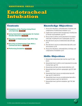
  • 1. 1 © 2012 Jones & Bartlett Learning, LLC (www.jblearning.com) Knowledge Objectives 1. Discuss the indications, contraindications, technique, and complications of endotracheal intubation. (p 2) 2. Explain how to perform direct laryngoscopy, including how to find the visual landmarks. (pp 4–9) 3. Discuss how to confirm correct placement of an endotra- cheal tube, including the use of capnography. (pp 9, 12) 4. Describe how to secure an endotracheal tube with tape. (p 14) 5. Describe how to secure an endotracheal tube with a com- mercial device. (p 14) 6. Discuss the indications, contraindications, technique, and complications of extubation. (p 17) Skills Objectives 1. Demonstrate endotracheal tube insertion. (pp 9–11, Skill Drill 1) 2. Demonstrate how to confirm correct placement of an endo- tracheal tube. (pp 9–12) 3. Demonstrate end-tidal carbon dioxide detection. (p 9) 4. Demonstrate how to perform capnography. (pp 12–13, Skill Drill 2) 5. Demonstrate how to secure an endotracheal tube with tape. (pp 14, 15, Skill Drill 3) 6. Demonstrate how to secure an endotracheal tube with a commercial device. (pp 14, 16, Skill Drill 4) 7. Demonstrate how to perform extubation. (pp 17–18, Skill Drill 5) Contents ■ ■ Intubation of the Trachea Using Direct Laryngoscopy Skill Drill 1 Skill Drill 1 ■ ■ Using Colorimetric Capnography for Carbon Dioxide Detection Skill Drill 2 Skill Drill 2 ■ ■ Securing an Endotracheal Tube With Tape Skill Drill 3 Skill Drill 3 ■ ■ Securing an Endotracheal Tube With a Commercial Device Skill Drill 4 Skill Drill 4 ■ ■ Performing Extubation Skill Drill 5 Skill Drill 5 ADDITIONAL SKILLS Endotracheal Intubation © Jones & Bartlett Learning, LLC NOT FOR SALE OR DISTRIBUTION © Jones & Bartlett Learning, LLC NOT FOR SALE OR DISTRIBUTION © Jones & Bartlett Learning, LLC NOT FOR SALE OR DISTRIBUTION © Jones & Bartlett Learning, LL NOT FOR SALE OR DISTRIBUT © Jones & Bartlett Learning, LLC NOT FOR SALE OR DISTRIBUTION © Jones & Bartlett Learning, LLC NOT FOR SALE OR DISTRIBUTION © Jones & Bartlett Learning, LLC NOT FOR SALE OR DISTRIBUTION © Jones & Bartlett Learning, LLC NOT FOR SALE OR DISTRIBUTION © Jones & Bartlett Learning, LLC NOT FOR SALE OR DISTRIBUTION © Jones & Bartlett Learning, LL NOT FOR SALE OR DISTRIBUT © Jones & Bartlett Learning, LLC NOT FOR SALE OR DISTRIBUTION © Jones & Bartlett Learning, LLC NOT FOR SALE OR DISTRIBUTION © Jones & Bartlett Learning, LLC NOT FOR SALE OR DISTRIBUTION © Jones & Bartlett Learning, LLC NOT FOR SALE OR DISTRIBUTION © Jones & Bartlett Learning, LLC NOT FOR SALE OR DISTRIBUTION © Jones & Bartlett Learning, LL NOT FOR SALE OR DISTRIBUT © Jones & Bartlett Learning, LLC NOT FOR SALE OR DISTRIBUTION © Jones & Bartlett Learning, LLC NOT FOR SALE OR DISTRIBUTION © Jones & Bartlett Learning, LLC NOT FOR SALE OR DISTRIBUTION © Jones & Bartlett Learning, LLC NOT FOR SALE OR DISTRIBUTION
  • 2. 2 Endotracheal Intubation © 2012 Jones & Bartlett Learning, LLC (www.jblearning.com) 6 Introduction For patients unable to maintain their own airway, endotra- cheal intubation provides not only airway management, but also a medication route for several prehospital drugs. The endotracheal (ET) tube can also be used for deep tracheal suctioning when needed.   Endotracheal intubation.   Endotracheal intubation. Table 1 Endotracheal Intubation Indications ■ ■ Present or impending respiratory failure ■ ■ Apnea ■ ■ Hypoxia ■ ■ Poor respiratory effort ■ ■ Suppression or absence of gag reflex ■ ■ Inability of patient to protect own airway Contraindications ■ ■ None in emergency situations ■ ■ Caution in unsuppressed gag reflex Advantages ■ ■ Provides a secure airway ■ ■ Protects against aspiration ■ ■ Provides a route for certain medications (as a last resort; absorption of medications is unpredictable at best) ■ ■ Direct visualization of anatomy and tube placement ■ ■ Ideal method for confirming placement ■ ■ May be performed in breathing or apneic patients Disadvantages ■ ■ Requires special equipment and training ■ ■ Bypasses physiologic function of upper airway ■ ■ Warming ■ ■ Filtering ■ ■ Humidifying Complications ■ ■ Bleeding ■ ■ Hypoxia ■ ■ Laryngeal swelling and trauma ■ ■ Laryngospasm ■ ■ Vocal cord damage ■ ■ Dental trauma ■ ■ Mucosal necrosis ■ ■ Barotrauma ■ ■ Misplacement ■ ■ Right mainstem bronchus ■ ■ Esophagus Endotracheal intubation involves placing a tube through the glottic opening and sealing the tube with a cuff inflated against the wall of the trachea. Endotracheal intubation pro- vides an airtight seal between the patient’s lungs and the ven- tilation device. A sealed cuff placed below the level of the vocal cords is the only form of definitive airway management. A solid understanding of the basics of endotracheal intuba- tion is needed when making urgent decisions about when and how to intubate a patient. Table 1 Table 1 lists indications, contraindications, advantages, disadvantages, and complica- tions of endotracheal intubation. By definition, endotracheal intubation means simply placing a tube into the trachea Figure 1 Figure 1 . In common usage, however, this term is used in a variety of contexts. For the purposes of this chapter, the phrase endotracheal intubation means the process of placing a tube into the trachea. It is a common error to consider the term endotracheal intubation as synonymous with direct laryngoscopy, the most common method of putting a tube in the trachea. Direct laryngoscopy is only one of many techniques used to place a tube into the trachea. The tube is passed into the trachea to provide externally controlled breathing through a bag-mask device or other ventilation device. 6 Equipment Endotracheal (ET) tubes range in size from 2.5 to 9.0 mm for the inside diameter, and the length ranges from 12 to 32 cm Figure 2 Figure 2 . Sizes ranging from 5.0 to 9.0 mm are cuffed to make an airtight seal with the tracheal wall. They have a proximal end 15/22-mm adapter that allows for ventilation with a standard device. There is also an inflation port with a pilot balloon at the proximal end. The distal cuff is inflated © Jones & Bartlett Learning, LLC NOT FOR SALE OR DISTRIBUTION © Jones & Bartlett Learning, LLC NOT FOR SALE OR DISTRIBUTION © Jones & Bartlett Learning, LLC NOT FOR SALE OR DISTRIBUTION © Jones & Bartlett Learning, LL NOT FOR SALE OR DISTRIBUT © Jones & Bartlett Learning, LLC NOT FOR SALE OR DISTRIBUTION © Jones & Bartlett Learning, LLC NOT FOR SALE OR DISTRIBUTION © Jones & Bartlett Learning, LLC NOT FOR SALE OR DISTRIBUTION © Jones & Bartlett Learning, LLC NOT FOR SALE OR DISTRIBUTION © Jones & Bartlett Learning, LLC NOT FOR SALE OR DISTRIBUTION © Jones & Bartlett Learning, LL NOT FOR SALE OR DISTRIBUT © Jones & Bartlett Learning, LLC NOT FOR SALE OR DISTRIBUTION © Jones & Bartlett Learning, LLC NOT FOR SALE OR DISTRIBUTION © Jones & Bartlett Learning, LLC NOT FOR SALE OR DISTRIBUTION © Jones & Bartlett Learning, LLC NOT FOR SALE OR DISTRIBUTION © Jones & Bartlett Learning, LLC NOT FOR SALE OR DISTRIBUTION © Jones & Bartlett Learning, LL NOT FOR SALE OR DISTRIBUT © Jones & Bartlett Learning, LLC NOT FOR SALE OR DISTRIBUTION © Jones & Bartlett Learning, LLC NOT FOR SALE OR DISTRIBUTION © Jones & Bartlett Learning, LLC NOT FOR SALE OR DISTRIBUTION © Jones & Bartlett Learning, LLC NOT FOR SALE OR DISTRIBUTION
  • 3. Endotracheal Intubation 3 © 2012 Jones & Bartlett Learning, LLC (www.jblearning.com) with a syringe attached to a one-way valve. The pilot bal- loon indicates whether the cuff is inflated or deflated once the distal end of the tube is inserted into the patient’s airway. Centimeter markings along the length of the tube provide a measurement of the depth of the tube. The distal end has a beveled tip to facilitate insertion. Tubes ranging from 2.5 to 5.0 mm are used in pediatric patients and are generally uncuffed. The narrowest portion of the pediatric airway is the cricoid ring. In children the cricoid ring is funnel-shaped and forms an anatomic seal with the ET tube, eliminating the need for a cuff. The proxi- mal end still has a 15/22-mm adapter for use with standard ventilating devices. The distal end has a beveled tip with distal end depth markings. Because there is no balloon cuff, there is no pilot balloon. Selecting the proper tube size is important. When the tube selected is too small for the patient, it increases the resistance to flow and, therefore, difficulty in ventilating. When the tube selected is too large, it can be difficult to insert and can cause trauma. The average-sized adult female uses a 7.0- to 8.0-mm tube. An average-sized adult male uses a 7.5- to 8.5-mm tube. A number of anatomic clues can help determine the proper tube size for adults and children. First, the internal diameter of the nostril is a good approximation of the diam- eter of the glottic opening. The diameter of the little finger or the size of the thumbnail is also a good approximation of airway size. There are many types of laryngoscope blades, each with its own advantages and disadvantages, depending on per- sonal preferences. The two most common blades are the straight (Miller) and the curved (Macintosh) blades Figure 3 Figure 3 . Many other blade designs are manufactured for specialty purposes, with the Wisconsin blade having gained much popularity, especially for use in intubating children. Various   Endotracheal tube.   Endotracheal tube.   A. A straight (Miller) blade. B. A laryngoscope and an assortment of curved (Macintosh) blades.   A. A straight (Miller) blade. B. A laryngoscope and an assortment of curved (Macintosh) blades. blade designs have been shown to be effective, although the curved and straight blades are the most commonly used and readily available. In most emergency situa- tions, it is unlikely that you will have immediate access to other blades. Blade choice is mainly a matter of personal preference and is more related to experience than to functional dif- ferences. Neverthe- less, the two blades are used differently. The straight blade is narrow and has a curved channel. Its tip is rounded and is designed to lift the epiglottis to provide the laryngoscopic view. The curved blade has a broad flange that is used to move the tongue out of the way. The tip of the curved blade is flat and broader and is designed to fit into the vallecula. Because of the hypoepiglottic ligament, upward pressure in the vallecula moves the epiglottis, providing the laryngo- scopic view. Blade sizes range from 0 to 4. Infants and children use sizes 0, 1, and 2, whereas 3 and 4 are considered adult sizes. In most adults of average size, a size 3 straight or curved blade provides the best visualization. For pediatric patients, blade sizes are often recommended based on patient age or height. Most providers choose the blade for adults based on experience and the size of the patient (size 3 for average-sized adults and size 4 for larger persons). Two other pieces of equipment have specific uses. The first is the stylet. It is common, especially in emergency situa- tions, to be unable to obtain a full view of the glottic opening. The stylet enables you to guide the tip of the tube over the arytenoid cartilage, even if you cannot see the entire glottic opening. The second is the Magill forceps. Magill forceps have two uses in the emergency setting. First, they are used © Jones Bartlett Learning, LLC NOT FOR SALE OR DISTRIBUTION © Jones Bartlett Learning, LLC NOT FOR SALE OR DISTRIBUTION © Jones Bartlett Learning, LLC NOT FOR SALE OR DISTRIBUTION © Jones Bartlett Learning, LL NOT FOR SALE OR DISTRIBUT © Jones Bartlett Learning, LLC NOT FOR SALE OR DISTRIBUTION © Jones Bartlett Learning, LLC NOT FOR SALE OR DISTRIBUTION © Jones Bartlett Learning, LLC NOT FOR SALE OR DISTRIBUTION © Jones Bartlett Learning, LLC NOT FOR SALE OR DISTRIBUTION © Jones Bartlett Learning, LLC NOT FOR SALE OR DISTRIBUTION © Jones Bartlett Learning, LL NOT FOR SALE OR DISTRIBUT © Jones Bartlett Learning, LLC NOT FOR SALE OR DISTRIBUTION © Jones Bartlett Learning, LLC NOT FOR SALE OR DISTRIBUTION © Jones Bartlett Learning, LLC NOT FOR SALE OR DISTRIBUTION © Jones Bartlett Learning, LLC NOT FOR SALE OR DISTRIBUTION © Jones Bartlett Learning, LLC NOT FOR SALE OR DISTRIBUTION © Jones Bartlett Learning, LL NOT FOR SALE OR DISTRIBUT © Jones Bartlett Learning, LLC NOT FOR SALE OR DISTRIBUTION © Jones Bartlett Learning, LLC NOT FOR SALE OR DISTRIBUTION © Jones Bartlett Learning, LLC NOT FOR SALE OR DISTRIBUTION © Jones Bartlett Learning, LLC NOT FOR SALE OR DISTRIBUTION
  • 4. 4 Endotracheal Intubation © 2012 Jones Bartlett Learning, LLC (www.jblearning.com) to remove obstructions from the airway under direct visual- ization. If you see a solid obstruction in the airway, hold the Magill forceps in your right hand and remove the obstruction. The Magill forceps can also be used to guide the tip of the ET tube through the glottic opening if you are unable to get the proper angle with simple manipulation of the tube. 6 Orotracheal Intubation by Direct 6 Laryngoscopy Orotracheal intubation by direct laryngoscopy involves inserting an ET tube through the mouth and into the trachea while visualizing the glottic opening with a laryngoscope; it is by far the most common method of performing endotracheal intubation in the emergency setting. It is important to always prepare and check the equip- ment before beginning the intubation attempt Table 2 Table 2 . However, it is easy to become complacent with this step. With practice, you will find that most intubations are rela- tively easy and uncomplicated and you will most likely not use all of the extra equipment that you assembled for the intubation attempt. Unfortunately, this usual experience may lull you into a false sense of security. When you do encounter Table 2 Preparing Equipment for Intubation Equipment What to Check, Prepare, and Assemble Ventilation equipment Have an assistant ventilate the patient while you are assembling, checking, and preparing your equipment. Check to make sure that the patient is being ventilated with 100% oxygen and that the pulse oximeter is reading 96% to 100%. Endotracheal tube, stylet, 10-mL syringe (or appropriate size for the type of tube you are using), water-soluble lubricant Select the proper size endotracheal tube (7.0 to 8.0 for adult female; 7.5 to 8.5 for adult male). Inject 10 mL of air into the cuff, and check that it holds air. Check to make sure that the 15/22-mm adapter is firmly inserted into the tube. Insert the stylet, and be sure that the tip is proximal to Murphy’s eye. Bend it to be sure that the stylet does not protrude. Bend the tube/stylet into a hockey stick configuration. Increase the angle of the bend if you anticipate a difficult intubation. Laryngoscope handle and blades It is best to have an assortment of blades available because some patients are easier to intubate with one than another. Check the blade that you plan to use. Be sure that it is free from any nicks in the metal (which could easily cause a laceration). Check the bulb to be sure that the light is “bright, white, steady, and tight.” The light should be bright enough that it is uncomfortable to look directly at it. It should be white, not yellow or dim. The light should not flicker, especially as the blade is moved on the handle. Most important, the bulb must be tightly screwed into the handle to prevent its loss in the airway. Magill forceps Have available to guide the tip of the tube into the trachea or to remove foreign body airway obstructions. Suction You will not need it in most patients, but when you need it, you need it fast! Towels Needed to position the patient’s head. Confirmation devices Stethoscope and an end-tidal carbon dioxide detector or an esophageal detector device. ET tube securing device Have the appropriate device (eg, tape or a commercial ET tube securing device) ready before you start. a difficult airway in a patient, you need to have the necessary extra equipment immediately available. Remember that you are preparing for possible difficulty, even though you hope that you have none. Therefore, it is best to have the extra equipment assembled because in a small percentage of cases you will be glad to have it when you need it. Standard Precautions Intubation is a procedure that may expose you to body fluids; therefore, proper precautions should be taken. In addition to gloves, you should wear a mask and eye shield because your face will be relatively close to the patient’s mouth and nose. The mask and eye shield will protect you if the patient vomits. Masks with eye protection included are inexpensive, easy to put on, and unobtrusive. They should be considered mandatory parts of your airway kit. Preoxygenation The importance of preoxygenation cannot be overstated. During an intubation attempt, the patient will undergo a period of forced apnea. The goal is to prevent hypoxia from occurring during this period in which the patient is not breathing or being ventilated. In general, patients in stable © Jones Bartlett Learning, LLC NOT FOR SALE OR DISTRIBUTION © Jones Bartlett Learning, LLC NOT FOR SALE OR DISTRIBUTION © Jones Bartlett Learning, LLC NOT FOR SALE OR DISTRIBUTION © Jones Bartlett Learning, LL NOT FOR SALE OR DISTRIBUT © Jones Bartlett Learning, LLC NOT FOR SALE OR DISTRIBUTION © Jones Bartlett Learning, LLC NOT FOR SALE OR DISTRIBUTION © Jones Bartlett Learning, LLC NOT FOR SALE OR DISTRIBUTION © Jones Bartlett Learning, LLC NOT FOR SALE OR DISTRIBUTION © Jones Bartlett Learning, LLC NOT FOR SALE OR DISTRIBUTION © Jones Bartlett Learning, LL NOT FOR SALE OR DISTRIBUT © Jones Bartlett Learning, LLC NOT FOR SALE OR DISTRIBUTION © Jones Bartlett Learning, LLC NOT FOR SALE OR DISTRIBUTION © Jones Bartlett Learning, LLC NOT FOR SALE OR DISTRIBUTION © Jones Bartlett Learning, LLC NOT FOR SALE OR DISTRIBUTION © Jones Bartlett Learning, LLC NOT FOR SALE OR DISTRIBUTION © Jones Bartlett Learning, LL NOT FOR SALE OR DISTRIBUT © Jones Bartlett Learning, LLC NOT FOR SALE OR DISTRIBUTION © Jones Bartlett Learning, LLC NOT FOR SALE OR DISTRIBUTION © Jones Bartlett Learning, LLC NOT FOR SALE OR DISTRIBUTION © Jones Bartlett Learning, LLC NOT FOR SALE OR DISTRIBUTION
  • 5. Endotracheal Intubation 5 © 2012 Jones Bartlett Learning, LLC (www.jblearning.com) 20° extension of the atlanto-occipital joint and 30° flexion of the neck at C6 and C7. In most supine patients, the sniffing position is achieved by extension of the head and elevation of the occiput 2.5 to 5 cm. The most practical guide to the amount of occipital ele- vation necessary in a given patient is to elevate the head until the ear is at the level of the sternum. Elevation of the head is best achieved with folded towels positioned under the head and/or neck Figure 5 Figure 5 . The advantage of using towels is that the thickness can easily be adjusted by changing the number of folds. In obese patients, however, padding under the head alone may not result in the sniffing position. You may need to pad also under the shoulders, neck, and head. When in doubt as to whether the patient is in a true sniffing position, looking at the person from the side usually provides the best view for evaluating the adequacy of the head tilt. After the patient is in the sniffing position, have the assistant stop ventilating. The intubation attempt should take no more than 30 seconds. The attempt should be stopped at 30 seconds, if the oxygen saturation falls significantly, or if the patient’s heart rate or rhythm changes dramatically. condition can undergo 2 to 3 minutes of apnea provided they are adequately preoxygenated. Many patients requiring intubation are physiologically compromised. To prepare them for the apnea that occurs during intubation, they must be preoxygenated and the amount of time between breaths significantly limited. Pulse oximetry has dramatically changed how patients are monitored before and during intubation. Pulse oximetry is performed by placing the probe of the pulse oximeter on the patient’s finger. Ideally, the patient should have an oxy- gen saturation of 100% for 2 to 3 minutes before intubation, and it should never fall below 95% during an intubation attempt. Because the patient’s baseline level before the crisis is unknown, you cannot rely entirely on the pulse oximeter reading, but nevertheless, it provides you with important trending information. The consequences of brief periods of hypoxia can be disastrous. Do not rely too heavily on the pulse oximeter reading because it may be falsely high, even if the patient is profoundly hypoxic. Although some of the sequelae of hypoxia are dramatic and immediate, most are subtle and occur gradually. Some of the poor neurologic outcomes fol- lowing aggressive airway management result from intuba- tion-induced hypoxia. These incidents can be avoided by providing adequate preintubation hyperoxygenation. Position the Patient Proper positioning of the patient is one of the main keys to successful laryngoscopy. To properly position a patient before performing intubation by direct laryngoscopy, you need to have knowledge of basic airway anatomy. There are three axes of the airway: the oral plane, the pharyn- geal plane, and the tracheal plane. Ordinarily these axes are at sharp angles, facilitating the entry of food into the esophagus and reducing the likelihood of food entering the airway. Although this is an obvious advantage for everyday life, the angles of these three axes make intubation difficult Figure 4 Figure 4 . Intubation is a procedure that may expose you to body fluids; therefore, proper precautions should be taken. In addition to gloves, you should wear a mask and eye shield because your face will be relatively close to the patient’s mouth and nose. The mask and eye shield help protect you if the patient vomits. Safety To facilitate visualization of the airway, you want to align the three axes to the extent possible. This is achieved by plac- ing the patient in the “sniffing” position. The sniffing position is so named because this is the position of the head when intentionally sniffing. The position involves approximately a   Three axes of the airway: oral, pharyngeal, and tracheal. A. Neutral position. B. Sniffing position.   Three axes of the airway: oral, pharyngeal, and tracheal. A. Neutral position. B. Sniffing position. B B A A © Jones Bartlett Learning, LLC NOT FOR SALE OR DISTRIBUTION © Jones Bartlett Learning, LLC NOT FOR SALE OR DISTRIBUTION © Jones Bartlett Learning, LLC NOT FOR SALE OR DISTRIBUTION © Jones Bartlett Learning, LL NOT FOR SALE OR DISTRIBUT © Jones Bartlett Learning, LLC NOT FOR SALE OR DISTRIBUTION © Jones Bartlett Learning, LLC NOT FOR SALE OR DISTRIBUTION © Jones Bartlett Learning, LLC NOT FOR SALE OR DISTRIBUTION © Jones Bartlett Learning, LLC NOT FOR SALE OR DISTRIBUTION © Jones Bartlett Learning, LLC NOT FOR SALE OR DISTRIBUTION © Jones Bartlett Learning, LL NOT FOR SALE OR DISTRIBUT © Jones Bartlett Learning, LLC NOT FOR SALE OR DISTRIBUTION © Jones Bartlett Learning, LLC NOT FOR SALE OR DISTRIBUTION © Jones Bartlett Learning, LLC NOT FOR SALE OR DISTRIBUTION © Jones Bartlett Learning, LLC NOT FOR SALE OR DISTRIBUTION © Jones Bartlett Learning, LLC NOT FOR SALE OR DISTRIBUTION © Jones Bartlett Learning, LL NOT FOR SALE OR DISTRIBUT © Jones Bartlett Learning, LLC NOT FOR SALE OR DISTRIBUTION © Jones Bartlett Learning, LLC NOT FOR SALE OR DISTRIBUTION © Jones Bartlett Learning, LLC NOT FOR SALE OR DISTRIBUTION © Jones Bartlett Learning, LLC NOT FOR SALE OR DISTRIBUTION
  • 6. 6 Endotracheal Intubation © 2012 Jones Bartlett Learning, LLC (www.jblearning.com) All laryngoscope handles and blades are held in the left hand. Some custom-made or specialty blades can be held in the right hand, but these are rare in emergency settings. Hold the laryngoscope in your left hand and the prepared, lubricated tube like a pencil in your right hand. Grasp the laryngoscope with your left hand and hold it as low down on the handle as possible. If the patient’s mouth is not open, the easiest technique is to place the side of your right-hand thumb just below the bottom lip and push the mouth open, or “scissor” your thumb and forefinger between the molars Figure 7 Figure 7 . Regardless of the blade being used, it should be inserted into the right side of the patient’s mouth. Use the flange of the blade to sweep the tongue to the left side of the mouth while the blade is moved into the midline. Moving the tongue from right to left is a critical step. If you simply insert the blade in the midline, the tongue will hang over both sides of the blade and you will see only the tongue Figure 8 Figure 8 . If you abort an intubation attempt, simply reoxygenate the patient for 2 to 3 minutes with 100% oxygen and try again. Repeated attempts do not harm the patient, but pro- longed attempts do. Blade Insertion The tongue is a sticky, amorphous structure that is a major hindrance to visualizing the airway. The proper use of the laryngoscope blade is critical to controlling the tongue; this is difficult to simulate in practice with manikins. Nevertheless, you need to develop excellent technique in manikins to help avoid difficulties in patients. Position yourself at the top of the patient’s head. If the patient is on a gurney, you can squat to put your head at the level of the patient’s face. If the patient is on the floor or ground, you may need to kneel and lean forward or lie down on the floor to get into the proper position Figure 6 Figure 6 .   Head elevation is best achieved with folded towels positioned under the head and/or neck.   Head elevation is best achieved with folded towels positioned under the head and/or neck.   If the patient is on the floor or ground, you may need to kneel and lean forward or lie down on the floor to get into the proper position.   If the patient is on the floor or ground, you may need to kneel and lean forward or lie down on the floor to get into the proper position.   Place the side of your right-hand thumb just below the bottom lip and push the mouth open, or scissor your thumb and forefinger between the molars.   Place the side of your right-hand thumb just below the bottom lip and push the mouth open, or scissor your thumb and forefinger between the molars.   The tongue is a sticky, amorphous structure that can be a major hindrance to visualizing the airway. Proper use of the laryngoscope is critical to controlling the tongue.   The tongue is a sticky, amorphous structure that can be a major hindrance to visualizing the airway. Proper use of the laryngoscope is critical to controlling the tongue. © Jones Bartlett Learning, LLC NOT FOR SALE OR DISTRIBUTION © Jones Bartlett Learning, LLC NOT FOR SALE OR DISTRIBUTION © Jones Bartlett Learning, LLC NOT FOR SALE OR DISTRIBUTION © Jones Bartlett Learning, LL NOT FOR SALE OR DISTRIBUT © Jones Bartlett Learning, LLC NOT FOR SALE OR DISTRIBUTION © Jones Bartlett Learning, LLC NOT FOR SALE OR DISTRIBUTION © Jones Bartlett Learning, LLC NOT FOR SALE OR DISTRIBUTION © Jones Bartlett Learning, LLC NOT FOR SALE OR DISTRIBUTION © Jones Bartlett Learning, LLC NOT FOR SALE OR DISTRIBUTION © Jones Bartlett Learning, LL NOT FOR SALE OR DISTRIBUT © Jones Bartlett Learning, LLC NOT FOR SALE OR DISTRIBUTION © Jones Bartlett Learning, LLC NOT FOR SALE OR DISTRIBUTION © Jones Bartlett Learning, LLC NOT FOR SALE OR DISTRIBUTION © Jones Bartlett Learning, LLC NOT FOR SALE OR DISTRIBUTION © Jones Bartlett Learning, LLC NOT FOR SALE OR DISTRIBUTION © Jones Bartlett Learning, LL NOT FOR SALE OR DISTRIBUT © Jones Bartlett Learning, LLC NOT FOR SALE OR DISTRIBUTION © Jones Bartlett Learning, LLC NOT FOR SALE OR DISTRIBUTION © Jones Bartlett Learning, LLC NOT FOR SALE OR DISTRIBUTION © Jones Bartlett Learning, LLC NOT FOR SALE OR DISTRIBUTION
  • 7. Endotracheal Intubation 7 © 2012 Jones Bartlett Learning, LLC (www.jblearning.com) in the position of the blade to aid in visualization of the glottic opening. If you are not able to identify any familiar structures, it is generally fruitless to continue, and it may be better to start over. With the curved blade, it is best to walk the blade down the tongue because you know that the val- lecula and the epiglottis lie at the base of the tongue. With the straight blade, insert the blade straight back until the tip touches the posterior pharyngeal wall. This is the proper depth of insertion. As you continue to work the tip of the blade into the proper position (lifting the epiglottis or in the vallecula), the glottic opening should come into view as you lift. Do not be concerned if you do not have a full view of the glottic opening. The vocal cords are the white fibrous bands that lie vertically within the glottic opening; they should be slightly open Figure 11 Figure 11 . When the angle of the pharynx and the larynx is particu- larly acute, it is often difficult to see the entire glottic opening. You can do two things to increase the percent- age of the glottic opening that you can see. You can use cricoid pressure (also called the Sellick maneuver) or the BURP maneuver. (Per the American Heart Associa- tion, cricoid pressure should not be used to prevent or treat gastric distention; however, it is still an acceptable method of improving the view during intubation.) Cricoid pressure moves the airway structures more posteriorly. If applied by an assistant during direct laryn- goscopy, it reduces the acuity of the angle between the pharynx and larynx and can improve the laryngoscopic view Figure 12 Figure 12 . Words ofWisdom If you are using a straight blade, insert the tip of the blade all the way to the posterior pharyngeal wall and then lift the jaw. If you are using a curved blade, insert the tip into the vallecula. With both types of blades, the goal is to lift the jaw. The most common beginning error is to pull the jaw back using the patient’s teeth as a leverage point Figure 9 Figure 9 . This can break the teeth, and it does not provide a view of the glottis. Lifting the patient’s jaw is accomplished by keep- ing your left wrist straight, elbow bent, and back straight Figure 10 Figure 10 . The correct motion is similar to holding a wine glass and offering a toast. Visualization of the Glottic Opening As you look down the blade, you should start to see some familiar airway landmarks. Identifying the epiglottis or the arytenoid cartilage is very important at this point. Identify- ing these structures enables you to make small adjustments   Prying against the upper teeth with the laryn- goscope can result in breaking and potential aspiration of the teeth. Do not do it!   Prying against the upper teeth with the laryn- goscope can result in breaking and potential aspiration of the teeth. Do not do it!   Keep your back and your left arm straight as you pull upward. This allows you to use the strength of your shoulders to lift the patient’s jaw.   Keep your back and your left arm straight as you pull upward. This allows you to use the strength of your shoulders to lift the patient’s jaw.   Laryngoscopic view of the vocal cords (white fibrous bands).   Laryngoscopic view of the vocal cords (white fibrous bands). © Jones Bartlett Learning, LLC NOT FOR SALE OR DISTRIBUTION © Jones Bartlett Learning, LLC NOT FOR SALE OR DISTRIBUTION © Jones Bartlett Learning, LLC NOT FOR SALE OR DISTRIBUTION © Jones Bartlett Learning, LL NOT FOR SALE OR DISTRIBUT © Jones Bartlett Learning, LLC NOT FOR SALE OR DISTRIBUTION © Jones Bartlett Learning, LLC NOT FOR SALE OR DISTRIBUTION © Jones Bartlett Learning, LLC NOT FOR SALE OR DISTRIBUTION © Jones Bartlett Learning, LLC NOT FOR SALE OR DISTRIBUTION © Jones Bartlett Learning, LLC NOT FOR SALE OR DISTRIBUTION © Jones Bartlett Learning, LL NOT FOR SALE OR DISTRIBUT © Jones Bartlett Learning, LLC NOT FOR SALE OR DISTRIBUTION © Jones Bartlett Learning, LLC NOT FOR SALE OR DISTRIBUTION © Jones Bartlett Learning, LLC NOT FOR SALE OR DISTRIBUTION © Jones Bartlett Learning, LLC NOT FOR SALE OR DISTRIBUTION © Jones Bartlett Learning, LLC NOT FOR SALE OR DISTRIBUTION © Jones Bartlett Learning, LL NOT FOR SALE OR DISTRIBUT © Jones Bartlett Learning, LLC NOT FOR SALE OR DISTRIBUTION © Jones Bartlett Learning, LLC NOT FOR SALE OR DISTRIBUTION © Jones Bartlett Learning, LLC NOT FOR SALE OR DISTRIBUTION © Jones Bartlett Learning, LLC NOT FOR SALE OR DISTRIBUTION
  • 8. 8 Endotracheal Intubation © 2012 Jones Bartlett Learning, LLC (www.jblearning.com) an ET tube, it is useful if you cannot obtain a full glottic view. The angle at its distal tip facilitates entry into the airway and enables you to “feel” the ridges of the tracheal wall. Once the gum bougie is placed deeply into the trachea, it becomes a guide for the ET tube. Simply slide the tube over the device and into the trachea. Remove the gum bougie, ventilate, and confirm placement. Tube Insertion Once you have visualized the glottic opening, the next step is to insert the ET tube. When you find the glottic opening, do not take your eyes off it. Losing sight of the glottic opening is a major cause of failed intubation. Have the tube ready in your right hand so that you can advance it immediately on identification of the vocal cords. A major mistake beginners make is to try to pass the tube down the barrel of the blade. The laryngoscope blade is not designed as a guide for the tube; it is a tool used only to visualize the glottic opening. Placing the tube down the blade obscures your view of the glottic opening Figure 15 Figure 15 . Pass the tube as far to the right as possible and at an angle that lets you watch the tip as you insert it through the vocal cords. Continue to insert the tube until the proximal end of the cuff is 1 to 2 cm past the vocal cords. If you take your eyes off the tip of the tube, even for a second, you significantly increase the likelihood of allowing the tube to slip into the esophagus. The BURP maneuver is an acronym for Backward, Upward, Rightward Pressure. If you are having difficulty see- ing the glottic opening, take your right hand and locate the lower third of the thyroid cartilage. By applying backward, upward, and rightward pressure, you can often move the larynx into view Figure 13 Figure 13 . Unfortunately, sometimes when you let go to pass the tube with your right hand, you will lose the view. If possible, once you have visualized the glottic opening, have an assistant hold the larynx in position as you pass the tube. The BURP maneuver can also be applied to the cricoid ring or the hyoid bone. The gum bougie is an ingenious device that can make intubation possible in some difficult situations, especially when you have limited glottic visualization. The gum bougie is a flexible device that is roughly a centimeter in diameter Figure 14 Figure 14 . It is rigid enough to be easily directed through the glottic opening, but flexible enough so that it does not cause damage to the tracheal walls. There is a slight angle at its distal tip. The gum bougie is inserted through the glottic opening under direct visualization. Because it is much smaller than   Cricoid pressure (also called the Sellick maneuver).   Cricoid pressure (also called the Sellick maneuver).   The BURP maneuver—displacing the larynx back, up, and to the right—may help visualize the vocal cords.   The BURP maneuver—displacing the larynx back, up, and to the right—may help visualize the vocal cords.   The gum bougie device. Source: Courtesy of Marianne Gausche-Hill, MD, FACEP, FAAP   The gum bougie device. Source: Courtesy of Marianne Gausche-Hill, MD, FACEP, FAAP   Do not place the tube down the blade; this obscures your view of the glottic opening.   Do not place the tube down the blade; this obscures your view of the glottic opening. © Jones Bartlett Learning, LLC NOT FOR SALE OR DISTRIBUTION © Jones Bartlett Learning, LLC NOT FOR SALE OR DISTRIBUTION © Jones Bartlett Learning, LLC NOT FOR SALE OR DISTRIBUTION © Jones Bartlett Learning, LL NOT FOR SALE OR DISTRIBUT © Jones Bartlett Learning, LLC NOT FOR SALE OR DISTRIBUTION © Jones Bartlett Learning, LLC NOT FOR SALE OR DISTRIBUTION © Jones Bartlett Learning, LLC NOT FOR SALE OR DISTRIBUTION © Jones Bartlett Learning, LLC NOT FOR SALE OR DISTRIBUTION © Jones Bartlett Learning, LLC NOT FOR SALE OR DISTRIBUTION © Jones Bartlett Learning, LL NOT FOR SALE OR DISTRIBUT © Jones Bartlett Learning, LLC NOT FOR SALE OR DISTRIBUTION © Jones Bartlett Learning, LLC NOT FOR SALE OR DISTRIBUTION © Jones Bartlett Learning, LLC NOT FOR SALE OR DISTRIBUTION © Jones Bartlett Learning, LLC NOT FOR SALE OR DISTRIBUTION © Jones Bartlett Learning, LLC NOT FOR SALE OR DISTRIBUTION © Jones Bartlett Learning, LL NOT FOR SALE OR DISTRIBUT © Jones Bartlett Learning, LLC NOT FOR SALE OR DISTRIBUTION © Jones Bartlett Learning, LLC NOT FOR SALE OR DISTRIBUTION © Jones Bartlett Learning, LLC NOT FOR SALE OR DISTRIBUTION © Jones Bartlett Learning, LLC NOT FOR SALE OR DISTRIBUTION
  • 9. Endotracheal Intubation 9 © 2012 Jones Bartlett Learning, LLC (www.jblearning.com) 13. Secure the ET tube Step 12 . 14. Place a bite block in the patient’s mouth Step 13 . Confirm Placement Watching the ET tube pass between the vocal cords is your first method of confirming tube placement. You must con- tinue the process of gathering information to assess the loca- tion of the tube. Remember that a misplaced tube that goes undetected is a fatal error. You must incorporate multiple assessment findings into the determination of where the tube is located. If the tube must be repositioned or moved, be sure to remove the air from the cuff first. After visualizing the tube passing through the glottis, note the depth of the markings on the tube. The average depth in adult males is 22 cm at the teeth, and the aver- age depth in adult females is 21 cm at the teeth. Look for condensation in the tube. Condensation indicates correct placement in the trachea. Auscultation over the lungs and epigastric area is the next step. Air entry into the stomach indicates esophageal placement, mandating immediate removal of the tube. Equal breath sounds should be heard over the apices of each lung and equal breath sounds and expansion over the bilateral bases. Unequal or absent breath sounds over the lung fields indicate esophageal placement, right mainstem placement, pneumothorax, or bronchial obstruction. Palpation of the balloon cuff at the sternal notch by compressing the pilot balloon is another indication of cor- rect placement. Pulse oximetry provides a measurement of oxygenation as long as other factors do not interfere with peripheral circulation. Carbon dioxide detectors (end-tidal carbon dioxide) may also be used. They detect the presence of carbon dioxide in expired air. These detectors may be colorimetric, digital, or digital/waveform. A capnographer attaches in between the ET tube and bag-mask device; it contains colorimetric paper, which should turn yellow dur- ing exhalation, indicating proper tube placement Figure 16 Figure 16 . Ventilate After watching the tube pass between the patient’s vocal cords, gently remove the blade, hold the tube with your right hand, and carefully remove the stylet. Fill the tube cuff with just enough air to stop the leaking sound around the tube and then detach the syringe from the inflation port. Have your assistant attach the ventilation device to the tube with an end-tidal carbon dioxide detector (note, if you are using an esophageal detection device, it is important to use it before the first breath is taken). As the first breaths are delivered, watch the patient’s chest rise. You should now listen to breath sounds as another way to confirm the tube location. You should listen to both lungs at both the apices and bases and to the stomach over the epigastrium. If the tube is properly positioned in the trachea, you will hear equal breath sounds bilaterally and a quiet epigastrium. Gurgling over the stomach suggests esophageal placement, and the tube should be immediately removed and the patient ventilated. Unilateral breath sounds gener- ally indicate a mainstem intubation. Place your stethoscope over the quiet side of the chest and slowly withdraw the tube until you hear breath sounds return. Ventilation should continue as indicated according to the patient’s size and clinical condition. It is prudent to slightly hyperventilate the patient for 30 seconds to 1 minute immediately after intubation to eliminate any accumulated carbon dioxide. A summary of the steps of orotracheal intubation by direct laryngoscopy is listed here, and the steps are shown in Skill Drill 1 Skill Drill 1 : 1. Use standard precautions (gloves and face shield). 2. Preoxygenate the patient whenever possible with a bag-mask device and 100% oxygen Step 1 . 3. Check, prepare, and assemble your equipment Step 2 . 4. Place the patient’s head in the sniffing position Step 3 . 5. Insert the blade into the right side of the patient’s mouth, and displace the tongue to the left Step 4 . 6. Gently lift the long axis of the laryngoscope handle until you can visualize the glottic opening and the vocal cords Step 5 . 7. Insert the ET tube through the right corner of the mouth, and place it between the vocal cords Step 6 . 8. Remove the laryngoscope from the patient’s mouth Step 7 . 9. Remove the stylet from the ET tube Step 8 . 10. Inflate the distal cuff of the ET tube with 5 to 10 mL of air, and detach the syringe Step 9 . 11. Attach the end-tidal carbon dioxide detector to the ET tube Step 10 . 12. Attach the bag-valve device, ventilate, and auscultate over the apices and bases of both lungs and over the epigastrium Step 11 .   Colorimetric capnographers. Source: Courtesy of Marianne Gausche-Hill, MD, FACEP, FAAP   Colorimetric capnographers. Source: Courtesy of Marianne Gausche-Hill, MD, FACEP, FAAP © Jones Bartlett Learning, LLC NOT FOR SALE OR DISTRIBUTION © Jones Bartlett Learning, LLC NOT FOR SALE OR DISTRIBUTION © Jones Bartlett Learning, LLC NOT FOR SALE OR DISTRIBUTION © Jones Bartlett Learning, LL NOT FOR SALE OR DISTRIBUT © Jones Bartlett Learning, LLC NOT FOR SALE OR DISTRIBUTION © Jones Bartlett Learning, LLC NOT FOR SALE OR DISTRIBUTION © Jones Bartlett Learning, LLC NOT FOR SALE OR DISTRIBUTION © Jones Bartlett Learning, LLC NOT FOR SALE OR DISTRIBUTION © Jones Bartlett Learning, LLC NOT FOR SALE OR DISTRIBUTION © Jones Bartlett Learning, LL NOT FOR SALE OR DISTRIBUT © Jones Bartlett Learning, LLC NOT FOR SALE OR DISTRIBUTION © Jones Bartlett Learning, LLC NOT FOR SALE OR DISTRIBUTION © Jones Bartlett Learning, LLC NOT FOR SALE OR DISTRIBUTION © Jones Bartlett Learning, LLC NOT FOR SALE OR DISTRIBUTION © Jones Bartlett Learning, LLC NOT FOR SALE OR DISTRIBUTION © Jones Bartlett Learning, LL NOT FOR SALE OR DISTRIBUT © Jones Bartlett Learning, LLC NOT FOR SALE OR DISTRIBUTION © Jones Bartlett Learning, LLC NOT FOR SALE OR DISTRIBUTION © Jones Bartlett Learning, LLC NOT FOR SALE OR DISTRIBUTION © Jones Bartlett Learning, LLC NOT FOR SALE OR DISTRIBUTION
  • 10. 10 Endotracheal Intubation © 2012 Jones Bartlett Learning, LLC (www.jblearning.com) Skill Drill Skill Drill 1 1 Intubation of the Trachea Using Direct Laryngoscopy Step 1 Use standard pre- cautions (gloves and face shield). Preoxygenate the patient whenever possible with a bag-mask device and 100% oxygen. Step 2 Check, prepare, and assemble your equipment. Step 3 Place the patient’s head in the sniffing position. Step 4 Insert the blade into the right side of the patient’s mouth, and displace the tongue to the left. Step 5 Gently lift the long axis of the laryngoscope handle until you can visualize the glottic opening and the vocal cords. Step 6 Insert the ET tube through the right corner of the mouth, and visualize its entry between the vocal cords. Step 7 Remove the laryngo- scope from the patient’s mouth. Step 8 Remove the stylet from the ET tube. Step 9 Inflate the distal cuff of the ET tube with 5 to 10 mL of air, and detach the syringe from the inflation port. © Jones Bartlett Learning, LLC NOT FOR SALE OR DISTRIBUTION © Jones Bartlett Learning, LLC NOT FOR SALE OR DISTRIBUTION © Jones Bartlett Learning, LLC NOT FOR SALE OR DISTRIBUTION © Jones Bartlett Learning, LL NOT FOR SALE OR DISTRIBUT © Jones Bartlett Learning, LLC NOT FOR SALE OR DISTRIBUTION © Jones Bartlett Learning, LLC NOT FOR SALE OR DISTRIBUTION © Jones Bartlett Learning, LLC NOT FOR SALE OR DISTRIBUTION © Jones Bartlett Learning, LLC NOT FOR SALE OR DISTRIBUTION © Jones Bartlett Learning, LLC NOT FOR SALE OR DISTRIBUTION © Jones Bartlett Learning, LL NOT FOR SALE OR DISTRIBUT © Jones Bartlett Learning, LLC NOT FOR SALE OR DISTRIBUTION © Jones Bartlett Learning, LLC NOT FOR SALE OR DISTRIBUTION © Jones Bartlett Learning, LLC NOT FOR SALE OR DISTRIBUTION © Jones Bartlett Learning, LLC NOT FOR SALE OR DISTRIBUTION © Jones Bartlett Learning, LLC NOT FOR SALE OR DISTRIBUTION © Jones Bartlett Learning, LL NOT FOR SALE OR DISTRIBUT © Jones Bartlett Learning, LLC NOT FOR SALE OR DISTRIBUTION © Jones Bartlett Learning, LLC NOT FOR SALE OR DISTRIBUTION © Jones Bartlett Learning, LLC NOT FOR SALE OR DISTRIBUTION © Jones Bartlett Learning, LLC NOT FOR SALE OR DISTRIBUTION
  • 11. Endotracheal Intubation 11 © 2012 Jones Bartlett Learning, LLC (www.jblearning.com) Skill Drill Skill Drill 1 1 Intubation of the Trachea Using Direct Laryngoscopy (continued) Step 10 Attach the end-tidal carbon dioxide detector to the ET tube. Step 11 Attach the bag-valve device, ventilate, and auscultate over the apices and bases of both lungs and over the epigastrium. Step 12 Secure the ET tube. Step 13 Place a bite block in the patient’s mouth. © Jones Bartlett Learning, LLC NOT FOR SALE OR DISTRIBUTION © Jones Bartlett Learning, LLC NOT FOR SALE OR DISTRIBUTION © Jones Bartlett Learning, LLC NOT FOR SALE OR DISTRIBUTION © Jones Bartlett Learning, LL NOT FOR SALE OR DISTRIBUT © Jones Bartlett Learning, LLC NOT FOR SALE OR DISTRIBUTION © Jones Bartlett Learning, LLC NOT FOR SALE OR DISTRIBUTION © Jones Bartlett Learning, LLC NOT FOR SALE OR DISTRIBUTION © Jones Bartlett Learning, LLC NOT FOR SALE OR DISTRIBUTION © Jones Bartlett Learning, LLC NOT FOR SALE OR DISTRIBUTION © Jones Bartlett Learning, LL NOT FOR SALE OR DISTRIBUT © Jones Bartlett Learning, LLC NOT FOR SALE OR DISTRIBUTION © Jones Bartlett Learning, LLC NOT FOR SALE OR DISTRIBUTION © Jones Bartlett Learning, LLC NOT FOR SALE OR DISTRIBUTION © Jones Bartlett Learning, LLC NOT FOR SALE OR DISTRIBUTION © Jones Bartlett Learning, LLC NOT FOR SALE OR DISTRIBUTION © Jones Bartlett Learning, LL NOT FOR SALE OR DISTRIBUT © Jones Bartlett Learning, LLC NOT FOR SALE OR DISTRIBUTION © Jones Bartlett Learning, LLC NOT FOR SALE OR DISTRIBUTION © Jones Bartlett Learning, LLC NOT FOR SALE OR DISTRIBUTION © Jones Bartlett Learning, LLC NOT FOR SALE OR DISTRIBUTION
  • 12. 12 Endotracheal Intubation © 2012 Jones Bartlett Learning, LLC (www.jblearning.com) resistance to bag-mask compliance may indicate gastric dis- tention, esophageal placement, or tension pneumothorax. After confirming proper tube placement, note and mark the ET tube with an ink line or piece of tape at the point where it emerges from the patient’s mouth; this will enable medical personnel involved in the subsequent care of the patient to determine whether the tube has slipped in or out. The average depth for adult patients is 21 to 25 cm. Evidence of a misplaced tube, regardless of when it was last checked, must be reconfirmed. Reconfirmation should also be performed after any major move and after any manip- ulation of the neck. Manipulation of the neck may displace the tube up to 5 cm. A capnometer performs the same function; however, instead of using colorimetric paper, it attaches in the same way as a capnographer, but provides an LED readout of the patient’s exhaled carbon dioxide, again indicating correct ET tube placement Figure 17 Figure 17 . Because carbon dioxide is not pres- ent in the esophagus, use of carbon dioxide detectors is yet another method of determining correct tube placement. The steps are as follows Skill Drill 2 Skill Drill 2 . 1. Detach the ventilation device from the ET tube Step 1 . 2. Attach an in-line capnographer or capnometer to the proximal adapter of the ET tube Step 2 . 3. Reattach the ventilation device to the ET tube, and resume ventilations Step 3 . 4. Monitor the capnographer or capnometer for appro- priate reading (appropriate color change or digital reading) Step 4 . The esophageal detector device (EDD) is yet another method of confirming proper ET tube placement. The EDD is a bulb or syringe with a 15/22-mm adapter. With the syringe model, the syringe is attached to the end of the ET tube and the plunger is withdrawn, creating negative pres- sure Figure 18 Figure 18 . If the tube is in the trachea (which has rigid, noncollapsible walls), air is easily drawn into the syringe and the plunger does not move when released. Unlike the trachea, however, the esophagus is a flaccid, easily collapsible tube. Thus, if the tube is in the esophagus, a vacuum is created as the EDD’s plunger is withdrawn, and the plunger moves back toward zero when released. With the bulb model, the bulb is squeezed and then attached to the end of the ET tube. If it remains collapsed or inflates slowly, the esophageal wall has occluded the distal tip of the tube, indicating that esophageal intubation has likely occurred. If the bulb briskly expands, the tube is properly positioned in the trachea Figure 19 Figure 19 . The final determination of correct tube placement is bag-mask ventilation compliance. The bag should be easy to compress with corresponding chest expansion. Increased   A capnometer. Source: Courtesy of Marianne Gausche-Hill, MD, FACEP, FAAP   A capnometer. Source: Courtesy of Marianne Gausche-Hill, MD, FACEP, FAAP   With the esophageal detector device syringe, the ability to freely withdraw air indicates placement of the tube in the trachea. Source: Courtesy of Marianne Gausche-Hill, MD, FACEP, FAAP   With the esophageal detector device syringe, the ability to freely withdraw air indicates placement of the tube in the trachea. Source: Courtesy of Marianne Gausche-Hill, MD, FACEP, FAAP   If the ET tube is in the trachea, the esophageal detector device bulb should briskly fill with air. Source: Courtesy of Marianne Gausche-Hill, MD, FACEP, FAAP   If the ET tube is in the trachea, the esophageal detector device bulb should briskly fill with air. Source: Courtesy of Marianne Gausche-Hill, MD, FACEP, FAAP © Jones Bartlett Learning, LLC NOT FOR SALE OR DISTRIBUTION © Jones Bartlett Learning, LLC NOT FOR SALE OR DISTRIBUTION © Jones Bartlett Learning, LLC NOT FOR SALE OR DISTRIBUTION © Jones Bartlett Learning, LL NOT FOR SALE OR DISTRIBUT © Jones Bartlett Learning, LLC NOT FOR SALE OR DISTRIBUTION © Jones Bartlett Learning, LLC NOT FOR SALE OR DISTRIBUTION © Jones Bartlett Learning, LLC NOT FOR SALE OR DISTRIBUTION © Jones Bartlett Learning, LLC NOT FOR SALE OR DISTRIBUTION © Jones Bartlett Learning, LLC NOT FOR SALE OR DISTRIBUTION © Jones Bartlett Learning, LL NOT FOR SALE OR DISTRIBUT © Jones Bartlett Learning, LLC NOT FOR SALE OR DISTRIBUTION © Jones Bartlett Learning, LLC NOT FOR SALE OR DISTRIBUTION © Jones Bartlett Learning, LLC NOT FOR SALE OR DISTRIBUTION © Jones Bartlett Learning, LLC NOT FOR SALE OR DISTRIBUTION © Jones Bartlett Learning, LLC NOT FOR SALE OR DISTRIBUTION © Jones Bartlett Learning, LL NOT FOR SALE OR DISTRIBUT © Jones Bartlett Learning, LLC NOT FOR SALE OR DISTRIBUTION © Jones Bartlett Learning, LLC NOT FOR SALE OR DISTRIBUTION © Jones Bartlett Learning, LLC NOT FOR SALE OR DISTRIBUTION © Jones Bartlett Learning, LLC NOT FOR SALE OR DISTRIBUTION
  • 13. Endotracheal Intubation 13 © 2012 Jones Bartlett Learning, LLC (www.jblearning.com) Corrective Measures If the ET tube is confirmed to be in the esophagus, it must be removed immediately. Be ready to vigorously suction as needed. The likelihood of emesis is increased, especially if gastric distention is present. Ideally, you should preoxygenate the patient before reintubation. The misplaced tube may be removed before or after proper tracheal placement is con- firmed. Leaving the first tube in place may serve as a guide or block for the second tube. If the tube is removed before reintubation, make sure you are ready to perform diligent and vigorous airway suctioning. Skill Drill Skill Drill 2 2 Using Colorimetric Capnography for Carbon Dioxide Detection Step 1 Detach the ventilation device from the ET tube. Step 2 Attach an in-line capnographer or cap- nometer to the proximal adapter of the ET tube. Step 3 Reattach the ventilation device to the ET tube, and resume ventilations. Step 4 Monitor the capnographer or capnom- eter for appropriate reading (appropriate color change or digital reading). © Jones Bartlett Learning, LLC NOT FOR SALE OR DISTRIBUTION © Jones Bartlett Learning, LLC NOT FOR SALE OR DISTRIBUTION © Jones Bartlett Learning, LLC NOT FOR SALE OR DISTRIBUTION © Jones Bartlett Learning, LL NOT FOR SALE OR DISTRIBUT © Jones Bartlett Learning, LLC NOT FOR SALE OR DISTRIBUTION © Jones Bartlett Learning, LLC NOT FOR SALE OR DISTRIBUTION © Jones Bartlett Learning, LLC NOT FOR SALE OR DISTRIBUTION © Jones Bartlett Learning, LLC NOT FOR SALE OR DISTRIBUTION © Jones Bartlett Learning, LLC NOT FOR SALE OR DISTRIBUTION © Jones Bartlett Learning, LL NOT FOR SALE OR DISTRIBUT © Jones Bartlett Learning, LLC NOT FOR SALE OR DISTRIBUTION © Jones Bartlett Learning, LLC NOT FOR SALE OR DISTRIBUTION © Jones Bartlett Learning, LLC NOT FOR SALE OR DISTRIBUTION © Jones Bartlett Learning, LLC NOT FOR SALE OR DISTRIBUTION © Jones Bartlett Learning, LLC NOT FOR SALE OR DISTRIBUTION © Jones Bartlett Learning, LL NOT FOR SALE OR DISTRIBUT © Jones Bartlett Learning, LLC NOT FOR SALE OR DISTRIBUTION © Jones Bartlett Learning, LLC NOT FOR SALE OR DISTRIBUTION © Jones Bartlett Learning, LLC NOT FOR SALE OR DISTRIBUTION © Jones Bartlett Learning, LLC NOT FOR SALE OR DISTRIBUTION
  • 14. 14 Endotracheal Intubation © 2012 Jones Bartlett Learning, LLC (www.jblearning.com) 3. Position the ET tube in the center of the patient’s mouth Step 3 . 4. Place the commercial device over the ET tube and secure Step 4 . 5. Reattach the bag-valve device, and auscultate again over the apices and bases of the lungs and over the epigastrium Step 5 . After the tube is secured, be sure that a bite block or oral airway is inserted into the patient’s mouth. If the patient bites the tube or has a seizure, there is a risk of occluding the airway. A rigid device that will not damage the teeth but that is hard enough to prevent biting the tube should be placed in the mouth. Finally, it is important to limit head movement in the intubated patient. With a firmly secured tube, the tip can move as much as 5 cm during head flexion and exten- sion. If you are going to move the patient, consider placing the patient on a rigid board and using a cervical collar and/or head immobilization device to reduce the likelihood of tube dislodgment during head movement. Following endotracheal intubation, thick pulmonary secretions or other fluids may occlude the ET tube, pre- venting effective ventilation. This situation requires tra- cheobronchial suctioning of an intubated patient, which is covered in Chapter 10, Airway Management. Words ofWisdom When determining the length of a suction catheter for endotracheal intubation, measure from the center of the lips to the earlobe to the xiphoid process Figure 20 Figure 20 . Words ofWisdom If breath sounds are heard only on the right side, the tube may be advanced too far into the right mainstem bronchus. Follow these steps to reposition the tube: ■ ■ Deflate the balloon cuff. ■ ■ While ventilation continues, SLOWLY retract the tube while simultaneously listening for breath sounds over the left side of the chest. ■ ■ STOP as soon as breath sounds are heard in the left side of the chest. ■ ■ Note the tube depth. ■ ■ Reinflate the balloon cuff. ■ ■ Secure the tube. ■ ■ Resume ventilations. Misplacement of an ET tube can be fatal if not corrected. It is imperative that you frequently check tube placement and quickly remedy any misplacement. Securing theTube The last, and very important step, is to secure the tube. Never take your hand off the ET tube before it has been secured with tape or a commercial device! Inadvertent extubation caused by the patient or someone else is common and very traumatic to the patient. A second intubation would not be as easy as the first because of swelling and possible bleeding. It is disheartening to accomplish a difficult intubation and then find that the tube has been accidentally dislodged. Be sure to secure the tube well to prevent this from happening. Also, it is a good idea to support the tube manually while you ventilate the patient to avoid a sudden jolt from the bag-mask device that could pull the tube from the trachea. Many commercial tube holders are available that have varying degrees of efficiency. If one is available in your facil- ity, become familiar with its use. You should know how to secure a tube using tape, because it is almost always available. The steps for securing an ET tube with tape are listed here and shown in Skill Drill 3 Skill Drill 3 : 1. Note the centimeter marking on the tube at the level of the patient’s teeth Step 1 . 2. Remove the bag-valve device from the ET tube Step 2 . 3. Move the ET tube to the corner of the patient’s mouth Step 3 . 4. Encircle the ET tube with tape, and secure the tape to the patient’s maxilla (using tincture of benzoin to facilitate tape adhesion) Step 4 . 5. Reattach the bag-valve device, and auscultate again over the apices and bases of the lungs and over the epigastrium Step 5 . The steps for securing an ET tube with a commercial device are listed here and shown in Skill Drill 4 Skill Drill 4 : 1. Note the centimeter marking on the tube at the level of the patient’s teeth Step 1 . 2. Remove the bag-valve device from the ET tube Step 2 .   When determining the length of a suction catheter for endotracheal suction, measure from the center of the lips to the earlobe to the xiphoid process.   When determining the length of a suction catheter for endotracheal suction, measure from the center of the lips to the earlobe to the xiphoid process. © Jones Bartlett Learning, LLC NOT FOR SALE OR DISTRIBUTION © Jones Bartlett Learning, LLC NOT FOR SALE OR DISTRIBUTION © Jones Bartlett Learning, LLC NOT FOR SALE OR DISTRIBUTION © Jones Bartlett Learning, LL NOT FOR SALE OR DISTRIBUT © Jones Bartlett Learning, LLC NOT FOR SALE OR DISTRIBUTION © Jones Bartlett Learning, LLC NOT FOR SALE OR DISTRIBUTION © Jones Bartlett Learning, LLC NOT FOR SALE OR DISTRIBUTION © Jones Bartlett Learning, LLC NOT FOR SALE OR DISTRIBUTION © Jones Bartlett Learning, LLC NOT FOR SALE OR DISTRIBUTION © Jones Bartlett Learning, LL NOT FOR SALE OR DISTRIBUT © Jones Bartlett Learning, LLC NOT FOR SALE OR DISTRIBUTION © Jones Bartlett Learning, LLC NOT FOR SALE OR DISTRIBUTION © Jones Bartlett Learning, LLC NOT FOR SALE OR DISTRIBUTION © Jones Bartlett Learning, LLC NOT FOR SALE OR DISTRIBUTION © Jones Bartlett Learning, LLC NOT FOR SALE OR DISTRIBUTION © Jones Bartlett Learning, LL NOT FOR SALE OR DISTRIBUT © Jones Bartlett Learning, LLC NOT FOR SALE OR DISTRIBUTION © Jones Bartlett Learning, LLC NOT FOR SALE OR DISTRIBUTION © Jones Bartlett Learning, LLC NOT FOR SALE OR DISTRIBUTION © Jones Bartlett Learning, LLC NOT FOR SALE OR DISTRIBUTION
  • 15. Endotracheal Intubation 15 © 2012 Jones Bartlett Learning, LLC (www.jblearning.com) Skill Drill Skill Drill 3 3 Securing an Endotracheal Tube With Tape Step 1 Note the centimeter marking on the tube at the level of the patient’s teeth. Step 2 Remove the bag-valve device from the ET tube. Step 3 Move the ET tube to the corner of the patient’s mouth. Step 4 Encircle the ET tube with tape, and secure the tape to the patient’s maxilla (using tincture of benzoin to facilitate tape adhesion). Step 5 Reattach the bag-valve device, and auscultate again over the apices and bases of the lungs and over the epigastrium. © Jones Bartlett Learning, LLC NOT FOR SALE OR DISTRIBUTION © Jones Bartlett Learning, LLC NOT FOR SALE OR DISTRIBUTION © Jones Bartlett Learning, LLC NOT FOR SALE OR DISTRIBUTION © Jones Bartlett Learning, LL NOT FOR SALE OR DISTRIBUT © Jones Bartlett Learning, LLC NOT FOR SALE OR DISTRIBUTION © Jones Bartlett Learning, LLC NOT FOR SALE OR DISTRIBUTION © Jones Bartlett Learning, LLC NOT FOR SALE OR DISTRIBUTION © Jones Bartlett Learning, LLC NOT FOR SALE OR DISTRIBUTION © Jones Bartlett Learning, LLC NOT FOR SALE OR DISTRIBUTION © Jones Bartlett Learning, LL NOT FOR SALE OR DISTRIBUT © Jones Bartlett Learning, LLC NOT FOR SALE OR DISTRIBUTION © Jones Bartlett Learning, LLC NOT FOR SALE OR DISTRIBUTION © Jones Bartlett Learning, LLC NOT FOR SALE OR DISTRIBUTION © Jones Bartlett Learning, LLC NOT FOR SALE OR DISTRIBUTION © Jones Bartlett Learning, LLC NOT FOR SALE OR DISTRIBUTION © Jones Bartlett Learning, LL NOT FOR SALE OR DISTRIBUT © Jones Bartlett Learning, LLC NOT FOR SALE OR DISTRIBUTION © Jones Bartlett Learning, LLC NOT FOR SALE OR DISTRIBUTION © Jones Bartlett Learning, LLC NOT FOR SALE OR DISTRIBUTION © Jones Bartlett Learning, LLC NOT FOR SALE OR DISTRIBUTION
  • 16. 16 Endotracheal Intubation © 2012 Jones Bartlett Learning, LLC (www.jblearning.com) Skill Drill Skill Drill 4 4 Securing an Endotracheal Tube With a Commercial Device Step 1 Note the centimeter marking on the tube at the level of the patient’s teeth. Step 2 Remove the bag-valve device from the ET tube. Step 3 Position the ET tube in the center of the patient’s mouth. Step 4 Place the commercial device over the ET tube and secure. Step 5 Reattach the bag-valve device, and auscultate again over the apices and bases of the lungs and over the epigastrium. © Jones Bartlett Learning, LLC NOT FOR SALE OR DISTRIBUTION © Jones Bartlett Learning, LLC NOT FOR SALE OR DISTRIBUTION © Jones Bartlett Learning, LLC NOT FOR SALE OR DISTRIBUTION © Jones Bartlett Learning, LL NOT FOR SALE OR DISTRIBUT © Jones Bartlett Learning, LLC NOT FOR SALE OR DISTRIBUTION © Jones Bartlett Learning, LLC NOT FOR SALE OR DISTRIBUTION © Jones Bartlett Learning, LLC NOT FOR SALE OR DISTRIBUTION © Jones Bartlett Learning, LLC NOT FOR SALE OR DISTRIBUTION © Jones Bartlett Learning, LLC NOT FOR SALE OR DISTRIBUTION © Jones Bartlett Learning, LL NOT FOR SALE OR DISTRIBUT © Jones Bartlett Learning, LLC NOT FOR SALE OR DISTRIBUTION © Jones Bartlett Learning, LLC NOT FOR SALE OR DISTRIBUTION © Jones Bartlett Learning, LLC NOT FOR SALE OR DISTRIBUTION © Jones Bartlett Learning, LLC NOT FOR SALE OR DISTRIBUTION © Jones Bartlett Learning, LLC NOT FOR SALE OR DISTRIBUTION © Jones Bartlett Learning, LL NOT FOR SALE OR DISTRIBUT © Jones Bartlett Learning, LLC NOT FOR SALE OR DISTRIBUTION © Jones Bartlett Learning, LLC NOT FOR SALE OR DISTRIBUTION © Jones Bartlett Learning, LLC NOT FOR SALE OR DISTRIBUTION © Jones Bartlett Learning, LLC NOT FOR SALE OR DISTRIBUTION
  • 17. Endotracheal Intubation 17 © 2012 Jones Bartlett Learning, LLC (www.jblearning.com) performed if there is a risk of continued or recurrent respira- tory failure. Keep in mind that post­ extubation vomiting and laryngospasm are possible. Field extubation is accomplished by first hyperoxygen- ating the patient. Discuss the procedure with the patient, and explain what you are going to do. If possible, it is best to have the patient sit up or lean slightly forward. Be sure to assemble and have available all equipment to suction, ven- tilate, and reintubate, if necessary. After you have confirmed that the patient remains responsive enough to protect his or her own airway, suction the oropharynx to remove any debris or secretions that may threaten the airway. Deflate the cuff on the ET tube at the beginning of an exhalation so that any accumulated secretions just above the cuff are not aspirated into the lungs. On the next exhalation, remove the tube in one steady motion, following the curvature of the airway. You may find it useful to hold a towel or emesis basin in front of the patient’s mouth, in case the patient gags or vomits. The steps for performing extubation are summarized here and shown in Skill Drill 5 Skill Drill 5 : 1. Hyperoxygenate the patient Step 1 . 2. Ensure that ventilation and suction equipment are immediately available Step 2 . 3. Confirm patient responsiveness Step 3 . 4. Lean the patient forward Step 4 . 5. Suction the oropharynx Step 5 . 6. Deflate the distal cuff of the ET tube Step 6 . 7. Remove the ET tube during a cough or when the patient exhales Step 7 . Following extubation, pro- vide 100% oxygen via a nonrebreathing mask. Have suction immediately available. Monitor breathing and oxygenation carefully. 6 Field Extubation Extubation is the process of removing the tube from an intubated patient. In the critical care environment, the deci- sion to extubate a patient is very complicated and depends on many factors. It may take days or weeks to wean a patient off of a ventilator and be confident that the person will be able to ventilate and maintain his or her own airway. The decision to extubate the critical care patient is beyond the scope of this chapter. In emergency medicine, extubation of patients is rarely performed. The major indication for extubation is when the patient’s level of consciousness improves and he or she begins gagging on the tube. In general, it is better to sedate the patient than to remove the tube, but this may not be an option in some systems or in patients who are in hemody- namically unstable condition. There are a number of risks in extubating patients in emergency situations. The most obvious is overestimating the ability of the patient to protect his or her own airway. Once you remove the tube, there is no guarantee that you will be able to replace it. Patients who are awake are at a high risk for laryngospasm on removal of an ET tube, and most patients experience some degree of upper airway swell- ing because of the trauma of having a tube in the throat. These two facts, complicated by the ever-present possibil- ity of vomiting, make reintubation challenging and maybe impossible. These risks must be factored against the benefit of removing the tube. Field extubation is indicated when the patient is able to protect and maintain the airway, the patient is not sedated, and you are confident that you will be able to ventilate and reintubate if necessary. Field extubation should never be © Jones Bartlett Learning, LLC NOT FOR SALE OR DISTRIBUTION © Jones Bartlett Learning, LLC NOT FOR SALE OR DISTRIBUTION © Jones Bartlett Learning, LLC NOT FOR SALE OR DISTRIBUTION © Jones Bartlett Learning, LL NOT FOR SALE OR DISTRIBUT © Jones Bartlett Learning, LLC NOT FOR SALE OR DISTRIBUTION © Jones Bartlett Learning, LLC NOT FOR SALE OR DISTRIBUTION © Jones Bartlett Learning, LLC NOT FOR SALE OR DISTRIBUTION © Jones Bartlett Learning, LLC NOT FOR SALE OR DISTRIBUTION © Jones Bartlett Learning, LLC NOT FOR SALE OR DISTRIBUTION © Jones Bartlett Learning, LL NOT FOR SALE OR DISTRIBUT © Jones Bartlett Learning, LLC NOT FOR SALE OR DISTRIBUTION © Jones Bartlett Learning, LLC NOT FOR SALE OR DISTRIBUTION © Jones Bartlett Learning, LLC NOT FOR SALE OR DISTRIBUTION © Jones Bartlett Learning, LLC NOT FOR SALE OR DISTRIBUTION © Jones Bartlett Learning, LLC NOT FOR SALE OR DISTRIBUTION © Jones Bartlett Learning, LL NOT FOR SALE OR DISTRIBUT © Jones Bartlett Learning, LLC NOT FOR SALE OR DISTRIBUTION © Jones Bartlett Learning, LLC NOT FOR SALE OR DISTRIBUTION © Jones Bartlett Learning, LLC NOT FOR SALE OR DISTRIBUTION © Jones Bartlett Learning, LLC NOT FOR SALE OR DISTRIBUTION
  • 18. 18 Endotracheal Intubation © 2012 Jones Bartlett Learning, LLC (www.jblearning.com) Skill Drill Skill Drill 5 5 Performing Extubation Step 1 Hyperoxygenate the patient. Step 2 Ensure that ventila- tion and suction equipment are immediately available. Step 3 Confirm patient responsiveness. Step 4 Lean the patient forward. Step 5 Suction the orophar- ynx. Step 6 Deflate the distal cuff of the ET tube. Step 7 Remove any device or tape which was used to secure the ET tube. Then, remove the ET tube as the patient coughs or begins to exhale. © Jones Bartlett Learning, LLC NOT FOR SALE OR DISTRIBUTION © Jones Bartlett Learning, LLC NOT FOR SALE OR DISTRIBUTION © Jones Bartlett Learning, LLC NOT FOR SALE OR DISTRIBUTION © Jones Bartlett Learning, LL NOT FOR SALE OR DISTRIBUT © Jones Bartlett Learning, LLC NOT FOR SALE OR DISTRIBUTION © Jones Bartlett Learning, LLC NOT FOR SALE OR DISTRIBUTION © Jones Bartlett Learning, LLC NOT FOR SALE OR DISTRIBUTION © Jones Bartlett Learning, LLC NOT FOR SALE OR DISTRIBUTION © Jones Bartlett Learning, LLC NOT FOR SALE OR DISTRIBUTION © Jones Bartlett Learning, LL NOT FOR SALE OR DISTRIBUT © Jones Bartlett Learning, LLC NOT FOR SALE OR DISTRIBUTION © Jones Bartlett Learning, LLC NOT FOR SALE OR DISTRIBUTION © Jones Bartlett Learning, LLC NOT FOR SALE OR DISTRIBUTION © Jones Bartlett Learning, LLC NOT FOR SALE OR DISTRIBUTION © Jones Bartlett Learning, LLC NOT FOR SALE OR DISTRIBUTION © Jones Bartlett Learning, LL NOT FOR SALE OR DISTRIBUT © Jones Bartlett Learning, LLC NOT FOR SALE OR DISTRIBUTION © Jones Bartlett Learning, LLC NOT FOR SALE OR DISTRIBUTION © Jones Bartlett Learning, LLC NOT FOR SALE OR DISTRIBUTION © Jones Bartlett Learning, LLC NOT FOR SALE OR DISTRIBUTION
  • 19. Prep Kit © 2012 Jones Bartlett Learning, LLC (www.jblearning.com) 6 Ready for Review ■ ■ Unresponsive patients or patients who cannot maintain their own airway should be considered candidates for endotracheal intubation, the insertion of an endotra- cheal tube into the trachea. ■ ■ In orotracheal intubation, the endotracheal tube is inserted into the trachea via the mouth. ■ ■ Once an endotracheal tube is inserted, you must con- firm tube placement. Methods include auscultation over the lungs and the epigastric area, performing pulse oximetry, and using carbon dioxide detectors. ■ ■ Carbon dioxide detectors detect the presence of carbon dioxide in expired air. These may be colorimetric, digi- tal, or digital/waveform. ■ ■ A capnographer attaches in between the endotracheal tube and bag-mask device; it contains colorimetric paper, which should turn yellow during exhalation, indicating proper tube placement. A capnometer per- forms the same function, but provides an LED readout of the patient’s exhaled carbon dioxide. ■ ■ The final determination of correct tube placement is bag-mask ventilation compliance. The bag should be easy to compress with corresponding chest expansion. ■ ■ Extubation should not be performed in the prehospital setting unless the patient is unreasonably intolerant of the tube. It is generally best to sedate the intubated patient who is becoming intolerant of the endotracheal tube. 6 VitalVocabulary atlanto-occipital joint The joint formed at the articulation of the atlas of the vertebral column and the occipital bone of the skull. BURP maneuver  Acronym for Backward, Upward, and Rightward Pressure. capnographer A device used to confirm endotracheal tube placement that contains colorimetric paper, which should turn yellow during exhalation, indicating proper tube placement. capnometer  A device used to confirm endotracheal tube placement that provides an LED readout of the patient’s exhaled carbon dioxide, indicating proper tube place- ment. cricoid pressure  The application of posterior pressure to the cricoid cartilage; used to improve the view during intubation; also called the Sellick maneuver. direct laryngoscopy  A technique to accomplish endotra- cheal intubation by visualizing the glottic opening with the aid of a laryngoscope. endotracheal intubation Placement of a tube into the tra- chea. endotracheal (ET) tube A tube designed to be placed into the trachea for the purpose of airway management, ven- tilatory control, and/or medication delivery. esophageal detector device (EDD) Bulb or syringe that is attached to the proximal end of the endotracheal tube; a device used to confirm proper endotracheal tube place- ment. extubation The process of removing the tube from an intu- bated patient. gum bougie A device that is placed between the vocal cords under direct laryngoscopy. The endotracheal tube is then advanced over the gum bougie and into the tra- chea. This device is useful when you are unable to obtain a full view of the glottic opening. laryngospasm  The spasmodic contraction of the vocal cords, accompanied by an enfolding of the arytenoid and aryepiglottic folds. Murphy’s eye The opening on the side of the endotracheal tube at its distal end; prevents occlusion of the tube with secretions. Sellick maneuver A technique in which pressure is applied on the cricoid cartilage; used to improve the view dur- ing intubation; also called cricoid pressure. Credits Unless otherwise indicated, all photographs and illustrations are under copyright of Jones Bartlett Learning, LLC, courtesy of Mary- land Institute for Emergency Medical Services Systems, or have been provided by the American Academy of Orthopaedic Surgeons. © Jones Bartlett Learning, LLC NOT FOR SALE OR DISTRIBUTION © Jones Bartlett Learning, LLC NOT FOR SALE OR DISTRIBUTION © Jones Bartlett Learning, LLC NOT FOR SALE OR DISTRIBUTION © Jones Bartlett Learning, LL NOT FOR SALE OR DISTRIBUT © Jones Bartlett Learning, LLC NOT FOR SALE OR DISTRIBUTION © Jones Bartlett Learning, LLC NOT FOR SALE OR DISTRIBUTION © Jones Bartlett Learning, LLC NOT FOR SALE OR DISTRIBUTION © Jones Bartlett Learning, LLC NOT FOR SALE OR DISTRIBUTION © Jones Bartlett Learning, LLC NOT FOR SALE OR DISTRIBUTION © Jones Bartlett Learning, LL NOT FOR SALE OR DISTRIBUT © Jones Bartlett Learning, LLC NOT FOR SALE OR DISTRIBUTION © Jones Bartlett Learning, LLC NOT FOR SALE OR DISTRIBUTION © Jones Bartlett Learning, LLC NOT FOR SALE OR DISTRIBUTION © Jones Bartlett Learning, LLC NOT FOR SALE OR DISTRIBUTION © Jones Bartlett Learning, LLC NOT FOR SALE OR DISTRIBUTION © Jones Bartlett Learning, LL NOT FOR SALE OR DISTRIBUT © Jones Bartlett Learning, LLC NOT FOR SALE OR DISTRIBUTION © Jones Bartlett Learning, LLC NOT FOR SALE OR DISTRIBUTION © Jones Bartlett Learning, LLC NOT FOR SALE OR DISTRIBUTION © Jones Bartlett Learning, LLC NOT FOR SALE OR DISTRIBUTION