Extra credit instructions
After reading the two articles posted on Learn, write a summary (2-3 pages ideally but can go certainly over that) of what you have read / learned about left-right patterning.
Try to be clear, concise and precise: 2 pages of good writing will be better than 4 of gibberish. I am looking for ideas / concepts / mechanisms linked in a coherent and logical order (you do not have to follow the order of the paragraphs of the paper). Do not patch statements one after the other with no transitions: this applies particularly to the “meeting review”. This one is a little trickier to address but I do not want to see “Mr. X said this” and “then Mrs. Y found that”. Try to say something like: “new research found …… “ or “this is corroborated by recent findings showing ….”.
HYPOTHESIS
The evolution and conservation of left-right patterning
mechanisms
Martin Blum‡, Kerstin Feistel, Thomas Thumberger* and Axel Schweickert
ABSTRACT
Morphological asymmetry is a common feature of animal body plans,
from shell coiling in snails to organ placement in humans. The
signaling protein Nodal is key for determining this laterality. Many
vertebrates, including humans, use cilia for breaking symmetry during
embryonic development: rotating cilia produce a leftward flow of
extracellular fluids that induces the asymmetric expression of Nodal.
By contrast, Nodal asymmetry can be induced flow-independently in
invertebrates. Here, we ask when and why flow evolved. We propose
that flow was present at the base of the deuterostomes and that it is
required to maintain organ asymmetry in otherwise perfectly
bilaterally symmetrical vertebrates.
KEY WORDS: Cilia, Evolution, Left-right asymmetry, Left-right
organizer, Leftward flow
Introduction
Symmetry is a guiding principle for the construction of animal body
plans. Apart from sponges, which are considered the most basal
branch of the animal phylogenetic tree (see Box 1), all other phyla
are characterized by one or several planes of symmetry along their
longitudinal axis. In radially symmetrical cnidarians, such as the
freshwater polyp Hydra, multiple planes of symmetry can be drawn.
All other major animal phyla belong to the bilateria, which are
marked by one plane of symmetry along the head to tail axis,
perpendicular to the dorsal-ventral axis. It has been suggested that
symmetry is used as a measurement of genetic fitness of a potential
mate in sexual selection (Brown et al., 2005). Asymmetry, in that
respect, is widely considered a defect. However, asymmetry is also
ubiquitously encountered in nature. This ranges from the chirality of
biomolecules, to functional asymmetries in symmetrical structures,
to the overt morphological asymmetries of organs.
In vertebrates, visceral and abdominal organs are asymmetrically
positioned with respect to the two main body axes (Fig. 1). This
arrangement, termed situs solitus (see Glossary, Box 2), is rarely
altered. Only ∼1/10,000 humans shows a mirror image o ...
Extra credit instructionsAfter reading the two articles posted.docx
1. Extra credit instructions
After reading the two articles posted on Learn, write a summary
(2-3 pages ideally but can go certainly over that) of what you
have read / learned about left-right patterning.
Try to be clear, concise and precise: 2 pages of good writing
will be better than 4 of gibberish. I am looking for ideas /
concepts / mechanisms linked in a coherent and logical order
(you do not have to follow the order of the paragraphs of the
paper). Do not patch statements one after the other with no
transitions: this applies particularly to the “meeting review”.
This one is a little trickier to address but I do not want to see
“Mr. X said this” and “then Mrs. Y found that”. Try to say
something like: “new research found …… “ or “this is
corroborated by recent findings showing ….”.
HYPOTHESIS
The evolution and conservation of left-right patterning
mechanisms
Martin Blum‡, Kerstin Feistel, Thomas Thumberger* and Axel
Schweickert
ABSTRACT
Morphological asymmetry is a common feature of animal body
plans,
from shell coiling in snails to organ placement in humans. The
signaling protein Nodal is key for determining this laterality.
Many
vertebrates, including humans, use cilia for breaking symmetry
during
embryonic development: rotating cilia produce a leftward flow
2. of
extracellular fluids that induces the asymmetric expression of
Nodal.
By contrast, Nodal asymmetry can be induced flow-
independently in
invertebrates. Here, we ask when and why flow evolved. We
propose
that flow was present at the base of the deuterostomes and that
it is
required to maintain organ asymmetry in otherwise perfectly
bilaterally symmetrical vertebrates.
KEY WORDS: Cilia, Evolution, Left-right asymmetry, Left-
right
organizer, Leftward flow
Introduction
Symmetry is a guiding principle for the construction of animal
body
plans. Apart from sponges, which are considered the most basal
branch of the animal phylogenetic tree (see Box 1), all other
phyla
are characterized by one or several planes of symmetry along
their
longitudinal axis. In radially symmetrical cnidarians, such as
the
freshwater polyp Hydra, multiple planes of symmetry can be
drawn.
All other major animal phyla belong to the bilateria, which are
marked by one plane of symmetry along the head to tail axis,
perpendicular to the dorsal-ventral axis. It has been suggested
that
symmetry is used as a measurement of genetic fitness of a
potential
mate in sexual selection (Brown et al., 2005). Asymmetry, in
that
3. respect, is widely considered a defect. However, asymmetry is
also
ubiquitously encountered in nature. This ranges from the
chirality of
biomolecules, to functional asymmetries in symmetrical
structures,
to the overt morphological asymmetries of organs.
In vertebrates, visceral and abdominal organs are
asymmetrically
positioned with respect to the two main body axes (Fig. 1). This
arrangement, termed situs solitus (see Glossary, Box 2), is
rarely
altered. Only ∼1/10,000 humans shows a mirror image of the
normal organ display (situs inversus; see Glossary, Box 2).
Other
vertebrate asymmetries, such as left and right handedness, vary
with
much higher frequencies in human populations and are not
covered
here. Asymmetric organ morphogenesis and placement is
initiated
during embryogenesis. In the early vertebrate neurula embryo,
three
genes – those encoding Nodal, its feedback inhibitor Lefty and
the
homeobox transcription factor Pitx2 – become asymmetrically
expressed in the left lateral plate mesoderm (LPM). This so-
called
Nodal cascade (see Box 3) is a conserved feature of vertebrate
left-
right (LR) axis formation. The functional importance of this
asymmetric expression has been demonstrated in all classes of
vertebrates (Yoshiba and Hamada, 2014). However, the
mechanism
4. of symmetry breakage, i.e. how Nodal, the first asymmetric
gene, is
initially induced in the left LPM, remains a matter of much
debate.
In recent years, various models have been put forward to
explain
how symmetry is first broken in early embryos. The flow model
(Fig. 2) claims that cilia on the LR organizer (LRO; see
Glossary,
Box 2) of early developing embryos produce a leftward fluid
flow in
the extracellular space (Hamada, 2008; Hamada et al., 2002;
Hirokawa et al., 2012). This flow occurs a few hours before
induction of the Nodal cascade; LROs, which feature a
surprising
diversity of morphologies, are transient structures that
disappear
once the Nodal cascade is induced (Blum et al., 2007, 2009b).
By
contrast, the early determinants/ion-flux model acknowledges
that
cilia may play a role in symmetry breakage in some species,
such as
mouse, but proposes that symmetry breakage is initiated much
earlier – in the zygote and during early cleavage divisions
(reviewed
by Levin, 2005; Vandenberg and Levin, 2009, 2010, 2013). In
this
model, early determinants, in particular ion channels, set up
voltage
gradients that lead to the asymmetric distribution of small
molecules. In particular, the candidate molecule serotonin has
been suggested to govern asymmetric Nodal cascade activation
in
the LPM at a much later stage (Fukumoto et al., 2005;
5. Vandenberg
et al., 2013).
Genetic and experimental data clearly support the flow model in
fish, amphibians and some mammals (mouse, rabbit, human)
(Hirokawa et al., 2012). However, despite considerable efforts
in
many laboratories over a long period of time, LROs have not
been
found in the chick embryo and seem to be absent in the pig
(Gros
et al., 2009), a finding on which the early model capitalizes. In
addition, the Nodal cascade is already present in mollusks;
through an
unknown mechanism, the spiral cleavage pattern observed in
these
animals places Nodal and Pitx2 asymmetrically in the early
larva.
This asymmetry governs shell coiling in much the same way as
organ
placement in vertebrates is under the control of the Nodal
cascade
(Grande and Patel, 2009; Kuroda et al., 2009; Patel, 2009).
Here, we examine the evolution of asymmetryand of leftward
flow.
We infer that the Nodal cascade was already present in the last
common ancestor of bilateria, the urbilateria (see Glossary, Box
2).
We argue that the gastrointestinal (GI) tract was the first
functionally
asymmetrical organ system, and that the gut tube was
asymmetrical in
urbilateria.Wehypothesizethataflow-
basedmechanismofsymmetry
breakage exists in the entire deuterostome lineage (Box 1). We
7. M
E
N
T
coherent hypothesis on the evolution and conservation of LR
patterning mechanisms that is testable and, we hope, will
provoke
investigations in different model organisms throughout the
animal kingdom.
Why did organ asymmetry evolve?
If one considers the functional relevance of organ asymmetry in
humans, the heart is certainly the most striking case. It is not
only
placed asymmetrically in the chest, but also is built
asymmetrically
such that the left and right atria and ventricles differ with
respect to
pumping performance and wiring to arteries and veins
(Ramsdell,
2005). In evolutionary terms, however, a primitive heart was
nothing more than a linear contractile muscle that pumped
hemolymph, facilitating the distribution of nutrients throughout
the body (Carroll et al., 2004). Such a pump did not require
asymmetric morphogenesis or asymmetric placement in the
body.
There are many examples of such primitive hearts in extant
animals,
including the cardiac tube in Drosophila. The fly heart, which
constitutes the entire cardiovascular system, is a simple
muscular
8. pump, working in an open circulation. Flies are even viable
without
their heart, supporting a role for the heart as a facilitator of
nutrient
circulation (Medioni et al., 2009). Lung asymmetries, too, seem
not
to be functionally relevant but might reflect space constraints in
the
thorax resulting from asymmetric heart placement. We therefore
hypothesize that the digestive tube, or GI tract, was the first
organ
system to undergo asymmetric organ morphogenesis during
evolution. Invertebrates, such as snails, feature GI tracts that,
from
mouth to anus, exceed the length of the main body axis (Fig.
3A), a
situation encountered in all extant vertebrates. In carnivores,
such as
cats, this ratio is ∼3:1. It increases from 6:1 in omnivores
(humans)
to ∼10:1 in herbivores such as horses, and is particularly high
in
ruminants [>20:1 (Nickel et al., 1995)].
It is not just length, but also compartmentalization of the GI
tract
that is seen universally in animals. The mouth, esophagus,
stomach
and gut each represent modules with distinct functions and
physiological specializations. Both length and modularization
are
functionally relevant for the efficient recovery of nutrients from
ingested food. Interestingly, compartmentalization of the
digestive
system is already present in protozoans such as the ciliate
Paramecium. Here, the length of the passage of food vacuoles
9. exceeds the longitudinal dimension of the cell, the phagosome-
lysosome system is extremely plastic, and acidification occurs
only
in certain parts of the system (Allen and Fok, 2000; Fok and
Allen,
1990). A compartmentalized GI tract that exceeds body length
will inevitably need to be packaged asymmetrically.
Furthermore,
achieving this in a reliable manner, as opposed to stochastic
placement within the body cavity, would no doubt be
advantageous
both with respect to nutrient recovery and for the prevention of
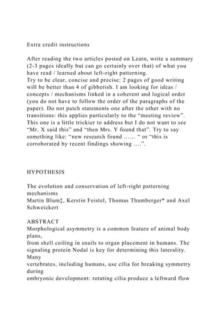
Fig. 1. Asymmetric organs. In humans, asymmetric organs are
found in the
chest (heart, lung) and abdomen (stomach, spleen, liver, small
and large
intestine). The apex of the heart, which is placed at the midline,
points to the left
side. Lungs differ with respect to lobation: two lobes are found
on the left and
three lobes on the right side. The stomach and spleen are
positioned on the
left, whereas the liver and appendix are found on the right. In
addition, the small
intestine and colon coil asymmetrically.
Box 1. Phylogenetic tree of the major animal clades
The lineage relationships between phyla, based on the Tree of
Life project (tolweb.org), is outlined. The last common ancestor
of protostomes and
deuterostomes, a hypothetical bilaterally symmetrical animal
called urbilateria, is also included (outlined in red). Phyla in
which Nodal cascade genes have
been identified are marked in red text. The presence of Nodal
10. cascade genes in both protostomes and deuterostomes suggests
that urbilateria also
possessed the Nodal cascade (Carroll et al., 2004).
1604
HYPOTHESIS Development (2014) 141, 1603-1613
doi:10.1242/dev.100560
D
E
V
E
L
O
P
M
E
N
T
malfunctions. The many problems associated with intestinal
malrotation in humans, which has been estimated to occur in as
many as 1/500 births, support this reasoning (Burn and Hill,
2009;
Stewart et al., 1976; Sutherland and Ware, 2009).
When did organ asymmetry evolve?
The Nodal cascade, which is responsible for asymmetric organ
morphogenesis and placement in the vertebrates, is found in
deuterostomes and protostomes alike (Chea et al., 2005). This
suggests that the last common ancestor was also characterized
by
11. the presence of a Nodal cascade. This hypothetical animal at the
base of all bilaterally symmetrical species has been dubbed
urbilateria (De Robertis and Sasai, 1996) (see Glossary, Box 2).
Although no fossil record of such an animal is known to date,
nor
likely to exist, it has been described as a creature that possessed
a
heart-like pump, body appendages, a light-sensing primitive
eye,
and compartmentalization of the nervous system and digestive
tract (Carroll et al., 2004). Based on our consideration of the
Nodal
cascade, we hypothesize that the urbilaterian GI tract was
asymmetrically arranged (Fig. 3B).
Among the protostomes, Nodal has so far only been described in
mollusks, which belong to the lophotrochozoa (Box 1), which
includes snails and slugs. Gain- and loss-of-function
experiments in
snails have unequivocally shown that Nodal cascade asymmetry
is
responsible for shell coiling, i.e. asymmetric organ placement
(Grande and Patel, 2009). Many species from phyla of the other
major protostome group, the ecdysozoa (Box 1), also display
marked morphological and functional asymmetries. The
nematode
C. elegans, for example, undergoes LR asymmetric rotation
within
the eggshell, cleaves asymmetrically, loses sensory rays in an
asymmetric manner and shows lateralization of the nervous
system
(summarized in Burdine and Caspary, 2013). In Drosophila, the
genital disc and the gut rotate asymmetrically, a process driven
by an
actin-based mechanism (Petzoldt et al., 2012). However, the
12. genomes of C. elegans and Drosophila melanogaster have been
sequenced without discovery of a Nodal homolog, indicating
that
Nodal might have been lost in ecdysozoa. Given the low degree
of
nucleotide and protein homology between snail and
deuterostome
Nodal (Grande and Patel, 2009), however, one should not render
a
premature judgment. In addition, both C. elegans and
Drosophila
melanogaster represent highly derived species in their
respective
phyla. Finally, it remains to be seen whether cnidarians have a
Nodal gene. Certainly, there are no apparent organ asymmetries
in
Hydra (Technau and Steele, 2011). However, asexual
reproduction
through budding in Hydra occurs asymmetrically along the body
column and thus creates an asymmetry (Bode, 2011). The
molecular
mechanisms of asymmetric bud morphogenesis have not been
elucidated (Böttger and Hassel, 2012; Meinhardt, 2012), but if
Hydra has a Nodal gene, it would be likely to act in this
process.
Flow is error-prone and expensive: why should it evolve?
In snails, asymmetric induction of the Nodal cascade occurs via
chiral
blastomere arrangement at the 8-cell stage, providing a strong
argument in favor of symmetry breakage through early
determinants
(Kuroda et al., 2009). We envisage a scenario reminiscent of
asymmetric cell division in C. elegans, whereby cell polarity
governs
asymmetric cell division (Li, 2013; Munro and Bowerman,
13. 2009;
Sawa, 2012). Remnants of spiral cleavage are still present in the
Box 3. The Nodal cascade
The Nodal signaling cascade is active in the left LPM (Hamada
et al.,
2002). Nodal, a member of the TGFβ growth factor family,
induces
transcription of three target genes: Nodal itself, providing a
positive-
feedback loop; Lefty, which encodes a Nodal inhibitor and acts
in a
negative-feedback loop; and Pitx2, which encodes a homeobox
transcription factor. The Nodal cascade spreads rapidly
throughout the
LPM using a self-enhancement and lateral-inhibition mechanism
(Nakamura et al., 2006). Upstream, the cascade is regulated by
members of the DAN family of proteins, such as Coco and
Cerberus,
which act as Nodal antagonists. Coco is a secreted protein that
is co-
expressed with Nodal in somitic LRO cells and is
downregulated as an
immediate target of cilia-driven leftward flow. Coco repression
liberates
Nodal to signal and/or transfer to the left LPM. Although Nodal
and Pitx2
are conserved, variations on the theme have been described in
vertebrates, mostly in chick, in which a highly divergent set of
asymmetrically transcribed genes exists (reviewed by Raya and
Izpisúa-Belmonte, 2006): the snail-related transcription factor
cSnR is
active in the right LPM, whereas Lefty is missing in the left
LPM. Instead,
caronte, a cerberus/Dan-related growth factor, is active in the
left paraxial
14. mesoderm. Curiously, the homeobox transcription factor Nkx3.2
is
asymmetrically expressed in the mouse and chick LPM, but on
opposite
sides [right in mouse versus left in chick (Schneider et al.,
1999)]. It
remains to be seen whether and how these chick-specific
asymmetries
relate to the node rotation observed in chick. Targets of the
Nodal
cascade can also differ. For example, the neuropeptide galanin
was
shown to be asymmetrically expressed, in a flow-dependent
manner, on
the left side of the mouse heart anlage. As galanin asymmetry
was not
detected in Xenopus embryonic heart, this species-specific
difference
might relate to the much higher complexity of the mammalian
four-
chambered heart (Schweickert et al., 2008). Galanin might have
been
co-opted to the Nodal cascade through evolution of Pitx2
binding sites in
its promoter, an option that has yet to be tackled
experimentally.
Box 2. Glossary
Archenteron. The primitive gut tube that forms during
gastrulation.
Sometimes also termed the gastrocoel (cavity of the gastrula
embryo), it
transiently harbors the left-right organizer in amphibian and
mammalian
embryos. The mouse left-right organizer, before its renaming as
the
15. node/posterior notochord, was also referred to as the
archenteron (Blum
et al., 2009a,b; Theiler, 1972). Relationships are less clear in
bony fish
and birds, in which the archenteron is ill-defined.
Ciliopathies. A collective term that describes a diverse group of
human
syndromes caused by ciliary dysfunction. These include Bardet-
Biedl
syndrome, polycystic kidney disease, nephronophthisis, Meckel-
Gruber
syndrome, Joubert syndrome and Senior-Løken syndrome, all of
which
are associated with laterality defects (Fliegauf et al., 2007).
Left-right organizer (LRO). A ciliated epithelium that either
represents
the posterior part of, or is located at the posterior end of, the
notochord.
LROs come in different shapes and sizes, ranging from flat to
indented,
dome-shaped to spherical. Cilia on the LRO are polarized and
rotate in a
clockwise fashion to produce a leftward fluid flow in the
extracellular
space.
Situs solitus. The stable asymmetric arrangement of organs in
the chest
(heart, lung) and abdomen (stomach, spleen, liver, colon,
intestine).
Situs inversus. An inversion in the asymmetric arrangement of
organs.
Such inversion is very rare and occurs in ∼1/10,000 humans.
Synapomorphy. A synapomorphic trait is a character shared by
two or
more taxa and in their most recent common ancestor.
Urbilateria. The term urbilateria has been coined by Eddy De
16. Robertis to
describe the common ancestor of all bilateral symmetrical
animals
(De Robertis, 2008; De Robertis and Sasai, 1996). Although, in
all
likelihood, a fossil urbilaterian will never be found given the
complex
circumstances of successful fossilization, cladistic (deductive)
logics
were applied to characterize urbilateria as a light-sensing,
motile, worm-
like creature with a heart-like pump, a regionalized gut and
nervous
system (Carroll et al., 2004).
1605
HYPOTHESIS Development (2014) 141, 1603-1613
doi:10.1242/dev.100560
D
E
V
E
L
O
P
M
E
N
T
vertebrates. In the frog Xenopus, for example, it has been
17. shown that
the first cleavage division is inherently chiral (Danilchik et al.,
2006).
If mollusks use spiral cleavage to induce Nodal asymmetrically,
why,
then, should leftward flow evolve? What could be the advantage
of
having such a complex machinery for the sole reason of
inducing
Nodal mRNA transcription on the left side of the neurula
embryo?
It is possible that, if it was more reliable or ‘cheaper’ than a
cell
polarity-based mechanism, flow might yield increased fitness.
A detailed consideration of leftward flow, however, provides
proof
of the contrary, and the spontaneous rate of situs inversus in
snails is
fortunately known. When snails such as the Burgundy snail
Helix
pomatia are raised in France for consumption, corkscrew-like
pincers
are used to extract the meat from the shell. These tools do not
easily fit
into shells with inverted torsion, allowing ready identification
of these
so-called snail-kings. A snail-king is discovered in ∼1/20,000
specimens, closely matching the rate of situs inversus in
humans
(Brunner, 1999). Therefore, if one assumes that human embryos
employ leftward flow for symmetry breakage, which is well
justified
on the basis of cilia mutants resulting in laterality syndromes
(Goetz
18. andAnderson,2010;NorrisandGrimes,2012;ShiraishiandIchikawa
,
2012), both mechanisms appear equally reliable at first glance.
Vertebrate LR axis specification using flow is, however,
exceedingly error-prone. It has been estimated that congenital
heart defects occur in up to 1% of live human births (Liu et al.,
2013), of which some 3% are considered to arise from defects in
the
LR pathway (Sutherland and Ware, 2009). Individual
ciliopathies
(see Glossary, Box 2), a sizable number of which are associated
with
LR defects (Fliegauf et al., 2007; Gerdes et al., 2009; Norris
and
Grimes, 2012; Oh and Katsanis, 2012), tend to be relatively rare
(≤10−4). If these syndromes are considered in combination, it
has
been estimated that ∼1/300 humans is affected by some form of
ciliopathy. Other mutations, for example those occurring in
Nodal
cascade genes, also result in human LR defects (Bisgrove et al.,
2003; Sutherland and Ware, 2009). In zebrafish and Xenopus,
depending on the clutch of eggs, LR defects can be observed in
up to
10% of wild-type embryos (Danos and Yost, 1995; Lohr et al.,
1997; Long et al., 2003), further supporting our conclusion that
LR
axis determination via leftward flow is, in fact, error-prone.
To make things worse, flow is expensive. The embryo invests a
lot of energy to specify and pattern the gastrula embryo for the
transient emergence of an LRO, which has no other function
than to
break symmetry. Ciliogenesis and cilia polarization within the
LRO
19. require an elaborate cooperation of growth and transcription
factors
and their respective target genes and processes. In addition,
once
flow has been sensed at the left margin of the LRO, a
complicated
transfer system has to be set in motion to transport (an)
asymmetric
cue(s) from the LRO to the left LPM (Fig. 2). Ablation
experiments
Fig. 2. Left-right organizers and the flow model of symmetry
breakage.
(A) Left-right organizers (LROs) come in different forms (Blum
et al., 2007).
In zebrafish, the LRO is known as Kupffer’s vesicle and is a
closed sphere.
In Xenopus, the gastrocoel roof plate (GRP) acts as the LRO
and is a flat
triangular to diamond-shaped epithelium. In mouse, the LRO
(the posterior
notochord/‘node’) is an indentation at the distal tip of the egg
cylinder. In all
cases the LRO is positioned at the posterior pole of the
notochord (gray).
Axes are indicated: a, anterior; p, posterior; l, left; r, right. (B)
Depiction of
leftward flow at the ciliated epithelium of an LRO. Motile and
polarized cilia
(positioned at the posterior pole of cells) rotate in a clockwise
fashion to
produce a leftward fluid flow in the extracellular space. Flow is
sensed by
unpolarized cilia on cells bordering the LRO. In mouse and
Xenopus these
cilia have been described as being immotile (Boskovski et al.,
20. 2013;
McGrath et al., 2003). These cells express both Nodal and the
Nodal
inhibitor Coco. As a result of flow, Coco becomes
downregulated on the left
side (Hojo et al., 2007; Nakamura et al., 2012; Schweickert et
al., 2010),
thereby derepressing and liberating Nodal protein. Also shown
is the
transfer of an unidentified asymmetric signal (likely to be Nodal
protein; blue
octagon labeled with question mark) to the left lateral plate
mesoderm
(LPM), where the Nodal cascade is induced. Nodal transfers
across the
somites and intermediate mesoderm (not shown) to the LPM,
where it
induces its own transcription and that of its feedback inhibitor
Lefty as well as
expression of Pitx2.
Fig. 3. Organ asymmetry evolved to store a regionalized and
long gut
tube. (A) The regionalized (as represented by the color
gradient)
gastrointestinal (GI) tract of a snail. Note that its length
exceeds that of the main
body axis. A compartmentalized GI tract that exceeds body
length will
inevitably be packaged asymmetrically. (B) We hypothesize that
the
urbilaterian GI tract was also regionalized and asymmetrically
arranged.
D, dorsal; V, ventral; other axes as Fig. 2.
1606
21. HYPOTHESIS Development (2014) 141, 1603-1613
doi:10.1242/dev.100560
D
E
V
E
L
O
P
M
E
N
T
in the freshwater fish medaka and in Xenopus have shown that,
despite LR defects, development proceeded normally when
Kupffer’s vesicle, which is the fish LRO, was manually
destroyed, or upon removal of the superficial mesoderm, which
is
the LRO precursor in the frog (Bajoghli et al., 2007; Blum et
al.,
2009a). Following Nodal cascade induction, LROs rapidly
integrate
into the notochord and somites (Brennan et al., 2002; Shook et
al.,
2004; Yamanaka et al., 2007). Snails certainly use an easier and
cheaper way of rendering Nodal asymmetric.
In search of a solution: evolutionary considerations
Faced with two apparently very distinct modes of symmetry
breakage – one that is flow-based and one that requires early
22. determinants – we now consider more basal chordates, which
turns
out to be a rewarding exercise.
Insights from echinoderms
To begin this evolutionary reflection, we look at echinoderms,
the
other major deuterostome phylum aside from the chordata (Box
1).
In particular, we focus on LR asymmetry in sea urchins, which
has
been studied in great depth. Although apparently radially
symmetrical, echinoderms belong to the bilateria and develop
via
a larval stage that is bilaterally symmetrical; radial symmetry of
the
adult only develops during metamorphosis (McClay, 2011). Sea
urchin larvae show asymmetrical expression of Nodal cascade
genes, interestingly in the primitive gut (the archenteron; see
Glossary, Box 2), although in only a single patch of staining
(for a
recent review see Molina et al., 2013). From the archenteron,
coelomic pouches or sacs bud off in a symmetrical fashion. The
coelomic tissue may be considered as a structure homologous to
the
LPM, as it splits the LPM horizontally in vertebrates. It is
important
for the future development of the sea urchin; the rudiment of the
adult animal, an imaginal disc-like structure (Molina et al.,
2013),
only develops from the coelom on one side. In this case, it is
the side
on which Nodal is not expressed. Asymmetric Nodal expression
in
the archenteron, however, is responsible for the development of
the
23. adult rudiment; when Nodal signaling was inhibited after
gastrulation, an ectopic rudiment formed, whereas ectopic
Nodal
expression prevented rudiment formation (Duboc et al., 2005).
These experiments also confirmed that the LR axis becomes
fixed
only after gastrulation, a conclusion that was derived previously
from the culture of LR-bisected embryos (Aihara and Amemiya,
2001; McCain and McClay, 1994).
Nodal cascade asymmetry in sea urchins has been described as
right-sided, in contrast to the left-sided cascade in the LPM of
vertebrates (Molina et al., 2013). In the absence of a notochord
or
neural tube, the definition of left and right relies exclusively on
the
position of the mouth, which is considered to open on the
ventral
side. In fact, the oral ectoderm of the sea urchin embryo, from
which
the mouth develops, expresses all of the genes that are typically
expressed on the dorsal side of vertebrates, such as chordin,
nodal
and goosecoid (Li et al., 2013). In addition, gene regulatory
networks between sea urchins and vertebrates are apparently
inverted with respect to the dorsal-ventral axis (Molina et al.,
2013). If one considers the possibility that the mouth of the sea
urchin larva might open on the dorsal side (i.e. due to the
apparent
inversion of the dorsal-ventral axis) then the left and right sides
would also flip, and the sea urchin larva would display a left
asymmetric Nodal cascade like all other deuterostomes (Blum et
al.,
2009b). How, then, is this asymmetry set up in the larva?
We speculate that echinoderms possess archenteron cilia that
24. produce a directed fluid flow in much the same way as chordate
archenteron cilia, and that it is this fluid flow that induces the
asymmetric Nodal cascade (Fig. 4). Two published observations
support this notion. First, archenteron cilia have been described
in
gastrula embryos of the feather star Comanthus japonica
(Holland,
1976), which belongs to another, more basal, class of
echinoderms.
The second lead is more indirect and relates to the coelomic
pouches
before the development of the adult rudiment. In larvae of the
sea
urchin Temnopleurus hardwickii, it has been reported that the
coelomic sacs undergo a tube-like extension when they separate
from the archenteron tip (the future esophagus) and organize
into a
ciliated epithelium with motile 9+2 cilia (Hara et al., 2003).
Remarkably, these cilia produce a directed fluid flow (Ruppert
and
Balser, 1986). In addition, the tubulin staining pattern observed
in
these larvae strongly suggests that the archenteron tip cells are
also
ciliated, at least at the (late) stage when the coelom buds off
(Hara
et al., 2003). It would be worthwhile investigating whether
polarized and motile LR cilia are indeed present earlier, before
asymmetric Nodal expression, and whether flow, as we predict,
induces the asymmetric Nodal cascade.
Interestingly, monocilia are also found covering the epidermis
of
neurula embryos of ascidians, which are considered a sister
group to
25. the vertebrates (Box 1). These cilia show similarities to LR
cilia, as
they are polarized and ∼5 µm in length, and it is the motility of
these
cilia that is responsible for the chiral, anticlockwise rotation of
the
embryo (neurula rotation). Interfering with this process alters
LR
asymmetry, for example asymmetric Nodal expression, in the
ascidian Halocynthia roretzi (Nishide et al., 2012; Thompson et
al.,
2012). It has been proposed that such epidermal motile cilia,
which
are commonly used for locomotion and swimming by most non-
chordate embryos (such as the sea urchin pluteus larva), might
have
been re-adapted for symmetry breakage (Nishide et al., 2012). It
is
therefore tempting to speculate that the gastrocoel/archenteron
cilia of vertebrates evolved because superficial cells, from
which
the archenteron derives and which bear motile cilia, became
internalized during gastrulation. As the longitudinal extension
of the
archenteron corresponds to the anterior-posterior axis, it is not
difficult to imagine that cilia became polarized to the posterior
pole of cells, using global anterior-posterior cues, which await
identification even in the vertebrates. If this were the case, this
polarization would inevitably result in a leftward fluid flow.
Molecular data support our reasoning: manipulation of Notch
signaling or of the ion pump ATP4 affects asymmetric Nodal
expression in the sea urchin archenteron (Bessodes et al., 2012).
A role of Notch in vertebrate LR axis determination has long
been
Fig. 4. Cilia in the sea urchin gastrula embryo. Schematic of the
26. dorsal half
of a late gastrula sea urchin embryo. We speculate that
archenteron (ac) cells
are ciliated (A), and that cilia are polarized and produce a
leftward fluid flow
(B, green arrow). Nodal-expressing cells at the archenteron tip
are in blue.
1607
HYPOTHESIS Development (2014) 141, 1603-1613
doi:10.1242/dev.100560
D
E
V
E
L
O
P
M
E
N
T
known (Krebs et al., 2003; Przemeck et al., 2003). In addition,
it has
recently been shown that Notch signaling governs the ratio and
distribution of motile and non-motile sensory cilia on the frog
LRO
(Boskovski et al., 2013). ATP4 plays a dual role in the frog: it
is
required for the induction of the cilia transcription factor Foxj1
27. and
for cilia polarization, under the control of non-canonical and
canonical Wnt signaling, respectively (Walentek et al., 2012).
In
conclusion, we speculate that polarized archenteron cilia in sea
urchins produce a leftward fluid flow that is responsible for the
asymmetric induction of Nodal.
Lessons from amphioxus
We now consider amphioxus (also known as lancelets), which
belong to the chordate subphylum of the cephalochordates (Box
1).
This subphylum is evolutionarily ancient: fossils were reported
from
the Burgess shale, i.e. they date back ∼500-550 million years to
the
Cambrian (Putnam et al., 2008). Remarkably, the body plan of
all of
the ∼45 extant species, as well as that of fossil amphioxus, is
asymmetrical (Fig. 5): the mouth and anus open on the left side,
the
midgut cecum, where digestion and absorption take place,
extends
along the right side, and the cone-shaped muscle segments
(myomeres) are asymmetrically arranged on the left and right
side
of the body. Although mostly buried in the sand, where they
live as
filter-feeders, animals occasionally swim in an undulating
manner
owing to the asymmetric muscle alignment (Liem et al., 2001).
Because of its position at the base of the chordates, rooting the
vertebrates in the phylogenetic tree (Box 1), the embryology of
amphioxus has been studied in great detail (Bertrand and
28. Escriva,
2011; Holland et al., 2004). The first asymmetry that was
described
concerns the alignment of the somites on the left and right sides
of the
notochord (Schubert et al., 2001). Somites and the notochord
represent evolutionary novelties of the chordates, which makes
this
observation all the more interesting. Remarkably, somites form
asymmetrically on both sides of the amphioxus notochord, with
the
left side being slightly advanced compared with its counterpart
on the
right. It has been a long-standing debate as to when this
asymmetry
first appeared during development. Cerfontaine claimed
asymmetries
from the first pair of somites onward (Cerfontaine, 1906),
whereas
Hatschek and Conklin agreed that the first 7-8 somites develop
in a
symmetrical manner (Hatschek, 1893; Conklin, 1932) (Fig. 5F).
However, Conklin noted that the viewing perspective plays a
role in
judging these asymmetries, which is why he was “not inclined
to
place much weight upon this observation” (Conklin, 1932).
More
recently, molecular studies using the homeobox gene Mox to
mark
the forming somites clearly showed asymmetry from the fifth
somite
onwards, without resolving the dispute surrounding the first
pairs
(Minguillón and Garcia-Fernandez, 2002).
29. How do somites become asymmetrical during early
somitogenesis?
Perhaps not too surprisingly, the Nodal cascade in its entirety is
conserved in amphioxus. Nodal, Lefty and Pitx2 genes have
been
cloned and their expression patterns described during early
development (Boorman and Shimeld, 2002; Yu et al., 2002,
2007).
In thelategastrula/early neurula amphioxusembryo, Nodal is
found in
two patches in the roof of the archenteron (Fig. 6) (Yu et al.,
2002).
Fig. 5. Asymmetry in amphioxus. (A) Subadult animal (before
differentiation of the gonads). Histological transverse (B) and
longitudinal (C-E) sections of
adult animals. The longitudinal sections were taken at the level
of the dorsal muscle, dorsal to the neural tube (nt; C), at the
level of the notochord (no; D)
and at the level of the neural tube (E). Note the asymmetric
body plan as reflected in the placement of the pharynx (ph),
cecum (ce) and testes (te), and the
alignment of muscles [myomeres (my)], nerve (n) and nerve
fibers (nf). ar, fixation artifact; Rf, Reissner’s fiber. (F)
Reproduction of original drawings from
Conklin’s 1932 description of embryogenesis in amphioxus
[reproduced with permission (Conklin, 1932)]. At 18 h post-
fertilization (18 hpf, top), the somites
are symmetrically aligned. However, by 24 hpf (bottom),
somitogenesis has become out of register, as is obvious from the
seventh somite onwards. The
fourth (top) and seventh (bottom) somites are boxed in red. d,
dorsal; l, left; r, right; v, ventral.
1608
30. HYPOTHESIS Development (2014) 141, 1603-1613
doi:10.1242/dev.100560
D
E
V
E
L
O
P
M
E
N
T
Remarkably, this expression of Nodal marks the pre-somitic
mesoderm, as these cells in the archenteron roof bud off to give
rise
to the somites shortly thereafter (Bertrand and Escriva, 2011;
Holland
etal.,2004;Yuetal.,2002).Concomitantwiththebuddingoffofthese
cells, Nodal expression becomes asymmetrical, with much
stronger
signals on the left than right side (Yu et al., 2002).
Interestingly, the
cells in between these pre-somitic Nodal-positive cells also bud
off to
giverisetothenotochord(Fig.6)(Yuetal.,2002).Ifonecomparesthis
arrangement in the archenteron roof of amphioxus with that in
the
gastrocoel roof plate (GRP; i.e. the LRO) of Xenopus, striking
similarities become apparent (Fig. 6). The fate of the frog GRP
cells is
31. identical to that of the equivalent cells of amphioxus: Nodal-
positive
lateral cells integrate into the somites, while central cells fold
off to
become part of the notochord (Fig. 6C) (Shook et al., 2004). In
addition, it is these very cells of notochordal fate that in the
frog are
ciliated and produce a leftward fluid flow, which is sensed by
the
somitic lateral Nodal-positive cells (Boskovski et al., 2013;
Schweickert et al., 2007). In one of the extant cephalochordates,
Branchiostoma belcheri tsingtauense, ciliated archenteron cells
have
indeed been described (Hirakow and Kajita, 1991),
substantiating the
similarities between Xenopus and amphioxus.
We therefore propose that the axial cells in the archenteron roof
of
amphioxus possess polarized LR cilia that produce a fluid flow
from
right to left (Fig. 6A,B). This reasoning is further supported by
the
recent description of the expression of a cerberus gene in
amphioxus (Le Petillon et al., 2013). Amphioxus cerberus is
homologous to frog Coco, mouse cerberus-like 2 (Dand5) and
zebrafish charon (dand5), which all encode Nodal inhibitors that
become downregulated in a flow-dependent manner on the left
side
of the LRO (Box 3) (Hojo et al., 2007; Nakamura et al., 2012;
Schweickert et al., 2010). As in vertebrates, amphioxus cerberus
is
initially expressed in a bilaterally symmetrical fashion, co-
expressed
with Nodal. During early somitogenesis, it becomes
32. downregulated
on the left, i.e. appears asymmetrically expressed on the right
(Le Petillon et al., 2013). In vertebrates this asymmetry is the
result
of cilia-driven leftward flow (Schweickert et al., 2010), lending
strong support to the hypothesis that leftward flow was already
present in the cephalochordates at the base of the vertebrate
tree. It
will be rewarding to investigate cilia in amphioxus. The
prediction
based on vertebrate LR cilia would be to find motile cilia that
are
∼5 µm long, polarized to the posterior pole and perhaps exhibit
a
mix of different axoneme types, such as the 9+0, 9+2 and 9+4
configurations described in both rabbit and mouse (Caspary et
al.,
2007; Feistel and Blum, 2006).
One striking difference between asymmetric Nodal expression
in
sea urchin and amphioxus embryos is the split Nodal domain in
the
latter (Fig. 6A,B). As these domains contain descendants of the
organizer, which expresses Nodal in a single domain in the
early
gastrula (Yu et al., 2002), this split merely reflects the fate of
the
organizer: axial Nodal-negative notochordal cells and lateral
(paraxial) Nodal-positive somitic cells. In sea urchins, the
hypothesized leftward flow would displace the entire Nodal
domain
totheleft.Inamphioxus,flowacrosstheepitheliumofthenotochordal
plate would render the Nodal domain asymmetrical, using all of
the
players of vertebrate LROs, but without the need for further
33. transfer
into the LPM, as the entire mesoderm derives from the
archenteron
roof (notochordal plate and somites; Fig. 6B) (Yu et al., 2002).
LR patterning during vertebrate evolution
Our line of argument predicts leftward flow as a synapomorphy
of the
deuterostomes. Its presence throughout the vertebrates is thus
no
surprise, although its apparent loss in some species awaits
explanation. Remarkably, cilia and flow are not found in chick
embryos; here, asymmetric cell migration renders Hensen’s
node (the
organizer) and, in due course Nodal expression, asymmetrical
(Gros
etal.,2009).The emu,asarepresentative ofamore primitive
bird,also
has an asymmetric Hensen’s node (Nagai et al., 2011). It is
therefore
conceivable that all birds may lack LR cilia and leftward flow.
The
analysis of more basal reptiles will thus be a worthwhile
enterprise.
Node asymmetry and a lack of notochordal cilia are not
restricted to
birds: the pig embryo resembles the chick blastodisc in many
aspects,
including node and Nodal asymmetry (Gros et al., 2009).
Before we suggest a solution for the riddle that chick and pig
apparently lack cilia and flow, we wish first to discuss a more
fundamental problem for the evolution of vertebrates. It is
difficult to
imagine that the body plan of the cephalochordates, in
particular
34. somite asymmetry, is compatible with the development of a
perfectly bilateral axial skeleton. The morphogenesis of
vertebrae
requires somites to develop in register, and the same holds true
for
Fig. 6. Homology between amphioxus and Xenopus gastrocoel
roof
plates. Schematics of the gastrocoel roof plates (GRPs) of
amphioxus (A,B)
and Xenopus (C). Dorsal views of late gastrula (A) and 11.5 h
neurula (B)
embryos are shown (left) together with transverse sections
(right) at the levels
indicated. Nodal mRNA expression (blue) in amphioxus
becomes asymmetric
during early neurulation. Cells that are Nodal positive at late
gastrula bud off
from the archenteron roof to form the somites. At later stages,
the epithelium in
between the Nodal-positive cells likewise buds off to become
the notochord
(not indicated). Drawn according to Yu et al. (Yu et al., 2002).
Cilia and flow at
the notochordal part of the archenteron are a hypothetical
prediction of the
authors. (C) Schematic transverse section through a stage 17
Xenopus
neurula embryo. Drawn according to Schweickert et al.
(Schweickert et al.,
2007). Note the striking homology between amphioxus and
Xenopus GRPs: in
both cases, lateral cells expressing Nodal are fated to become
somites while
central cells fold off to form (amphioxus) or integrate into
(frog) the notochord.
35. no, notochord; np, neural plate; som, somite.
1609
HYPOTHESIS Development (2014) 141, 1603-1613
doi:10.1242/dev.100560
D
E
V
E
L
O
P
M
E
N
T
muscles that connect to the vertebral column and allow for
synchronous locomotion. As mentioned above, cephalochordates
swim in an oscillating manner (Liem et al., 2001) due to
asymmetric
myomeres that are connected to the notochord, i.e. the
functional
equivalent of the vertebral column in amphioxus. Vertebrates
thus
have two options: (1) to abandon flow and invent a new way to
induce Nodal asymmetrically in the left LPM; (2) to shield
somites
from the influence of flow and transfer the asymmetric signal
[Nodal
protein, in all likelihood (Yoshiba and Hamada, 2014)] across
36. the
somitic tissue without leaving an imprint.
We favor the second option, particularly because a shielding
mechanism that acts in both mouse (a flow species) and chick (a
no-
flow species) has been described. When embryos were depleted
of
retinoic acid, either genetically (mouse) or through drug
treatment
(chick), somite formation became out of register, with a delay
of
somitogenesis on the right side, precisely as is observed in
amphioxus (Vermot and Pourquié, 2005; Vermot et al., 2005).
Another observation fits with this reasoning: in mouse, it has
been
shown that retinoic acid-containing vesicles arise at the apical
surface of the LRO and that vesicles transfer to the left side of
the
LRO with flow (Tanaka et al., 2005). The evolution and the
mechanism of such shielding, which we predict was not present
in
cephalochordates, await elucidation.
Finally, it should be noted that vertebrates have minimized the
problem of shielding the somites from leftward flow, as most of
the
mesoderm does not bud off from the archenteron as it does in
amphioxus. The LRO cells, however, still stick out. They
produce and
sense the flow, create the asymmetric signal and send the signal
to the
LPM. In addition, where these cells have been studied it has
been
shown that they are of mesodermal fate and integrate into the
notochord [frog and mouse (Brennan et al., 2002; Shook et al.,
37. 2004;
Yamanaka et al., 2007)] as well as into somites [frog and fish
(Long
et al., 2003; Shook et al., 2004)]. Histology and scanning
electron
microscopy of frog neurula embryos clearly showed that the
sensory
lateral GRPcells of somitic fateare alreadya part ofthesomiteand
that
justtheirapicalsurfacesticksout,andonlyuntilflowhasbeenreceived
(Schweickert et al., 2007, 2010). In the zebrafish LRO, the
Nodal gene
southpaw is co-expressed with the presomitic marker spadetail
(Long
et al., 2003), suggesting that a somitic fate of flow-sensing cells
is
conserved from amphioxusto at least fish and amphibians. We
believe
that it is these cells that require shielding from flow.
A role for early determinants in vertebrate symmetry
breaking?
The scenario outlined above is of course hypothetical, as cilia-
driven leftward flow has not yet been described in echinoderms
or
amphioxus. If flow only evolved in the vertebrates, a shielding
mechanism would have had to evolve at the same time.
Furthermore, flow should have been present at the base of the
vertebrates. In line with this, primitive fish (sturgeon), which
gastrulate like amphibians, have a ciliated GRP as in the frog
(Blum
et al., 2009b; Bolker, 1993). The LRO/Kupffer’s vesicle in bony
fish is also ciliated. We thus suggest that leftward flow
represents the
ancestral mode of symmetry breakage in vertebrates.
Species that display flow at an LRO are characterized by the
38. expression of the cilia transcription factor Foxj1 in the
precursor tissue
of the LRO, i.e. the primary embryonic organizer or node.
Expression
of Foxj1 correlates with motile cilia in all cases investigated so
far. In
zebrafish and Xenopus, loss of Foxj1 function deletes motile
cilia,
whereas ectopic Foxj1 expression gives rise to ectopic motile
cilia
(Stubbs et al., 2008; Yu et al., 2008). We have previously
shown that
Foxj1 mRNA is expressed in the Hensen’s node of pig (Gros et
al.,
2009). The pig, like chick, lacks cilia at the posterior
notochord, which
otherwise resembles the ciliated LRO of the rabbit (Feistel and
Blum,
2006). Foxj1 expression in the chick embryo has not yet been
published; however, the chick Hensen’s node does express the
dynein
heavy chain gene Dnah11 (previously known as left-right
dynein, lrd)
(Essner et al., 2002), which is induced by Foxj1 (Stubbs et al.,
2008)
and is required for the motility of LRO cilia (Supp et al., 1997).
Based
onthesedata,wesuggestthatchickandpigbothinheritedthemolecula
r
Foxj1/Dnah11 module,which in other vertebrates sets up
leftward flow
at the LRO. It has been suggested that, in mouse, as few as two
cilia are
sufficient to produce and sense LRO flow (Shinohara et al.,
39. 2012), so it
is possible that chick and pig might have a tiny LRO with just a
few
motile and sensory cilia, which thus far have gone unnoticed.
We
consider this possibility unlikely, however, because many
laboratories
(including our own) have looked in vain for chick cilia in the
past.
Instead, we suggest that chick and pig have lost the
functionality of the
Foxj1/Dnah11module,perhapsthroughlossofapromoterelementin
a
Foxj1 target gene or through an epigenetic mechanism.
A precedent for such a loss is set by the limbless snakes, an
adaptation of the tetrapod body plan to a new form of
locomotion.
The python lineage represents a transitional stage that retains a
pelvic girdle and rudimentary hindlimbs. During development, a
hindlimb bud still forms but it lacks a functional apical
ectodermal
ridge (AER) and therefore does not elongate. Recombination
with a
chick AER or simply providing the AER signaling molecule
FGF
induces leg bud outgrowth, demonstrating that the molecular
machinery for limb outgrowth, with the sole exception of the
inducing AER signal, is present in the python (Cohn and Tickle,
1999; Graham and McGonnell, 1999). Thus, loss of a structure
or
process is not necessarily accompanied by the loss of the
genetic
modules required to set it into motion and, vice versa, the
presence
of a genetic module without the respective structure or process
40. represents a tell-tale sign of a function now lost during
evolution.
If chick and pig lack a functional Foxj1 module and hence do
not
use cilia, how do they break symmetry and induce the Nodal
cascade
on the left side? The fact that the ion pump ATP4 acts
hierarchically
upstream of asymmetric cell migration in the chick has been
taken as
support for the early determinants/ion-flux model of symmetry
breakage, acting before the onset of gastrulation in a cilia/LRO-
independentmanner(Grosetal.,2009;Vandenbergand Levin,2013).
ATP4 is an important player in the ion-flux model, and
asymmetric
expression of ATP4 in the 4-cell Xenopus embryo has been
described.
This asymmetry has been proposed to set up a voltage gradient
that
drives serotonin through gap junctions to the right side of the
embryo,
where it repressesthe Nodal cascade to break symmetryearlyon
(fora
recent review see Vandenberg and Levin, 2013). However,
recent
work, mostly in Xenopus but also in mouse, has examined the
role of
the centralcomponents oftheion-fluxmodelinLRO-
basedsymmetry
breakage. For example, ATP4 was shown to be symmetrically
expressed in frog embryos and to control ciliogenesis and cilia
polarization (Walentek et al., 2012). Serotonin was also found
in a
symmetrical fashion and was shown to be required for
specification of
41. the LRO precursor in the frog, the so-called superficial
mesoderm
(Beyer et al., 2012a). Finally, gap junction communication has
been
implicatedinthetransferofasymmetriccue(s)fromtheLROtotheleft
LPM in frog and mouse (Beyeret al., 2012b; Saund et al., 2012;
Viotti
et al., 2012). Together, these recent findings render the ion-flux
model
of symmetry breakage unlikely.
Symmetry breaking in chick and pig
How then do chick and pig, which have no functional LRO and
do not
use early determinants, break symmetry? Our last hypothesis,
which
1610
HYPOTHESIS Development (2014) 141, 1603-1613
doi:10.1242/dev.100560
D
E
V
E
L
O
P
M
E
N
T
42. suggests a solution to this riddle, rests on descriptive
embryology and
on a continuation of the evolutionary considerations outlined
above.
Let us recall the specifics of Nodal asymmetry in echinoderms
and
cephalochordates: there is a single asymmetric domain in the
sea
urchin archenteron but a split domain in the amphioxus
gastrocoel,
which only becomes asymmetric during late gastrulation (Figs 4
and 6). We hypothesize that, at least in the chordates, these
domains
are direct descendants of the previous Nodal domain in the
organizer
tissue at the onset of gastrulation. In many cases, Nodal
expression is
transiently off during involution of the organizer tissue. A
continuity
can occasionally be seen in mouse, i.e. expression in the node,
which
splits in its anteriormost aspect (Blum et al., 2007). The main
difference in the chordates compared with echinoderms is the
fate of
Nodal-positive organizer cells, which develop into notochord
and
somites in chordates (Spemann and Mangold, 1924). Following
involution (and specification of the notochord as the axial
mesodermal component) the Nodal domain splits down the
middle,
with central cells being Nodal negative and lateral cells
retaining
Nodal expression (Fig. 4). In the case of LRO flow, the central,
Nodal-
negative cells bear motile, polarized cilia, whereas the lateral,
43. Nodal-positive cells sense flow.
Remarkably, chick and pig embryos do not exhibit a split Nodal
domain. Rather, the organizer/node Nodal domain is displaced
to
the left side in its entirety, in chick through leftward migration
of
cells at Hensen’s node (Cui et al., 2009; Gros et al., 2009).
Recently, Viebahn and colleagues performed histological
analyses
of chicken nodes at different stages of development, showing
that
the right shoulder of the node differs from the left shoulder
following node rotation (Tsikolia et al., 2012). In particular,
they
describe that notochordal cells emerge via the thickened right
shoulder of the node (Tsikolia et al., 2012). A continuity
between
the right part of the node and the notochord was already
described
by Wetzel (Wetzel, 1929), the discoverer of node asymmetry in
chick. Based on these observations, we hypothesize that
leftward
node rotation leaves the organizer Nodal domain undivided,
because the notochord emerges on the right side of Hensen’s
node
(Fig. 7). Such a mechanism of leftward Nodal displacement
should be very robust and perhaps less error-prone than LRO
flow.
In agreement with this notion, no spontaneously occurring
alterations of Nodal cascade gene expression in chick and pig
embryos have been reported in the literature, in contrast to frog
and fish. How could leftward node rotation have evolved? We
can
suggest no solution for this fascinating question at this time.
44. Unraveling the precise role of ATP4 in the context of node
rotation
might be particularly rewarding as, to date, this ion pump
represents the only shared component between LRO flow and
node rotation.
Conclusions
Our examination of LR asymmetry in an evolutionary context
has
led us to three conclusions covering the base of bilateria, the
deuterostome tree and species-specific differences between
vertebrates. (1) Based on cladistic logics, we hypothesize that
urbilateria used the Nodal cascade for asymmetric
morphogenesis
of the gut tube. The Nodal cascade itself might have been lost in
ecdysozoa, and the elucidation of molecular mechanisms
underlying LR asymmetries in these species might lead to the
identification of novel homologies between protostomes and
deuterostomes, with the potential to sharpen the evolutionary
view
on the origin of animal LR asymmetry. (2) We speculate that a
cilia-driven mechanism of symmetry breakage exists throughout
the deuterostomes. We predict in which tissue and at what
embryonic stage LR cilia and leftward flow should be present in
sea urchin and amphioxus embryos, but descriptive and
functional
experiments will be required to prove or refute this hypothesis.
Not
working on these species ourselves, we hope to have provided
some leads for future investigations. (3) Finally, we highlight
that
traits, characters and processes can get lost during evolution if
other mechanisms are able to compensate. The absence of
leftward
flow in chick and pig, and perhaps in all birds and also in other
mammals, might represent such a case. Under the control of at
45. least
one common determinant, i.e. the ion pump ATP4, a new
process
has evolved to guide an as yet poorly understood mechanism for
breaking symmetry based on asymmetric cell movements. In
addition, the leftward displacement of an undivided organizer
Nodal domain can activate the asymmetric LPM signaling
cascade
in much the same way as if induced by flow, but this mechanism
should be cheaper, more robust and less error-prone. In the
future,
descriptive and functional work in both established and novel
model organisms, combined with evolutionary thinking and
reasoning, should hopefully provide a promising path through
which we can expand our understanding of the origin and
diversification of animal LR asymmetry.
Acknowledgements
We thank our colleagues in the left-right and EvoDevo
communities for numerous
discussions over the years, in particular Sebastian Shimeld,
Reinhard Schröder,
Julien Vermot, Philipp Vick and Christoph Viebahn. Bernd
Schmid produced some
of the artwork reproduced here. The amphioxus histology was
performed by the late
Otto Pflugfelder when Professor of Zoology at Hohenheim
University in the 1950s
and 1960s. We particularly thank Reinhard Hilbig for locating
and photographing
these slides.
Competing interests
The authors declare no competing financial interests.
Fig. 7. Divergent modes of symmetry breakage in vertebrates.
46. (A) Ancestral flow-based mode of chordates. The early gastrula
organizer
(node), which expresses Nodal (blue), differentiates into the
notochord and
somites during early neurulation. The notochord does not
express Nodal and
thus splits the Nodal-positive domain down the middle. Nodal-
positive cells
later integrate into the somites. (B) Divergent mechanism in
chick and pig. The
organizer (node) rotates during early gastrulation, which
renders the node left-
asymmetric. The notochord emerges over the right shoulder of
the node and
does not split the Nodal domain. Nodal thus becomes displaced
to the left
without the need for flow.
1611
HYPOTHESIS Development (2014) 141, 1603-1613
doi:10.1242/dev.100560
D
E
V
E
L
O
P
M
E
N
T
47. Funding
K.F. was supported by a Margarete-von-Wrangell Fellowship,
funded by the
European Social Fund and by the Ministry Of Science, Research
and the Arts in
Baden-Württemberg. Work in the M.B. laboratory was funded
by grants from the
Deutsche Forschungsgemeinschaft.
References
Aihara, M. and Amemiya, S. (2001). Left-right positioning of
the adult rudiment in
sea urchin larvae is directed by the right side. Development
128, 4935-4948.
Allen, R. D. and Fok, A. K. (2000). Membrane trafficking and
processing in
Paramecium. Int. Rev. Cytol. 198, 277-318.
Bajoghli, B., Aghaallaei, N., Soroldoni, D. and Czerny, T.
(2007). The roles of
Groucho/Tle in left-right asymmetry and Kupffer’s vesicle
organogenesis. Dev.
Biol. 303, 347-361.
Bertrand, S. and Escriva, H. (2011). Evolutionary crossroads in
developmental
biology: amphioxus. Development 138, 4819-4830.
Bessodes, N., Haillot, E., Duboc, V., Röttinger, E., Lahaye, F.
and Lepage, T.
(2012). Reciprocal signaling between the ectoderm and a
mesendodermal left-
right organizer directs left-right determination in the sea urchin
48. embryo. PLoS
Genet. 8, e1003121.
Beyer, T., Danilchik, M., Thumberger, T., Vick, P., Tisler, M.,
Schneider, I.,
Bogusch, S., Andre, P., Ulmer, B., Walentek, P. et al. (2012a).
Serotonin
signaling is required for Wnt-dependent GRP specification and
leftward flow in
Xenopus. Curr. Biol. 22, 33-39.
Beyer, T., Thumberger, T., Schweickert, A. and Blum, M.
(2012b). Connexin26-
mediated transfer of laterality cues in Xenopus. Biol. Open 1,
473-481.
Bisgrove, B. W., Morelli, S. H. and Yost, H. J. (2003). Genetics
of human laterality
disorders: insights from vertebrate model systems. Annu. Rev.
Genomics Hum.
Genet. 4, 1-32.
Blum, M., Andre, P., Muders, K., Schweickert, A., Fischer, A.,
Bitzer, E.,
Bogusch, S., Beyer, T., van Straaten, H. W. M. and Viebahn, C.
(2007). Ciliation
and gene expression distinguish between node and posterior
notochord in the
mammalian embryo. Differentiation 75, 133-146.
Blum, M., Beyer, T., Weber, T., Vick, P., Andre, P., Bitzer, E.
and Schweickert, A.
(2009a). Xenopus, an ideal model system to study vertebrate
left-right asymmetry.
Dev. Dyn. 238, 1215-1225.
49. Blum, M., Weber, T., Beyer, T. and Vick, P. (2009b). Evolution
of leftward flow.
Semin. Cell Dev. Biol. 20, 464-471.
Bode, H. (2011). Axis formation in hydra. Annu. Rev. Genet.
45, 105-117.
Bolker, J. A. (1993). The mechanism of gastrulation in the
white sturgeon. J. Exp.
Zool. 266, 132-145.
Boorman, C. J. and Shimeld, S. M. (2002). Pitx homeobox
genes in Ciona and
amphioxus show left-right asymmetry is a conserved chordate
character and
define the ascidian adenohypophysis. Evol. Dev. 4, 354-365.
Boskovski, M. T., Yuan, S., Pedersen, N. B., Goth, C. K.,
Makova, S., Clausen, H.,
Brueckner, M. and Khokha, M. K. (2013). The heterotaxy gene
GALNT11
glycosylates Notch to orchestrate cilia type and laterality.
Nature 504, 456-459.
Böttger, A. and Hassel, M. (2012). Hydra, a model system to
trace the emergence
of boundaries in developing eumetazoans. Int. J. Dev. Biol. 56,
583-591.
Brennan, J., Norris, D. P. and Robertson, E. J. (2002). Nodal
activity in the node
governs left-right asymmetry. Genes Dev. 16, 2339-2344.
Brown, W. M., Cronk, L., Grochow, K., Jacobson, A., Liu, C.
K., Popović, Z. and
Trivers, R. (2005). Dance reveals symmetry especially in young
men. Nature
50. 438, 1148-1150.
Brunner, H. (1999). Rechts oder links in der Natur und
anderswo. Weinheim:
Wiley-VCH.
Burdine, R. D. and Caspary, T. (2013). Left-right asymmetry:
lessons from Cancun.
Development 140, 4465-4470.
Burn, S. F. and Hill, R. E. (2009). Left-right asymmetry in gut
development: what
happens next? Bioessays 31, 1026-1037.
Carroll, S. B., Grenier, J. and Weatherbee, S. (2004). From
DNA to Diversity.
Hoboken, NJ: John Wiley & Sons.
Caspary, T., Larkins, C. E. and Anderson, K. V. (2007). The
graded response to
Sonic Hedgehog depends on cilia architecture. Dev. Cell 12,
767-778.
Cerfontaine, P. (1906). Recherche sur le développement de
l’Amphioxus. Arch. de
Biol. T22, 229-418.
Chea, H. K., Wright, C. V. and Swalla, B. J. (2005). Nodal
signaling and the
evolution of deuterostome gastrulation. Dev. Dyn. 234, 269-
278.
Cohn, M. J. and Tickle, C. (1999). Developmental basis of
limblessness and axial
patterning in snakes. Nature 399, 474-479.
51. Conklin, E. G. (1932). The embryology of amphioxus. J.
Morphol. 54, 69-151.
Cui, C., Little, C. D. and Rongish, B. J. (2009). Rotation of
organizer tissue
contributes to left-right asymmetry. Anat. Rec. (Hoboken) 292,
557-561.
Danilchik, M. V., Brown, E. E. and Riegert, K. (2006). Intrinsic
chiral properties of
the Xenopus egg cortex: an early indicator of left-right
asymmetry? Development
133, 4517-4526.
Danos, M. C. and Yost, H. J. (1995). Linkage of cardiac left-
right asymmetry and
dorsal-anterior development in Xenopus. Development 121,
1467-1474.
De Robertis, E. M. (2008). Evo-devo: variations on ancestral
themes. Cell 132,
185-195.
De Robertis, E. M. and Sasai, Y. (1996). A common plan for
dorsoventral patterning
in Bilateria. Nature 380, 37-40.
Duboc, V., Röttinger, E., Lapraz, F., Besnardeau, L. and
Lepage, T. (2005). Left-
right asymmetry in the sea urchin embryo is regulated by nodal
signaling on the
right side. Dev. Cell 9, 147-158.
Essner, J. J., Vogan, K. J., Wagner, M. K., Tabin, C. J., Yost,
H. J. and
Brueckner, M. (2002). Left–right development: conserved
function for embryonic
52. nodal cilia. Nature 418, 37-38.
Feistel, K. and Blum, M. (2006). Three types of cilia including
a novel 9+4 axoneme
on the notochordal plate of the rabbit embryo. Dev. Dyn. 235,
3348-3358.
Fliegauf, M., Benzing, T. and Omran, H. (2007). When cilia go
bad: cilia defects
and ciliopathies. Nat. Rev. Mol. Cell Biol. 8, 880-893.
Fok, A. K. and Allen, R. D. (1990). The phagosome-lysosome
membrane system
and its regulation in Paramecium. Int. Rev. Cytol. 123, 61-74.
Fukumoto, T., Kema, I. P. and Levin, M. (2005). Serotonin
signaling is a very early
step in patterning of the left-right axis in chick and frog
embryos. Curr. Biol. 15,
794-803.
Gerdes, J. M., Davis, E. E. and Katsanis, N. (2009). The
vertebrate primary cilium
in development, homeostasis, and disease. Cell 137, 32-45.
Goetz, S. C. and Anderson, K. V. (2010). The primary cilium: a
signalling centre
during vertebrate development. Nat. Rev. Genet. 11, 331-344.
Graham, A. and McGonnell, I. (1999). Developmental
evolution: this side of
paradise. Curr. Biol. 9, R630-R632.
Grande, C. and Patel, N. H. (2009). Nodal signalling is involved
in left-right
asymmetry in snails. Nature 457, 1007-1011.
53. Gros, J., Feistel, K., Viebahn, C., Blum, M. and Tabin, C. J.
(2009). Cell
movements at Hensen’s node establish left/right asymmetric
gene expression in
the chick. Science 324, 941-944.
Hamada, H. (2008). Breakthroughs and future challenges in left-
right patterning.
Dev. Growth Differ. 50 Suppl. 1, S71-S78.
Hamada, H., Meno, C., Watanabe, D. and Saijoh, Y. (2002).
Establishment of
vertebrate left-right asymmetry. Nat. Rev. Genet. 3, 103-113.
Hara, Y., Kuraishi, R., Uemura, I. and Katow, H. (2003).
Asymmetric formation and
possible function of the primary pore canal in plutei of
Temnopleurus hardwicki.
Dev. Growth Differ. 45, 295-308.
Hatschek, B. (1893). The Amphioxus and its Development.
London: Swan
Sonnenschein.
Hirakow, R. and Kajita, N. (1991). Electron microscopic study
of the development of
amphioxus, Branchiostoma belcheri tsingtauense: the gastrula.
J. Morphol. 207,
37-52.
Hirokawa, N., Tanaka, Y. and Okada, Y. (2012). Cilia, KIF3
molecular motor and
nodal flow. Curr. Opin. Cell Biol. 24, 31-39.
Hojo, M., Takashima, S., Kobayashi, D., Sumeragi, A.,
54. Shimada, A.,
Tsukahara, T., Yokoi, H., Narita, T., Jindo, T., Kage, T. et al.
(2007). Right-
elevated expression of charon is regulated by fluid flow in
medaka Kupffer’s
vesicle. Dev. Growth Differ. 49, 395-405.
Holland, N. D. (1976). The fine structure of the embryo during
the gastrula stage of
Comanthus japonica (Echinodermata: Crinoidea). Tissue Cell 8,
491-510.
Holland, L. Z., Laudet, V. and Schubert, M. (2004). The
chordate amphioxus: an
emerging model organism for developmental biology. Cell. Mol.
Life Sci. 61,
2290-2308.
Krebs, L. T., Iwai, N., Nonaka, S., Welsh, I. C., Lan, Y., Jiang,
R., Saijoh, Y.,
O’Brien, T. P., Hamada, H. and Gridley, T. (2003). Notch
signaling regulates left-
right asymmetry determination by inducing Nodal expression.
Genes Dev. 17,
1207-1212.
Kuroda,R.,Endo,B.,Abe,M.andShimizu,M.(2009).Chiralblastome
rearrangement
dictates zygotic left-right asymmetry pathway in snails. Nature
462, 790-794.
Le Petillon, Y., Oulion, S., Escande, M.-L., Escriva, H. and
Bertrand, S. (2013).
Gene expression patterns. Gene Expr. Patterns 13, 377-383.
Levin, M. (2005). Left-right asymmetry in embryonic
55. development: a comprehensive
review. Mech. Dev. 122, 3-25.
Li, R. (2013). The art of choreographing asymmetric cell
division. Dev. Cell 25,
439-450.
Li, E., Materna, S. C. and Davidson, E. H. (2013). New
regulatory circuit controlling
spatial and temporal gene expression in the sea urchin embryo
oral ectoderm
GRN. Dev. Biol. 382, 268-279.
Liem, K. F., Bemis, W. E., Walker,W. F. and Grande, L. (2001).
Functional Anatomy
of the Vertebrates: An Evolutionary Perspective. San Diego,
CA: Hartcourt College
Publishers.
Liu, X., Tobita, K., Francis, R. J. B. and Lo, C. W. (2013).
Imaging techniques for
visualizing and phenotyping congenital heart defects in murine
models. Birth
Defects Res. C Embryo Today 99, 93-105.
Lohr, J. L., Danos, M. C. and Yost, H. J. (1997). Left-right
asymmetry of a nodal-
related gene is regulated by dorsoanterior midline structures
during Xenopus
development. Development 124, 1465-1472.
Long, S., Ahmad, N. and Rebagliati, M. (2003). The zebrafish
nodal-related gene
southpaw is required for visceral and diencephalic left-right
asymmetry.
Development 130, 2303-2316.
56. 1612
HYPOTHESIS Development (2014) 141, 1603-1613
doi:10.1242/dev.100560
D
E
V
E
L
O
P
M
E
N
T
http://dx.doi.org/10.1016/S0074-7696(00)98007-0
http://dx.doi.org/10.1016/S0074-7696(00)98007-0
http://dx.doi.org/10.1016/j.ydbio.2006.11.020
http://dx.doi.org/10.1016/j.ydbio.2006.11.020
http://dx.doi.org/10.1016/j.ydbio.2006.11.020
http://dx.doi.org/10.1242/dev.066720
http://dx.doi.org/10.1242/dev.066720
http://dx.doi.org/10.1371/journal.pgen.1003121
http://dx.doi.org/10.1371/journal.pgen.1003121
http://dx.doi.org/10.1371/journal.pgen.1003121
http://dx.doi.org/10.1371/journal.pgen.1003121
http://dx.doi.org/10.1016/j.cub.2011.11.027
http://dx.doi.org/10.1016/j.cub.2011.11.027
http://dx.doi.org/10.1016/j.cub.2011.11.027
http://dx.doi.org/10.1016/j.cub.2011.11.027
http://dx.doi.org/10.1242/bio.2012760
http://dx.doi.org/10.1242/bio.2012760
61. insights into the functional evolution of Mox genes, somites,
and the asymmetry of
amphioxus somitogenesis. Dev. Biol. 246, 455-465.
Molina, M. D., de Crozé, N., Haillot, E. and Lepage, T. (2013).
Nodal: master and
commander of the dorsal. Curr. Opin. Genet. Dev. 23, 445-453.
Munro, E. and Bowerman, B. (2009). Cellular symmetry
breaking during
Caenorhabditis elegansdevelopment.Cold Spring Harb. Perspect.
Biol. 1, a003400.
Nagai, H., Mak, S.-S., Weng, W., Nakaya, Y., Ladher, R. and
Sheng, G. (2011).
Embryonic development of the emu, Dromaius novaehollandiae.
Dev. Dyn. 240,
162-175.
Nakamura, T., Mine, N., Nakaguchi, E., Mochizuki, A.,
Yamamoto, M.,
Yashiro, K., Meno, C. and Hamada, H. (2006). Generation of
robust left-right
asymmetry in the mouse embryo requires a self-enhancement
and lateral-
inhibition system. Dev. Cell 11, 495-504.
Nakamura, T., Saito, D., Kawasumi, A., Shinohara, K., Asai, Y.,
Takaoka, K.,
Dong, F., Takamatsu, A., Belo, J. A., Mochizuki, A. et al.
(2012). Fluid flow and
interlinked feedback loops establish left-right asymmetric decay
of Cerl2 mRNA.
Nat. Commun. 3, 1322.
Nickel, R., Schummer, A. and Seiferle, E. (1995). Lehrbuch der
62. Anatomie der
Haustiere, 9th edn (ed. J. Frewein, H. Gasse, R. Leiser, H.
Roos, H. Thomé,
B. Vollmerhaus and H. Waibl). Stuttgart: Parey Publishers.
Nishide, K., Mugitani, M., Kumano, G. and Nishida, H. (2012).
Neurula rotation
determines left-right asymmetry in ascidian tadpole larvae.
Development 139,
1467-1475.
Norris, D. P. and Grimes, D. T. (2012). Mouse models of
ciliopathies: the state of
the art. Dis. Model. Mech. 5, 299-312.
Oh, E. C. and Katsanis, N. (2012). Cilia in vertebrate
development and disease.
Development 139, 443-448.
Patel, N. H. (2009). Developmental biology: asymmetry with a
twist. Nature 462,
727-728.
Petzoldt, A. G., Coutelis, J.-B., Géminard, C., Spéder, P.,
Suzanne, M.,
Cerezo, D. and Noselli, S. (2012). DE-Cadherin regulates
unconventional
Myosin ID and Myosin IC in Drosophila left-right asymmetry
establishment.
Development 139, 1874-1884.
Przemeck, G. K. H., Heinzmann, U., Beckers, J. and Hrabé de
Angelis, M.
(2003). Node and midline defects are associated with left-right
development in
Delta1 mutant embryos. Development 130, 3-13.
63. Putnam, N. H., Butts, T., Ferrier, D. E. K., Furlong, R. F.,
Hellsten, U.,
Kawashima, T., Robinson-Rechavi, M., Shoguchi, E., Terry, A.,
Yu, J.-K. et al.
(2008). The amphioxus genome and the evolution of the
chordate karyotype.
Nature 453, 1064-1071.
Ramsdell, A. F. (2005). Left-right asymmetry and congenital
cardiac defects: getting
to the heart of the matter in vertebrate left-right axis
determination. Dev. Biol.
288, 1-20.
Raya, A. and Izpisúa-Belmonte, J. C. (2006). Left-right
asymmetry in the
vertebrate embryo: from early information to higher-level
integration. Nat. Rev.
Genet. 7, 283-293.
Ruppert, E. E. and Balser, E. J. (1986). Nephridia in the larvae
of hemichordates
and echinoderms. Biol. Bull. 171, 188-196.
Saund, R. S., Kanai-Azuma, M., Kanai, Y., Kim, I., Lucero, M.
T. and Saijoh, Y.
(2012). Gut endoderm is involved in the transfer of left-right
asymmetry from the
node to the lateral plate mesoderm in the mouse embryo.
Development 139,
2426-2435.
Sawa, H. (2012). Control of cell polarity and asymmetric
division in C. elegans. Curr.
Top. Dev. Biol. 101, 55-76.
64. Schneider, A., Mijalski, T., Schlange, T., Dai, W., Overbeek,
P., Arnold, H.-H. and
Brand, T. (1999). The homeobox gene it NKX3.2 is a target of
left-right signalling
and is expressed on opposite sides in chick and mouse embryos.
Curr. Biol. 9,
911-914.
Schubert, M., Holland, L. Z., Stokes, M. D. and Holland, N. D.
(2001). Three
amphioxus Wnt genes (AmphiWnt3, AmphiWnt5, and
AmphiWnt6) associated
with the tail bud: the evolution of somitogenesis in chordates.
Dev. Biol. 240,
262-273.
Schweickert, A., Weber, T., Beyer, T., Vick, P., Bogusch, S.,
Feistel, K. and
Blum, M. (2007). Cilia-driven leftward flow determines
laterality in Xenopus. Curr.
Biol. 17, 60-66.
Schweickert, A., Deissler, K., Britsch, S., Albrecht, M.,
Ehmann, H., Mauch, V.,
Gaio, U. and Blum, M. (2008). Left-asymmetric expression of
Galanin in the linear
heart tube of the mouse embryo is independent of the nodal co-
receptor gene
cryptic. Dev. Dyn. 237, 3557-3564.
Schweickert, A., Vick, P., Getwan, M., Weber, T., Schneider, I.,
Eberhardt, M.,
Beyer, T., Pachur, A. and Blum, M. (2010). The nodal inhibitor
Coco is a critical
target of leftward flow in Xenopus. Curr. Biol. 20, 738-743.
65. Shinohara, K., Kawasumi, A., Takamatsu, A., Yoshiba, S.,
Botilde, Y.,
Motoyama, N., Reith, W., Durand, B., Shiratori, H. and
Hamada, H. (2012).
Two rotating cilia in the node cavity are sufficient to break left-
right symmetry in the
mouse embryo. Nat. Commun. 3, 622.
Shiraishi, I. and Ichikawa, H. (2012). Human heterotaxy
syndrome – from molecular
genetics to clinical features, management, and prognosis. Circ.
J. 76, 2066-2075.
Shook, D. R., Majer, C. and Keller, R. (2004). Pattern and
morphogenesis of
presumptive superficial mesoderm in two closely related
species, Xenopus laevis
and Xenopus tropicalis. Dev. Biol. 270, 163-185.
Spemann, H. and Mangold, H. (1924). Über induktion von
Embryonalanlagen
durch Implantation artfremder Organisatoren. Roux’ Arch.
Entw. Mech. 100,
599-638.
Stewart, D. R., Colodny, A. L. and Daggett, W. C. (1976).
Malrotation of the bowel
in infants and children: a 15 year review. Surgery 79, 716-720.
Stubbs, J. L., Oishi, I., Izpisúa-Belmonte, J. C. and Kintner, C.
(2008). The
forkhead protein Foxj1 specifies node-like cilia in Xenopus and
zebrafish
embryos. Nat. Genet. 40, 1454-1460.
66. Supp, D. M., Witte, D. P., Potter, S. S. and Brueckner, M.
(1997). Mutation of an
axonemal dynein affects left-right asymmetry in inversus
viscerum mice. Nature
389, 963-966.
Sutherland, M. J. and Ware, S. M. (2009). Disorders of left-
right asymmetry:
heterotaxy and situs inversus. Am. J. Med. Genet. C Semin.
Med. Genet. 151C,
307-317.
Tanaka, Y., Okada, Y. and Hirokawa, N. (2005). FGF-induced
vesicular release of
Sonic hedgehog and retinoic acid in leftward nodal flow is
critical for left-right
determination. Nature 435, 172-177.
Technau, U. and Steele, R. E. (2011). Evolutionary crossroads
in developmental
biology: Cnidaria. Development 138, 1447-1458.
Theiler, K. (1972). The House Mouse. Berlin, Heidelberg, New
York: Springer-
Verlag.
Thompson, H., Shaw, M. K., Dawe, H. R. and Shimeld, S. M.
(2012). The formation
and positioning of cilia in Ciona intestinalis embryos in relation
to the generation
and evolution of chordate left-right asymmetry. Dev. Biol. 364,
214-223.
Tsikolia, N., Schröder, S., Schwartz, P. and Viebahn, C. (2012).
Paraxial left-
sided nodal expression and the start of left-right patterning in
67. the early chick
embryo. Differentiation 84, 380-391.
Vandenberg, L. N. and Levin, M. (2009). Perspectives and open
problems in the
early phases of left-right patterning. Semin. Cell Dev. Biol. 20,
456-463.
Vandenberg, L. N. and Levin, M. (2010). Far from solved: a
perspective on what we
know about early mechanisms of left-right asymmetry. Dev.
Dyn. 239, 3131-3146.
Vandenberg, L. N. and Levin, M. (2013). A unified model for
left-right asymmetry?
Comparison and synthesis of molecular models of embryonic
laterality. Dev. Biol.
379, 1-15.
Vandenberg, L. N., Lemire, J. M. and Levin, M. (2013).
Serotonin has early, cilia-
independent roles in Xenopus left-right patterning. Dis. Model.
Mech. 6, 261-268.
Vermot, J. and Pourquié, O. (2005). Retinoic acid coordinates
somitogenesis and
left-right patterning in vertebrate embryos. Nature 435, 215-
220.
Vermot, J., Gallego Llamas, J., Fraulob, V., Niederreither, K.,
Chambon, P. and
Dollé, P. (2005). Retinoic acid controls the bilateral symmetry
of somite formation
in the mouse embryo. Science 308, 563-566.
Viotti, M., Niu, L., Shi, S.-H. and Hadjantonakis, A.-K. (2012).
68. Role of the gut
endoderm in relaying left-right patterning in mice. PLoS Biol.
10, e1001276.
Walentek, P., Beyer, T., Thumberger, T., Schweickert, A. and
Blum, M. (2012).
ATP4a is required for Wnt-dependent Foxj1 expression and
leftward flow in
Xenopus left-right development. Cell Rep. 1, 516-527.
Wetzel, R. (1929). Untersuchungen am Hühnchen. Die
Entwicklung des Keims
während der ersten beiden Bruttage. Roux’ Arch. Entw. Mech.
119, 188-321.
Yamanaka, Y., Tamplin, O. J., Beckers, A., Gossler, A. and
Rossant, J. (2007).
Live imaging and genetic analysis of mouse notochord
formation reveals regional
morphogenetic mechanisms. Dev. Cell 13, 884-896.
Yoshiba, S. and Hamada, H. (2014). Roles of cilia, fluid flow,
and Ca(2+) signaling
in breaking of left-right symmetry. Trends Genet. 30, 10-17.
Yu, J.-K., Holland, L. Z. and Holland, N. D. (2002). An
amphioxus nodal gene
(AmphiNodal) with early symmetrical expression in the
organizer and mesoderm
and later asymmetrical expression associated with left-right axis
formation. Evol.
Dev. 4, 418-425.
Yu, J.-K., Satou, Y., Holland, N. D., Shin-I, T., Kohara, Y.,
Satoh, N., Bronner-
Fraser, M. and Holland, L. Z. (2007). Axial patterning in
69. cephalochordates and
the evolution of the organizer. Nature 445, 613-617.
Yu, X., Ng, C. P., Habacher, H. and Roy, S. (2008). Foxj1
transcription factors are
master regulators of the motile ciliogenic program. Nat. Genet.
40, 1445-1453.
1613
HYPOTHESIS Development (2014) 141, 1603-1613
doi:10.1242/dev.100560
D
E
V
E
L
O
P
M
E
N
T
http://dx.doi.org/10.1242/dev.048967
http://dx.doi.org/10.1242/dev.048967
http://dx.doi.org/10.1016/S0092-8674(03)00511-7
http://dx.doi.org/10.1016/S0092-8674(03)00511-7
http://dx.doi.org/10.1016/S0092-8674(03)00511-7
http://dx.doi.org/10.1016/j.gde.2009.07.004
http://dx.doi.org/10.1016/j.gde.2009.07.004
http://dx.doi.org/10.1016/j.gde.2009.07.004
http://dx.doi.org/10.1387/ijdb.113483hm
http://dx.doi.org/10.1387/ijdb.113483hm