Chapter 4-stomach
•Download as PPTX, PDF•
2 likes•905 views

stomach anatomy and histology gastric

Recommended
More Related Content
What's hot
What's hot (20)
Similar to Chapter 4-stomach
Similar to Chapter 4-stomach (20)
More from Mohanad Mohanad
More from Mohanad Mohanad (20)
Recently uploaded
Recently uploaded (20)
Chapter 4-stomach
- 2. The stomach The stomach is a dilated part of the alimentary canal between the esophagus and the small intestine. It is a muscular sac. It is a J-shaped.
- 3. The stomach It occupies the left upper quadrant, epigastric, and umbilical regions, and much of it lies under cover of the ribs. Stomach located at level of T10 and L3 vertebral. Position of the stomach varies with body habitués.
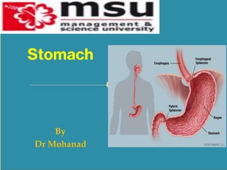
- 4. The stomach is divided into four regions: 1. The cardia, which surrounds the opening of the esophagus into the stomach. 2. The fundus of stomach, which is the area above the level of the cardial orifice. 3. The body of stomach, which is the largest region of the stomach. 4. The pyloric part, which is divided into the pyloric antrum and pyloric canal and is the distal end of the stomach. The stomach
- 5. The Stomach
- 6. The stomach Openings: Gastroesophageal: To esophagus Pyloric: To duodenum
- 7. The stomach Sphincters The cardiac sphincter (lower esophagus sphincter) closes off the top end of the stomach. The pyloric sphincter closes off the bottom.
- 8. The stomach Other features of the stomach include: The greater curvature, which is a point of attachment for the gastrosplenic ligament and the greater omentum The lesser curvature, which is a point of attachment for the lesser omentum.
- 9. The stomach Read about: Lesser omentum Greater omentum Layers of peritoneum attached to the stomach: Lesser omentum: attaches the liver to the lesser curvature. Greater omentum: attaches the greater curvature to the posterior body wall.
- 12. Arterial blood supply: 3 Branches Left Gastric Artery Supplies the cardia of the stomach and distal esophagus Splenic Artery Gives rise to 2 branches which help supply the greater curvature of the stomach Left Gastroepiploic Short GastricArteries Common Hepatic or Proper Hepatic Artery 2 major branches Right Gastric- supples a portion of the lesser curvature Gastroduodenal artery -helps supply greater curvature in conjunction with Left Gastroepiploic Artery Stomach Blood Supply
- 16. The main innervations are Left and Right Vagus Nerves. Stomach Innervations
- 17. Stomach Innervations Parasympathetic innervation of Stomach- Vagus Nerve 90% of fiber in vagal trunk is afferent (info transmitting from stomach to CNS). Sympathetic innervation of Stomach- Splanchnic Nerve Derived from spinal segement T5-T10
- 18. Stomach Microscopic Anatomy Four layers of stomach wall Mucosa Submucos Muscle Serosa
- 20. Stomach Microscopic Anatomy Mucosa: The first main layer. It consists of an epithelium. Lined by simple columnar epithelium The lamina propria composed of loose connective tissue It has gastric glands in it underneath, and a thin layer of smooth muscle called the muscularis mucosae.
- 21. Stomach Microscopic Anatomy Mucosa: This luminal surface is interrupted at intervals by gastric pits. Gastric pits formed by folded mucosa. Opening into these gastric pits are one or more gastric glands that have functional significance.
- 22. Stomach Microscopic Anatomy Mucosa: Four major types of secretory epithelial cells cover the surface of the stomach and extend down into gastric pits and glands: Mucous cells: secrete an alkaline mucus that protects the epithelium against shear stress and acid Parietal cells: secrete hydrochloric acid Chief cells: secrete pepsinogen, a proteolytic enzyme G cells: secrete the hormone gastrin
- 23. Stomach Microscopic Anatomy Mucosa: Mucous cells Parietal cells Chief cells G cells
- 24. Stomach Microscopic Anatomy Submucosa: o This layer lies over the mucosa. o It consists of fibrous connective tissue, separating the mucosa from the next layer. o The Meissner's plexus is in this layer.
- 25. Stomach Microscopic Anatomy Muscularis Externa: The muscularis externa in the stomach differs from that of other GI organs in that it has three layers of smooth muscle instead of two
- 26. Stomach Microscopic Anatomy Muscularis Externa: Inner Oblique Layer Middle Circular Layer Outer Longitudinal Layer
- 27. Stomach Microscopic Anatomy Serosa: This layer is over the muscularis externa, consisting of layers of connective tissue continuous with the peritoneum.
- 29. THE MUCOSAL BARRIER The mucosa of the stomach is exposed to the highly corrosive acidity of gastric juice. Gastric enzymes that can digest protein can also digest the stomach itself. The stomach is protected from self-digestion by the mucosal barrier. This barrier has several components. First, the stomach wall is covered by a thick coating of bicarbonate-rich mucus. This mucus forms a physical barrier, and its bicarbonate ions neutralize acid. Second, the epithelial cells of the stomach’s mucosa meet at tight junctions, which block gastric juice from penetrating the underlying tissue layers. Finally, stem cells located where gastric glands join the gastric pits quickly replace damaged epithelial mucosal cells, when the epithelial cells are shed. In fact, the surface epithelium of the stomach is completely replaced every 3 to 6 days.