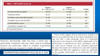
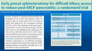
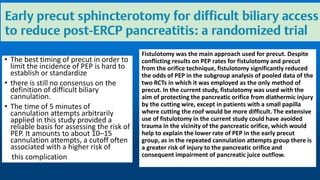
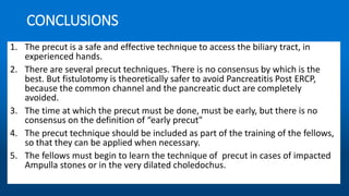
We review the most important articles above the differents Precut techiques: Fistulotomy, Papillotomy and Transpancreatic Sphincterotomy. The techique is safe and effective. And a brief comment about my experience in Fistulotomy, "No Post ERCP Pancreatitis because No touch the papilllary orifice"
22. Figure 2. The needle-knife tip is positioned at the junction of
the upper third and lower two thirds of the papillary mound. The
endoscopist is positioned to begin cutting in the downward
direction (modified technique).
Figure 3. The needle-knife cut is complete, with the ‘‘safety margin’’
between the cut and the papilla (outlined with a white line).
Early institution of pre-cutting for difficult biliary cannulation:a prospective study comparing conventional vs. a modified technique
23.
24.
25.
26. Comparison of early pre-cutting vs standard technique for
biliary cannulation in endoscopic retrograde
cholangiopancreatography: A personal experience
Kannikar Laohavichitra, Thawatchai Akaraviputh
World J Gastroenterol 2007 July 21; 13(27): 3734-3737
From January 2003 to December 2004
123 procedures (29.6%) with early pre-cutting
technique in case of difficult cannulation
If biliary access failed after 10 min or if more
than 3 pancreatic injections with contrast were
made (whichever occurred first), the early pre-
cutting technique with needle knife catheter
(MicroKnife XL, Boston Scientific, Natrick, USA
Endocut mode in the ERBE system
pure cutting mode
27. Comparison of early pre-cutting vs standard technique for
biliary cannulation in endoscopic retrograde
cholangiopancreatography: A personal experience
28.
29.
30.
31.
32.
33.
34.
35.
36. Evaluation of Early Precut with Needle-
Knife in Difficult Biliary Cannulation
During ERCP
Dig Dis Sci (2013) 58:3606–3610
Jian-hong Zhu • Qiang Liu • De-qing Zhang
Fig. 1 a–d Serial images show
the precut begins at the
ampullary upper lip and extends
upwards on the papillary mound
A total of 24 cases were abandoned due to
periampullary diverticulum or small papilla
from June 2007 to May 2011, and 56 of these
cases were carried out with precut biliary sphincterotomy
after the standard sphincterotomy had failed
37. Fig. 2 a–d The impacted stone
spontaneously passes after a
suprapaplillary fistulotomy was
performed
Evaluation of Early Precut with
Needle-Knife in Difficult Biliary
Cannulation During ERCP
Most precuts were performed without pancreatic
stent placement, starting from the papillary orifice
and proceeding at the 11 o’clock position upward over
the papillary mound (Fig. 1). In some clinical situations
where there was impacted stone at the ampulla of
Vater, a suprapapillary fistulotomy was performed
(Fig. 2), the needle was inserted towards 11 o’clock
orientation a few millimeters above the papillary
orifice, and the cutting was extended downwards
towards the orifice. If precut failed, the procedure was
abandoned.
38. Here, there are several points which need to be
noted when we are performing duodenum or papillary
precuts. First, PBS surgery is an injurious form of
manipulation, and brings with it a certain level of ignorance.
If those responsible for performance do not pay attention,
this will increase the risk of complications. Therefore,
operators must fully understand and carefully observe the
papillary organs and keep looking for formations or tumors.
Second, PBS should only be performed by highly trained
and well-experienced endoscopists, while, in addition, it is
particularly to be used only when the operator’s
professional intuition plus the patient’s condition means it is
required. Third, the incision should not be made too large
and the cutting should be gradually opened throughout the
precut process. It is worth pointing out that the blade cuts
in one direction only. In the event that a bile duct is cut, the
cutting should be immediately stopped. Only after standard
treatment is performed should one elongate the incision
Fourth, operators must be familiar with abdominal
anatomy. When the common bile duct is opened, it usually
appears white. At this point, sphincterotomy can be
attempted; if still problematic, the incision can be started
again at the white orifice, after which one should clearly
see the light pink epithelium. Fifth, during the course of
precut treatment, blood oozing from the incision could
make the field of vision murky and affect the cutting
operation. In this case, one may either spray the affected
area or wait for the blood to clot. Second cutting must be
avoided at all costs. And sixth, operators should be
proficient and capable of responding to unforeseen
complications or an emergency during a surgery
Evaluation of Early Precut with
Needle-Knife in Difficult Biliary
Cannulation During ERCP
39. Early precut fistulotomy for biliary access: time to
change the paradigm of “the later, the better”?
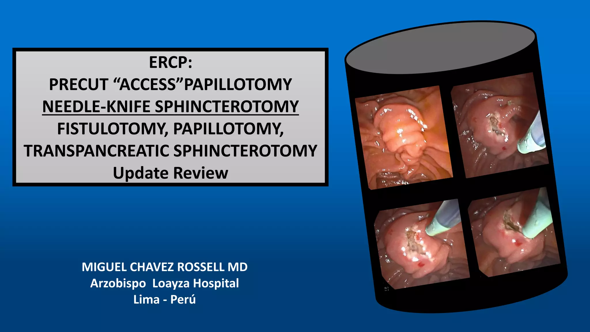
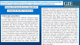
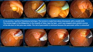
Needle-knife fistulotomy. NKF was performed by using a needle-knife
(Olympus KD-11Q; Olympus Corporation). After making a puncture in
the papilla above the orifice, the incision was made upward or
downward (depending on the position of the initial puncture), along the
axis of the bile duct, while maintaining at least a 3-mm distance from
the papillary orifice. The cut was slowly extended until the CBD was
exposed, followed by a small incision in the muscle. The CBD was then
cannulated directly with the closed needle-knife or with a papillotome
(wire guided) if the needle-knife did not slide. Once deep cannulation
was achieved, a cholangiogram was obtained by using low-osmolality,
nonionic contrast (Ultravist [iopromide]; Bayer Schering Pharma, Berlin,
Germany), and the necessary therapeutic maneuvers were performed.
• GASTROINTESTINAL
ENDOSCOPY
Volume 80, No. 4 :
2014
• Luís Lopes, MD,
Mário Dinis-Ribeiro,
41. Different Strategies for Transpancreatic Septotomy
and Needle Knife Infundibulotomy Due to the
Presence of Unintended Pancreatic Cannulation in
Difficult Biliary Cannulation
Gut and Liver, Vol. 9, No. 4, July 2015, pp. 534-539
Yoon Jung Lee, Yun Kyung Park, Min Ji Lee, Kyu Taek Lee,
The most widely practiced precut technique is needle
knife infundibulotomy, which is valuable if biliary
cannulation is not achieved for any duct. The
advantage of this technique is that it can minimize the
risk of perforation and pancreatitis, due to the
predefined upper extent of the cut and untouched
pancreatic duct.
needle knife infundibulotomy (NKI)
The papillary configuration was classified as follows:
normal, bulging, redundant, impacted stone, papillitis,
peripapillary diverticulum, abnormal location or small
orifice. The bulging type showed marked swelling of
the oral protrusion of papilla. The redundant type
showed the longitudinal shape of the papilla and the
orifice located in the lower portion of the papilla.
Abnormal location signified the deviated axis of
papilla from the 11 to 1 o’clock position or an unusual
location of the papilla which was leaning toward the
third portion of the duodenum.
When the biliary cannulation was not achieved within 5
minutes for any duct, NKI was performed. This technique
involves making a puncture into the papilla above the orifice to
avoid injuring the pancreatic duct and to minimize the rate of
post-ERCP pancreatitis, which is the most serious complication
of such procedures, and then cutting in the 11 to 12 o’clock
direction along the ridge of the common bile duct using a
needle knife papillotome. After a fistula to the bile duct was
made, the standard papillotome was repositioned to the biliary
duct and the sphincterotomy was extended
Precut sphincterotomy with a needle knife is performed either
by a precut papillotomy or by infundibulotomy. In the
papillotomy technique, the incision starts from the papillary
orifice, whereas in the fistulotomy technique, the incision starts
above the papillary orifice.
42. Different Strategies for Transpancreatic
Septotomy and Needle Knife Infundibulotomy
Due to the Presence of Unintended Pancreatic
Cannulation in Difficult Biliary Cannulation
In practice, the bulging type of papilla was a
good candidate for an infundibulotomy or the
modified techniques of needle knife precut.
So, future studies should examine the
adoption of precut method based on papillary
configuration in difficult biliary cannulation.
43. Different Strategies for Transpancreatic
Septotomy and Needle Knife Infundibulotomy
Due to the Presence of Unintended Pancreatic
Cannulation in Difficult Biliary Cannulation
52. Utility of needle-knife fistulotomy as an initial method of biliary cannulation to prevent post-ERCP
pancreatitis in a highly selected at-risk group: a single-arm prospective feasibility study
November 2016 Volume 84, Issue 5, Pages 808–813
53. Figure 1. Needle-knife fistulotomy technique. A, Major
duodenal papilla; B, small mucosal incision using a needle-
knife papillotome is performed in the 11 to 12 o’clock position
on the top of the infundibulum, avoiding touching the orifice;
C, sphincter of Oddi muscle of the bile duct is visible after the
incision; D, biliary cannulation is achieved by pricking the
stigma with a needle-knife.
Seven of the 62 patients were excluded due to periampullary
diverticulum (PAD) type I (n Z 1) or II (n Z 4),
stone located at the papilla orifice (n Z 1), and
choledochoduodenal fistula (n Z 1).
Thus, 55 patients were enrolled in this prospective study, all
of whom underwent ERCP and NKF.
54. Study participants
Between August 2013 and May 2015, 62 consecutive
patients were diagnosed as having CBD stones and
underwent ERCP for stone removal. They had at least one
of the following patient-related risk factors for PEP: young
age (<60 years), female, or normal CBD diameter (9 mm).
All patients were naive for ERCP and had no history of
pancreatitis. A prospective database was created for these
62 consecutive patients. Seven of the 62 patients were
excluded due to periampullary diverticulum (PAD) type I (n
Z 1) or II (n Z 4), stone located at the papilla orifice (n Z 1),
and choledochoduodenal fistula (n Z 1). Thus, 55 patients
were enrolled in this prospective study, all of whom
underwent ERCP and NKF.
The shape of the papilla was classified as hemispherical,
papillary, or flat according to the external appearance of
the major papilla
60. • The best timing of precut in order to
limit the incidence of PEP is hard to
establish or standardize
• there is still no consensus on the
definition of difficult biliary
cannulation.
• The time of 5 minutes of
cannulation attempts arbitrarily
applied in this study provided a
reliable basis for assessing the risk of
PEP. It amounts to about 10–15
cannulation attempts, a cutoff often
associated with a higher risk of
this complication
Fistulotomy was the main approach used for precut. Despite
conflicting results on PEP rates for fistulotomy and precut
from the orifice technique, fistulotomy significantly reduced
the odds of PEP in the subgroup analysis of pooled data of the
two RCTs in which it was employed as the only method of
precut. In the current study, fistulotomy was used with the
aim of protecting the pancreatic orifice from diathermic injury
by the cutting wire, except in patients with a small papilla
where cutting the roof would be more difficult. The extensive
use of fistulotomy in the current study could have avoided
trauma in the vicinity of the pancreatic orifice, which would
help to explain the lower rate of PEP in the early precut
group, as in the repeated cannulation attempts group there is
a greater risk of injury to the pancreatic orifice and
consequent impairment of pancreatic juice outflow.
61.
62.
63.
64.
65.
66.
67.
68.
69.
70. CONCLUSIONS
1. The precut is a safe and effective technique to access the biliary tract, in
experienced hands.
2. There are several precut techniques. There is no consensus by which is the
best. But fistulotomy is theoretically safer to avoid Pancreatitis Post ERCP,
because the common channel and the pancreatic duct are completely
avoided.
3. The time at which the precut must be done, must be early, but there is no
consensus on the definition of “early precut"
4. The precut technique should be included as part of the training of the fellows,
so that they can be applied when necessary.
5. The fellows must begin to learn the technique of precut in cases of impacted
Ampulla stones or in the very dilated choledochus.
71. In my practice, I perform Fistulotomy technique. The incision is made from above downward, with a needle knife.
The incision begin in the Midportion in the Ampulla of Vater, layer by layer , and it was stopped when pinkish mucosa
of the bile duct or bile juice was visible. The risk of pancreatitis is minimum because the pancreatic orifice is untouched