List of common ophthalmic abbreviations
•
0 likes•1,453 views

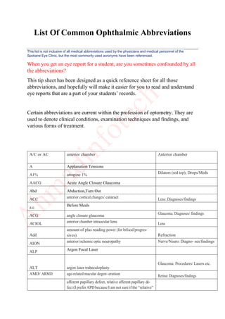
List Of Common Ophthalmic Abbreviations This list is not inclusive of all medical abbreviations used by the physicians and medical personnel of the Spokane Eye Clinic, but the most commonly used acronyms have been referenced. When you get an eye report for a student, are you sometimes confounded by all the abbreviations? This tip sheet has been designed as a quick reference sheet for all those abbreviations, and hopefully will make it easier for you to read and understand eye reports that are a part of your students’ records. Certain abbreviations are current within the profession of optometry. They are used to denote clinical conditions, examination techniques and findings, and various forms of treatment.
![APD, RAPD
is really needed)
APD by reverse testing/con- sensual response External
ARC Abnormal Retinal Correspondence
ARN Acute Retinal Necrosis
ARx autorefraction Refraction
ASC Anterior Subcapsular Cataract
AT, PFAT artificial tears, preservative- free artificial tears Drops/Meds
AV anterior vitreous
AVx anterior vitrectomy Glaucoma: Procedures/ Lasers etc.
Ax
axis
[+/- number sphere] + [number cylinder] x [0-180
axis]
Refraction
B Bilateral
b.i.d. or b.d. Twice Daily
BD Base Down
BCVA bestcorrectedvisualacuity Exam
BDR background diabetic retinopathy Retina: Diagnoses/findings
BI Base In
BILT IOOA Bilateral Inferior Oblique Over Action
BIO Binocular indirect ophthalmoscopy
BK band keratopathy Cornea: Diagnoses/ findings
BLR Bilateral Lateral Rectus
BMR Bilateral Medial Rectus
BO Base Out
BOT No Tears or Breaks in Retina
BRAO branch retinal artery occlusion Retina: Diagnoses/findings
BRVO branch retinal vein occlusion Retina: Diagnoses/findings
BSL Bandage Soft Contact Lens
BSV Binocular single vision
BV Binocular vision
BVD Back vertex distance
BVP Back vertex power
BU Base Up
C/D cup/disc ratio Fundus exam
C/F cell/flare (graded 1+ to 4+) Anterior chamber
C/S conjunctiva/sclera Slit lamp exam
C1 Cyclogyl (cyclopentolate) 1% Dilators (red top); Drops/Meds
CACG Chronic Angle Closure Glaucoma
Cat Cataract
CB ciliary body Anatomy
CBB ciliary body band Gonioscopy
CC Cortical Clouding, Cataract](data:image/gif;base64,R0lGODlhAQABAIAAAAAAAP///yH5BAEAAAAALAAAAAABAAEAAAIBRAA7)
Recommended
More Related Content
What's hot
What's hot (20)
Similar to List of common ophthalmic abbreviations
Similar to List of common ophthalmic abbreviations (20)
Recently uploaded
Recently uploaded (20)
List of common ophthalmic abbreviations
- 1. List Of Common Ophthalmic Abbreviations This list is not inclusive of all medical abbreviations used by the physicians and medical personnel of the Spokane Eye Clinic, but the most commonly used acronyms have been referenced. When you get an eye report for a student, are you sometimes confounded by all the abbreviations? This tip sheet has been designed as a quick reference sheet for all those abbreviations, and hopefully will make it easier for you to read and understand eye reports that are a part of your students’ records. Certain abbreviations are current within the profession of optometry. They are used to denote clinical conditions, examination techniques and findings, and various forms of treatment. A/C or AC anterior chamber Anterior chamber A Applanation Tensions A1% atropine 1% Dilators (red top); Drops/Meds AACG Acute Angle Closure Glaucoma Abd Abduction,Turn Out ACC anterior cortical changes/ cataract Lens: Diagnoses/findings a.c. Before Meals ACG angle closure glaucoma Glaucoma: Diagnoses/ findings ACIOL anterior chamber intraocular lens Lens Add amount of plus reading power (for bifocal/progres- sives) Refraction AION anterior ischemic optic neuropathy Nerve/Neuro: Diagno- ses/findings ALP Argon Focal Laser ALT argon laser trabeculoplasty Glaucoma: Procedures/ Lasers etc. AMD/ ARMD age-related macular degen- eration Retina: Diagnoses/findings afferent papillary defect, relative afferent papillary de- fect (IpreferAPDbecauseI am not sure if the “relative”
- 2. APD, RAPD is really needed) APD by reverse testing/con- sensual response External ARC Abnormal Retinal Correspondence ARN Acute Retinal Necrosis ARx autorefraction Refraction ASC Anterior Subcapsular Cataract AT, PFAT artificial tears, preservative- free artificial tears Drops/Meds AV anterior vitreous AVx anterior vitrectomy Glaucoma: Procedures/ Lasers etc. Ax axis [+/- number sphere] + [number cylinder] x [0-180 axis] Refraction B Bilateral b.i.d. or b.d. Twice Daily BD Base Down BCVA bestcorrectedvisualacuity Exam BDR background diabetic retinopathy Retina: Diagnoses/findings BI Base In BILT IOOA Bilateral Inferior Oblique Over Action BIO Binocular indirect ophthalmoscopy BK band keratopathy Cornea: Diagnoses/ findings BLR Bilateral Lateral Rectus BMR Bilateral Medial Rectus BO Base Out BOT No Tears or Breaks in Retina BRAO branch retinal artery occlusion Retina: Diagnoses/findings BRVO branch retinal vein occlusion Retina: Diagnoses/findings BSL Bandage Soft Contact Lens BSV Binocular single vision BV Binocular vision BVD Back vertex distance BVP Back vertex power BU Base Up C/D cup/disc ratio Fundus exam C/F cell/flare (graded 1+ to 4+) Anterior chamber C/S conjunctiva/sclera Slit lamp exam C1 Cyclogyl (cyclopentolate) 1% Dilators (red top); Drops/Meds CACG Chronic Angle Closure Glaucoma Cat Cataract CB ciliary body Anatomy CBB ciliary body band Gonioscopy CC Cortical Clouding, Cataract
- 3. cc with refractive correction Exam CCT/Pachy central corneal thickness/ pachymetry CE cataract extraction CF @ XX ft counts fingers (specify distance) Exam CHBL Check / Change / Bandage Lens CL/SCL/ HCL contact lens/soft .…/Hard .… Other CL THERA Contact Lens Therapeutic Contacts CM Cyclomydril (for peds patients) Dilators (red top); Drops/Meds CME cystoid macular edema Retina: Diagnoses/findings CNV choroidal neovascularization Retina: Diagnoses/findings CNVM choroidal neovascular membrane Retina: Diagnoses/findings COAG chronic open angle glaucoma Glaucoma: Diagnoses/ findings CORAB Corneal Abrasion C/O Complications c/o Complain of CPC cyclophotocoagulation Glaucoma: Procedures/ Lasers etc. CPM Continue Present Management CRA Chorioretinal Atrophy CRAO central retinal artery occlusion Retina: Diagnoses/findings CRVO central retinal vein occlusion Retina: Diagnoses/findings CRx cycloplegic refraction Refraction Cryo cryotherapy Glaucoma: Procedures/ Lasers etc. CSCR central serous chorioretin- opathy Retina: Diagnoses/findings CSDME Clinically Significant Diabetic Macular Edema CSM central, steady, maintained Exam CSME clinically significant macular edema (for diabetes) Retina: Diagnoses/findings CSR Central Serous Retinopathy CT Cover Test CVF confrontation visual fields External CWS cotton wool spot Retina: Diagnoses/findings Cyl cylinder Refraction D Diopter D&Q deep and quiet Anterior chamber D/C Discontinue d/c Discharge D/M/V/P disc/macula/vessels/periphery Fundus exam DALK deep anterior lamellar ker- atoplasty Cornea : Procedures/ Lasers etc. DBH dot blot heme (hemorrhage) Retina: Diagnoses/findings DD Disc Diameters DF Descemet’s fold Cornea: Diagnoses/find- ings DFE dilated fundus exam Fundus exam Dilators (red top) Drops/Meds DLK diffuse lamellar keratitis Cornea : Procedures/ Lasers etc.
- 4. DM Diabetes Mellitus DME Diabetic Macular Edema DMR Double Maddox Rod DMVP Disc,Macula,Vessels,and Periphery DSEK Endothelial Keratoplasty DV, dVA Distance Visual Acuity E’ Esophoria at Near E Endolaser E (Circled) Erythromycin E(T) intermittent esotropia distance Alignment E esophoria Alignment ECCE extracapsular cataract extraction Procedures/Lasers etc. ECP Endoscopic Cyclophotocoagulation ED epithelial defect Cornea: Diagnoses/find- ings EKC epidemic keratoconjunctivitis Cornea: Diagnoses/find- ings EOG electrooculogram Tests EOM extraocular muscles/ movement ERG electroretinogram Tests ERM epiretinal membrane Retina: Diagnoses/findings ET Esotropia for Distance Alignment ET` Esotropia for Near EUA Exam under Anesthesia F+F fixes and follows Exam F/U Follow-up FB Foreign Body FHx Family History Flt Flat FANG fluorescein angiography Tests Focal focal laser photocoagulation Retina: Procedures/ Lasers etc. FTMH Full Thickness Macular Hole Fuchs Fuchs Endothelial Corneal dystrophy GCA Giant cell arteritis Nerve/Neuro: Diagnoses/findings GDX Diagnostic Glaucoma Test gl Glasses GLC Glaucoma GLREF Glasses Refraction GRREM Growth Removal GT Glasses trouble gtt(s) Drop(s) GVF Goldmann visual field Tests HA homatropine Dilators (red top); Drops/Meds HA Headache HARC Harmonious abnormal retinal correspondence
- 5. Hx, H/O History of HM @ XX ft hand motion (specify dis- tance) Exam HoT hypotropia (add an apostrophe to indi- cate at near – eg. ET’ means esotropia at near) Alignment HSV Herpes Simplex Virus HT hypertropia Alignment HVF Humphrey visual field (usu- ally 30-2; need to specify if 10-2 or red target, etc) Tests I iris Iris ICCE intracapsular cataract extrac- tion Procedures/Lasers etc. IK interstitial keratitis Cornea: Diagnoses/ findings I.P.D. Interpupillary Distance IO inferior oblique Anatomy IOFB Intraocular Foreign Body IOL Intra-ocular lens ION ischemic optic neuropathy Nerve/Neuro: Diagnoses/findings IOP intraocular pressure External IR inferior Anatomy IRMA intraretinal microvascular anomalies Retina: Diagnoses/findings JOAG juvenile open angle glau- coma Glaucoma: Diagnoses/ findings K Cornea , keratometry slit lamp exam KC or KCN keratoconus Cornea: Diagnoses/ findings KCS keratoconjunctivitis sicca Cornea: Diagnoses/ findings KP keratic precipitate Cornea: Diagnoses/ findings L lens Lens L/L lids/lashes Slit lamp exam Lag lid lag External LASEK laser epithelial keratomileusis Cornea : Procedures/ Lasers etc. LASIK laser in situ keratomileusis, also laser-assisted in situ keratomileusis (Hofstetter) Cornea : Procedures/ Lasers etc. LCA Leber’s congenital amaurosis Nerve/Neuro: Diagnoses/findings LF levator function External LHON Leber’s hereditary optic neuropathy Nerve/Neuro: Diagnoses/findings LI/LPI laser iridotomy/laser pe- ripheral iridotomy Glaucoma: Procedures/ Lasers etc. LOE Loss of Eye LOV Loss of Vision LTG Low Tension Glaucoma LP c projection light perception with projec- tion Exam LP s projection light perception without projection Exam LP light perception Exam
- 6. LR lateral rectus Anatomy LVA Low vison aid M1 Mydriacyl (tropicamide) 1% Dilators (red top); Drops/Meds M Membrane Dissection MACCK Macular Degeneration Check MACEV Macular Degeneration Evaluation MG myasthenia gravis Nerve/Neuro: Diagnoses/findings MGD meibomian gland dysfunction Lids: Diagnoses/findings MGP meibomian gland plugging Lids: Diagnoses/findings MH Macular Hole MP/Mx membrane peel/mem- branectomy Retina: Procedures/ Lasers etc. MR medial rectus , Maddox rod Anatomy MRD1 margin to reflex distance 1 External MRD2 margin to reflex distance 2 External MRx manifest refraction Refraction MS multiple sclerosis Nerve/Neuro: Diagnoses/findings M.Wing Maddox Wing N2.5/N10 Neo-Synephrine (phenyleph- rine) 2.5% or 10% Dilators (red top); Drops/Meds NAION nonarteritic ischemic optic neuropathy Nerve/Neuro: Diagnoses/findings NAG Narrow Angle Glaucoma NCT Non-contact tonometer NI no improvement Exam Nl,nl Normal NLD Nasolacrimal Duct/ Obstruction NLDO Obstructed Nasolacrimal Duct NLP no light perception Exam NP New Patient NPA Near Point of Accommodation NPC Near Point of Convergence NPDR non-proliferative diabetic retinopathy Retina: Diagnoses/findings NRC Normal Retinal Correspondence NRx near refraction Refraction NS nuclear sclerosis,Cataract Lens: Diagnoses/findings NTG/LTG normal/low tension glau- coma Glaucoma: Diagnoses/ findings NV or nVA Near Vision NVA neovascularization of the angle Gonioscopy NVD neovascularization of the disc Retina: Diagnoses/findings NVE neovascularization elsewhere Retina: Diagnoses/findings NVG neovascular glaucoma Glaucoma: Diagnoses/ findings NVI neovascularization of the iris Iris NWT Normal wearing time OAG Open Angle Glaucoma
- 7. OCT NFL OCT of nerve fiber layer (optic nerve evaluation) Tests OCT optical coherence tomog- raphy Tests oculent Eye Ointment OD right eye Anatomy OHT ocular hypertension Glaucoma: Diagnoses/ findings ON Optic Nerve ONH Optic Nerve Head Or,Orx Over-Refraction OS Oculus sinister (left eye) Anatomy OT Ocular Tension OU Oculus uterque (both eyes) Anatomy PAS peripheral anterior synechiae Gonioscopy PBK pseudophakic bullous ker- atopathy Cornea: Diagnoses/ findings PCC posterior cortical changes Lens: Diagnoses/findings PCIOL posterior chamber Lens PCT Prism Cover Test PCO posterior capsular opacity (post-cataract patients) Lens: Diagnoses/findings PD pupillary distance or prism diopter Refraction PDG Pigment Dispersion Glaucoma PDR proliferative diabetic retin- opathy Retina: Diagnoses/findings PDT photodynamic therapy Retina: Procedures/ Lasers etc. PED Pigment Epithelial Detachment PEE punctate epithelial erosion Cornea: Diagnoses/ findings PEK punctate epithelial keratopa- thy/keratitis OR photo-electronic kerato- scope (Hofstetter) Cornea: Diagnoses/ findings PF palpebral fissure External PG Pigmentary Glaucoma PH pinhole visual acuity Exam Phaco/ ACIOL or Phaco/ PCIOL phaco with anterior chamber intraocular lens or posterior chamber intraocu- lar lens Procedures/Lasers etc. Phaco phacoemulsification Procedures/Lasers etc. PHNI pinhole no improvement Exam PION posterior ischemic optic neuropathy Nerve/Neuro: Diagno- ses/findings PK penetrating keratoplasty Cornea: Diagnoses/find- ings PKP penetrating keratoplasty Cornea : Procedures/ Lasers etc. PL Perception of light POAG/ OAG primary open angle glau- coma/open angle glaucoma Glaucoma: Diagnoses/ findings POHS presumed ocular histoplas- mosis Retina: Diagnoses/findings PPA peripapillary atrophy Nerve/Neuro: Diagnoses/findings PPV/Vx parsplanavitrectomy/vitrec- tomy Retina: Procedures/ Lasers etc.
- 8. PR Pneumatic Retinopexy PRK photorefractive keratectomy Cornea : Procedures/ Lasers etc. PRP pan-retinal photocoagulation Retina: Procedures/ Lasers etc. PRN As Needed PS posterior synechiae (desig- nate location/clock hours) Iris PSC posterior subcapsular cataract Lens: Diagnoses/findings Pt Patient PVD posterior vitreous detach- ment Retina: Diagnoses/findings PXE pseudoxanthoma elasticum Nerve/Neuro: Diagnoses/findings PXF pseudoexfoliation Glaucoma: Diagnoses/ findings q.2h. Every 2 Hours q.h. Every Hour q.i.d. Four Times Daily q.s. Quantity Sufficient qd Daily, Once a Day qhs Nightly qo Every Other R/R round/reactive Iris R retinoscopy Refraction RAPD Relative Afferent Pupillary Defect Rc cycloplegic retinoscopy Refraction RD retinal detachment Retina: Diagnoses/findings REF REFRACTION Ret. Retinoscopy
- 9. RNFL Retinal Nerve fibre Layer RP retinitis pigmentosa Retina: Diagnoses/findings RPE retinal pigment epithelium fundus exam RK Radial Keratotomy Rx Prescription SB scleral buckle Retina: Procedures/ Lasers etc. sc without refractive correction Exam SCH Subconjunctival Hemorrhage SiO Silicone IOL SL Schwalbe’s line Gonioscopy SLE slit lamp exam Slit lamp exam SLK superior limbic keratocon- junctivitis Cornea: Diagnoses/ findings SLT selective laser trabeculo- plasty Glaucoma: Procedures/ Lasers etc. SO superior oblique Anatomy SOAG secondary open angle glaucoma Glaucoma: Diagnoses/ findings Sph sphere Refraction SPK superficial punctate ker- atopathy/keratitis Cornea: Diagnoses/ findings SR superior rectus Anatomy SRF Subretinal Fluid SRNV subretinal neovascularization Retina: Diagnoses/findings SRNVM subretinal neovascular membrane Retina: Diagnoses/findings SS scleral spur Gonioscopy ST Schiotz tension Sub heme Subconjunctival Hemorrhage Sx Surgery T tonometry Tonometry Ta applanation (Goldmann) tonometry Tonometry t.i.d. Three Times Daily Tapp Pressure by Applanation TBU Tear Break Up TM trabecular meshwork Gonioscopy TON traumatic optic neuropathy Nerve/Neuro: Diagnoses/findings Tono Tonometer Pressure Check
- 10. topo Topography Tp pneumotonometer tonometry Trab trabeculectomy Glaucoma: Procedures/ Lasers etc. TRD tractional detachment Retina: Diagnoses/findings Ttono Pressure by Tonopen Tx Treatment U/S Ultrasound UDFE undilated fundus exam fundus exam UGH uveitis glaucoma hyphema syndrome Glaucoma: Diagnoses/ findings um Micron Ung Ointment V,Va,VA Vision or visual acuity Exam VAc or VAcc Visual Acuity with Correction Vas or VAsc Visual Acuity without Correction VF visual field Tests VH vitreous hemorrhage Retina: Diagnoses/findings VMT vitreomacular traction Retina: Diagnoses/findings W4D Worth 4 Dots WC/LS warmcompresses/lidscrubs Drops/Meds WD Working Distance WRx wearing Rx (currently worn eyeglass/contact lens pre- scription) Refraction X(T) intermittent exotropia Alignment X,XP exophoria Alignment XT exotropia Alignment YAG Yytrium Aluminum Garnet Laser Δ prism diopter Refraction Note :- This list is not inclusive of all medical abbreviations used by the physicians and medical personnel of the Spokane Eye Clinic, but the most commonly used acronyms have been referenced.