SlideShare a Scribd company logo
1 of 53
LECTURE 99From Neurons to the Nervous System to the Brain
The neuron's place as the primary functional unit of the nervous
system was first recognized in the late 19th century by the
Spanish anatomist Santiago Ramón y Cajal (1852-1934), a
neuroscientist and pathologist specializing in neuroanatomy
and, especially, the central nervous system.
In 1888 Ramón y Cajal published a paper about the pigeon
cerebellum. In this paper, he stated that he could not find
evidence for cross connections (anastomosis) between axons
and dendrites and called each nervous element "an absolutely
autonomous canton." This became known as the neuron
doctrine, one of the central tenets of modern neurobiology.
Above is his 1899 drawing of neurons in the pigeon cerebellum.
This Lecture focuses on the chemistry of neurons, specialized
cells that transmit chemical and electrical signals to facilitate
communication between the brain and the body. In learning
about a new field, one can get befuddled very quickly with the
jargon used by experts in the field and get lost, not being able
“to see the forest for the trees.” So, before each section, I give
a summary of Key Points and Key Terms you will need to
understand that section. At the expense of “over kill,” I have
repeated earlier “points” and “terms” at subsequent points in the
Lecture so that you won’t have to “backtrack” to figure out
what is going on.
Neurons are specialized cells that transmit chemical and
electrical signals in the brain. They are the basic building
blocks of the central nervous system.
Key Points:
· Neurons are specialized cells that transmit chemical and
electrical signals in the brain; they are the basic building blocks
of the central nervous system.
· The primary components of the neuron are the soma (cell
body), the axon (a long slender projection that conducts
electrical impulses away from the cell body), dendrites (tree-
like structures that receive messages from other neurons), and
synapses (specialized junctions between neurons).
· Some axons are covered with myelin, a fatty material that acts
as an insulator and conductor to speed up the process of
communication.
· Sensory neurons are neurons responsible for converting
external stimuli from the environment into corresponding
internal stimuli.
· Motor neurons are neurons located in the central nervous
system (CNS); they project their axons outside of the CNS to
directly or indirectly control muscles.
· Interneurons act as the “middle men” between sensory and
motor neurons, which convert external stimuli to internal
stimuli and control muscle movement, respectively.
Key Terms:
· glial cell: Non-neuronal cells that provide structure and
support to neurons.
· synapse: The junction between the terminal of a neuron and
either another neuron or a muscle or gland cell, over which
nerve impulses pass.
· myelin: A white, fatty material composed of lipids and
lipoproteins that surrounds the axons of nerves and facilitates
swift communication.
· nodes of Ranvier: Periodic gaps in the myelin sheath where
the signal is recharged as it moves along the axon.
The neuron is the basic building block of the brain and central
nervous system. The brain is made up entirely of neurons and
glial cells. Nearly 86 billion neurons work together within the
nervous system to communicate with the rest of the body.
Fun facts: The Milky Way galaxy has an estimated 100 billion
stars. It is estimated that there are 10 trillion galaxies in the
observable universe. Multiplying that by the Milky Way's
estimated 100 billion stars results in a large number indeed:
1,000,000,000,000,000,000,000,000
stars.
Given this astronomically-large value, the probability that there
is (at least) one star with a planet “just the right distance” from
that star is quite high. Whether there are large bodies of H2O
on this planet is another matter but, if so, assuming the same
distribution of elements as on planet Earth, the chemistry of
Carbon can kick in and life on that planet is possible.
Neurons are responsible for consciousness and thought to pain
and hunger. You need almost as many neurons to function as
there are stars in our Milky Way.
Structures of a Neuron
In addition to having all the normal components of a cell
(nucleus, organelles, etc.), neurons also contain unique
structures for receiving and sending the electrical signals that
make neuronal communication possible.
The structure of a neuron: The above image shows the basic
structural components of an average neuron, including the
dendrite, cell body, nucleus, Node of Ranvier, myelin sheath,
Schwann cell, and axon terminal.
Dendrite
Dendrites are branch-like structures extending away from the
cell body, and their job is to receive messages from other
neurons and allow those messages to travel to the cell body.
Although some neurons do not have any dendrites, other types
of neurons have multiple dendrites. Dendrites can have small
protrusions called dendritic spines, which further increase
surface area for possible connections with other neurons.
Cell Body
Like other cells, each neuron has a cell body (or soma) that
contains a nucleus, smooth and rough endoplasmic reticulum,
Golgi apparatus, mitochondria, and other cellular components.
Axon
An axon is a tube-like structure that carries an electrical
impulse from the cell body (or from another cell’s dendrites) to
the structures at opposite end of the neuron—axon terminals,
which can then pass the impulse to another neuron. The cell
body contains a specialized structure, the axon hillock, which
serves as a junction between the cell body and the axon.
Synapse
The synapse is the chemical junction between the axon
terminals of one neuron and the dendrites of the next. It is a gap
where specialized chemical interactions can occur, rather than
an actual structure.
Function of a Neuron
The specialized structure and organization of neurons allows
them to transmit signals in the form of electric impulses from
the brain to the body and back. Individually, neurons can pass a
signal all the way from their own dendrites to their own axon
terminals. At a higher level, neurons are organized in long
chains, allowing them to pass signals very quickly from one to
the other. One neuron’s axon will connect chemically to another
neuron’s dendrite at the synapse between them. Electrically
charged chemicals flow from the first neuron’s axon to the
second neuron’s dendrite, and that signal will then flow from
the second neuron’s dendrite, down its axon, across a synapse,
into a third neuron’s dendrites, and so on.
This is the basic chain of neural signal transmission, which is
how the brain sends signals to the muscles to make them move,
and how sensory organs send signals to the brain. It is important
that these signals can happen quickly, and they do.
Think of how fast you drop a hot potato—before you even
realize it is hot. This is because the sense organ (in this case,
the skin) sends the signal “This is hot!” to neurons with very
long axons that travel up the spine to the brain. If this didn’t
happen quickly, people would burn themselves.
Other Structures
Dendrites, cell bodies, axons, and synapses are the basic parts
of a neuron, but other important structures and materials
surround neurons to make them more efficient.
Myelin Sheath
Some axons are covered with myelin, a fatty material that wraps
around the axon to form the myelin sheath. This external
coating functions as insulation to minimize dissipation of the
electrical signal as it travels down the axon. Myelin’s presence
on the axon greatly increases the speed of conduction of the
electrical signal, because the fat prevents any electricity from
“leaking out”. This insulation is important, as the axon from a
human motor neuron can be as long as a meter—from the base
of the spine to the toes. Periodic gaps in the myelin sheath are
called nodes of Ranvier. At these nodes, the signal is
“recharged” as it travels along the axon.
Glial Cells
The myelin sheath is not actually part of the neuron. Myelin is
produced by glial cells (or simply glia, or “glue” in Greek),
which are non-neuronal cells that provide support for the
nervous system. Glia function to hold neurons in place (hence
their Greek name), supply them with nutrients, provide
insulation, and remove pathogens and dead neurons. In the
central nervous system, the glial cells that form the myelin
sheath are called oligodendrocytes; in the peripheral nervous
system, they are called Schwann cells.
Neuron in the central nervous system: This neuron diagram also
shows the oligodendrocyte, myelin sheath, and nodes of
Ranvier.
Types of Neurons
There are three major types of neurons: sensory neurons, motor
neurons, and interneurons. All three have different functions,
but the brain needs all of them to communicate effectively with
the rest of the body (and vice versa).
Sensory Neurons
Sensory neurons are neurons responsible for converting external
stimuli from the environment into corresponding internal
stimuli. They are activated by sensory input, and send
projections to other elements of the nervous system, ultimately
conveying sensory information to the brain or spinal cord.
Unlike the motor neurons of the central nervous system (CNS),
whose inputs come from other neurons, sensory neurons are
activated by physical stimuli (such as visible light, sound, heat,
physical contact, etc.) or by chemical signals (such as smell and
taste).
Most sensory neurons are pseudounipolar, meaning they have an
axon that branches into two extensions—one connected to
dendrites that receive sensory information and another that
transmits this information to the spinal cord.
Motor Neurons
Motor neurons are neurons located in the central nervous
system, and they project their axons outside of the CNS to
directly or indirectly control muscles. The interface between a
motor neuron and muscle fiber is a specialized synapse called
the neuromuscular junction. The structure of motor neurons
is multipolar, meaning each cell contains a single axon and
multiple dendrites. This is the most common type of neuron.
Interneurons
Interneurons are neither sensory nor motor; rather, they act as
the “middle men” that form connections between the other two
types. Located in the CNS, they operate locally, meaning their
axons connect only with nearby sensory or motor neurons.
Interneurons can save time and therefore prevent injury by
sending messages to the spinal cord and back instead of all the
way to the brain. Like motor neurons, they are multipolar in
structure.
Stages of the Action Potential
Neural impulses occur when a stimulus depolarizes a cell
membrane, prompting an action potential which sends an “all or
nothing” signal.
Key Points:
· The neurons (or excitable nerve cells) of the nervous system
conduct electrical impulses, or signals, that serve as
communication between sensory receptors, muscles and glands,
and the brain and spinal cord.
· An action potential occurs when an electrical signal disrupts
the original balance of Na+ and K+ within a cell membrane,
briefly depolarizing the concentrations of each.
· An electrical impulse travels along the axon via depolarized
voltage-gated ion channels in the membrane, and can either
“jump” along a myelinated area or travel continuously along an
unmyelinated area.
· While an action potential is being generated by a cell, no other
action potential may be generated until the cell’s channels
return to their resting state.
· Action potentials generated by neural impulses are “all or
nothing,” meaning the signal reaches the threshold for
communication or it doesn’t. No signal is stronger or weaker
than another.
Key Terms:
· polarity: The spatial differences in the shape, structure, and
function of cells. Almost all cell types exhibit some sort of
polarity, which enables them to carry out specialized functions.
· action potential: A short-term change in the electrical
potential that travels along a cell, such as a nerve or muscle
fiber, and allows nerves to communicate.
· neural impulse: The signal transmitted along a nerve fiber,
either in response to a stimulus (such as touch, pain, or heat), or
as an instruction from the brain (such as causing a muscle to
contract).
· resting potential: The nearly latent membrane potential of
inactive cells.
Neural Impulses in the Nervous System
The central nervous system (CNS) goes through a three-step
process when it functions: sensory input, neural processing, and
motor output. The sensory input stage is when the neurons (or
excitable nerve cells) of the sensory organs are excited
electrically. Neural impulses from sensory receptors are sent to
the brain and spinal cord for processing. After the brain has
processed the information, neural impulses are then conducted
from the brain and spinal cord to muscles and glands, which is
the resulting motor output.
A neuron affects other neurons by releasing a neurotransmitter
that binds to chemical receptors. The effect upon the
postsynaptic (receiving) neuron is determined not by the
presynaptic (sending) neuron or by the neurotransmitter itself,
but by the type of receptor that is activated.
A neurotransmitter can be thought of as a key, and a receptor as
a lock: the key unlocks a certain response in the postsynaptic
neuron, communicating a particular signal. However, in order
for a presynaptic neuron to release a neurotransmitter to the
next neuron in the chain, it must go through a series of changes
in electric potential.
Stages of Neural Impulses
“Resting potential ” is the name for the electrical state when a
neuron is not actively being signaled. A neuron at resting
potential has a membrane with established amounts of sodium
(Na+) and potassium (K+) ions on either side, leaving the inside
of the neuron negatively charged relative to the outside.
The action potential is a rapid change in polarity that moves
along the nerve fiber from neuron to neuron. In order for a
neuron to move from resting potential to action potential—a
short-term electrical change that allows an electrical signal to
be passed from one neuron to another—the neuron must be
stimulated by pressure, electricity, chemicals, or another form
of stimuli. The level of stimulation that a neuron must receive
to reach action potential is known as the threshold of excitation,
and until it reaches that threshold, nothing will happen.
Different neurons are sensitive to different stimuli, although
most can register pain.
The action potential has several stages.
1. Depolarization: A stimulus starts the depolarization of the
membrane. Depolarization is caused when positively charged
sodium ions rush into a nerve cell. As these positive ions rush
in, the membrane of the stimulated cell reverses its polarity so
that the outside of the membrane is negative relative to the
inside.
2. Repolarization. Once the electric gradient has reached the
threshold of excitement, the “downswing” of repolarization
begins. The channels that let the positive sodium ion channels
through close up, while channels that allow positive potassium
ions open, resulting in the release of positively charged
potassium ions from the neuron. This expulsion acts to restore
the localized negative membrane potential of the cell, bringing
it back to its normal voltage.
3. Refractory Phase. The refractory phase takes place over a
short period of time after the depolarization stage. Shortly after
the sodium gates open, they close and go into an inactive
conformation. The sodium gates cannot be opened again until
the membrane is repolarized to its normal resting potential. A
sodium-potassium pump returns sodium ions to the outside and
potassium ions to the inside. During the refractory phase this
particular area of the nerve cell membrane cannot be
depolarized. Therefore, the neuron cannot reach action potential
during this “rest period.”
Action potentials: A neuron must reach a certain threshold in
order to begin the depolarization step of reaching the action
potential.
This process of depolarization, repolarization, and recovery
moves along a nerve fiber from neuron to neuron like a very
fast wave. While an action potential is in progress, another
cannot be generated under the same conditions. In unmyelinated
axons (axons that are not covered by a myelin sheath), this
happens in a continuous fashion because there are voltage-gated
channels throughout the membrane. In myelinated axons (axons
covered by a myelin sheath), this process is described as
saltatory because voltage-gated channels are only found at the
nodes of Ranvier, and the electrical events seem to “jump” from
one node to the next.
Saltatory conduction is faster than continuous conduction. The
diameter of the axon also makes a difference, as ions diffusing
within the cell have less resistance in a wider space. Damage to
the myelin sheath from disease can cause severe impairment of
nerve-cell function. In addition, some poisons and drugs
interfere with nerve impulses by blocking sodium channels in
nerves. More on this point in the next Lecture.
All-or-none Signals
The amplitude of an action potential is independent of the
amount of current that produced it. In other words, larger
currents do not create larger action potentials. Therefore, action
potentials are said to be all-or-none signals, since either they
occur fully or they do not occur at all. The frequency of action
potentials is correlated with the intensity of a stimulus. This is
in contrast to receptor potentials, whose amplitudes are
dependent on the intensity of a stimulus.
Reuptake
Reuptake refers to the reabsorption of a neurotransmitter by a
presynaptic (sending) neuron after it has performed its function
of transmitting a neural impulse. Reuptake is necessary for
normal synaptic physiology because it allows for the recycling
of neurotransmitters and regulates the neurotransmitter level in
the synapse, thereby controlling how long a signal resulting
from neurotransmitter release lasts.
Mechanics of the Action Potential
The synapse is the site at which a chemical or electrical
exchange occurs between the presynaptic and postsynaptic cells.
Key Points:
· Receptors are pores that admit chemical or electrical signals
into the postsynaptic cell. There are two main types of receptor:
ligand-gated ion channels, which receive neurostransmitters,
and g-protein coupled receptors, which do not.
· There are two types of possible reactions at the synapse: a
chemical reaction or an electrical reaction.
· During a chemical reaction, neurotransmitters trigger the
opening of ligand-gated ion channels on the membrane of the
postsynaptic cell, resulting in a modification of the cell’s
interior chemical composition and, in some cases, physical
structure.
· In an electrical reaction, the electrical charge of one cell is
influenced by another.
· Although electrical synapses yield faster reactions, chemical
synapses result in stronger, more complex changes to the
postsynaptic cell.
Key Terms:
· vesicle: A membrane-bound compartment found in a cell.
· action potential: A short-term change in the electrical
potential that travels along a cell, such as a nerve or muscle
fiber, and allows nerves to communicate.
· depolarization: The act of depriving of polarity, or the result
of such action; reduction to an unpolarized condition.
· membrane potential: The voltage across the cell membrane,
with the inside relative to the outside.
Synapses
The synapse is the junction where neurons trade information. It
is not a physical component of a cell but rather a name for the
gap between two cells: the presynaptic cell (giving the signal)
and the postsynaptic cell (receiving the signal). There are two
types of possible reactions at the synapse—chemical or
electrical. During a chemical reaction, a chemical called a
neurotransmitter is released from one cell into another. In an
electrical reaction, the electrical charge of one cell is influenced
by the charge an adjacent cell.
The electrical response of a neuron to multiple synaptic inputs:
Synaptic responses summate in order to bring the postsynaptic
neuron to the threshold of excitation, so it can fire an action
potential (represented by the peak on the chart).
All synapses have a few characteristics in common:
· Presynaptic cell: a specialized area within the axon of the
giving cell that transmits information to the dendrite of the
receiving cell.
· Synaptic cleft: the small space at the synapse that receives
neurotransmitters.
· G-protein coupled receptors: receptors that sense molecules
outside the cell and thereby activate signals within it.
· Ligand-gated ion channels: receptors that are opened or closed
in response to the binding of a chemical messenger.
· Postsynaptic cell: a specialized area within the dendrite of the
receiving cell that contains receptors designed to process
neurotransmitters.
The Electrical Synapse
The stages of an electrical reaction at a synapse are as follows:
1. Resting potential. The membrane of a neuron is normally at
rest with established concentrations of sodium ions (Na+) and
potassium ions (K+) on either side. The membrane potential (or,
voltage across the membrane) at this state is -70 mV, with the
inside being negative relative to the outside.
2. Depolarization. A stimulus begins the depolarization of the
membrane. Depolarization, also referred to as the “upswing,”
occurs when positively charged sodium ions (Na+) suddenly
rush through open sodium gates into a nerve cell. If the
membrane potential reaches -55 mV, it has reached the
threshold of excitation. Additional sodium rushes in, and the
membrane of the stimulated cell actually reverses its polarity so
that the outside of the membrane is negative relative to the
inside. The change in voltage stimulates the opening of
additional sodium channels (called a voltage-gated ion channel),
providing what is known as a positive feedbackloop. Eventually,
the cell potential reaches +40 mV, or the action potential.
3. Repolarization. The “downswing” of repolarization is caused
by the closing of sodium ion channels and the opening of
potassium ion channels, resulting in the release of positively
charged potassium ions (K+) from the nerve cell. This expulsion
acts to restore the localized negative membrane potential of the
cell.
4. Refractory Phase. The refractory phase is a short period of
time after the repolarization stage. Shortly after the sodium
gates open, they close and go into an inactive conformation
where the cell’s membrane potential is actually even lower than
its baseline -70 mV. The sodium gates cannot be opened again
until the membrane has completely repolarized to its normal
resting potential, -70 mV. The sodium-potassium pump returns
sodium ions to the outside and potassium ions to the inside.
During the refractory phase this particular area of the nerve cell
membrane cannot be depolarized; the cell cannot be excited.
The Chemical Synapse
The process of a chemical reaction at the synapse has some
important differences from an electrical reaction. Chemical
synapses are much more complex than electrical synapses,
which makes them slower, but also allows them to generate
different results. Like electrical reactions, chemical reactions
involve electrical modifications at the postsynaptic membrane,
but chemical reactions also require chemical messengers, such
as neurotransmitters, to operate.
Neuron & chemical synapse: This image shows electric
impulses traveling between neurons; the inset shows a chemical
reaction occurring at the synapse.
A basic chemical reaction at the synapse undergoes a few
additional steps:
1. The action potential (which occurs as described above)
travels along the membrane of the presynaptic cell until it
reaches the synapse. The electrical depolarization of the
membrane at the synapse causes channels to open that are
selectively permeable, meaning they specifically only allow the
entry of positive sodium ions (Na+).
2. The ions flow through the presynaptic membrane, rapidly
increasing their concentration in the interior.
3. The high concentration activates a set of ion-sensitive
proteins attached to vesicles, which are small membrane
compartments that contain a neurotransmitter chemical.
4. These proteins change shape, causing the membranes of some
“docked” vesicles to fuse with the membrane of the presynaptic
cell. This opens the vesicles, which releases their
neurotransmitter contents into the synaptic cleft, the narrow
space between the membranes of the pre- and postsynaptic cells.
5. The neurotransmitter diffuses within the cleft. Some of it
escapes, but the rest of it binds to chemical receptor molecules
located on the membrane of the postsynaptic cell.
6. The binding of neurotransmitter causes the receptor molecule
to be activated in some way. Several types of activation are
possible, depending on what kind of neurotransmitter was
released. In any case, this is the key step by which the synaptic
process affects the behavior of the postsynaptic cell.
7. Due to thermal shaking, neurotransmitter molecules
eventually break loose from the receptors and drift away.
8. The neurotransmitter is either reabsorbed by the presynaptic
cell and repackaged for future release, or else it is broken down
metabolically.
Differences Between Electrical and Chemical Synapses
· Electrical synapses are faster than chemical synapses because
the receptors do not need to recognize chemical messengers.
The synaptic delay for a chemical synapse is typically about 2
milliseconds, while the synaptic delay for an electrical synapse
may be about 0.2 milliseconds.
· Because electrical synapses do not involve neurotransmitters,
electrical neurotransmission is less modifiable than chemical
neurotransmission.
· The response is always the same sign as the source. For
example, depolarization of the presynaptic membrane will
always induce a depolarization in the postsynaptic membrane,
and vice versa for hyperpolarization.
· The response in the postsynaptic neuron is generally smaller
in amplitude than the source. The amount of attenuation of the
signal is due to the membrane resistance of the presynaptic and
postsynaptic neurons.
· Long-term changes can be seen in electrical synapses. For
example, changes in electrical synapses in the retina of your
eyeare seen during light and dark adaptations of the retina.
Neurotransmitters
Neurotransmitters are chemicals that transmit signals from a
neuron across a synapse to a target cell.
Key Points:
· Neurotransmitters dictate communication between cells by
binding to specific receptors and depolarizing or
hyperpolarizing the cell.
· Inhibitory neurotransmitters cause hyperpolarization of the
postsynaptic cell; excitatory neurotransmitters cause
depolarization of the postsynaptic cell.
· Too little of a neurotransmitter may cause the over
accumulation of proteins, leading to disorders like Alzheimer’s
disease. Too much of a neurotransmitter may block receptors
required for proper brain function, leading to disorders like
schizophrenia.
· The three neurotransmitter systems in the brain are
cholinergic, amino acids, and biogenic amines.
Key Terms
· reuptake: The reabsorption of a neurotransmitter by a neuron
after the transmission of a neural impulse across a synapse.
· vesicle: A membrane-bound compartment found in a cell.
· action potential: A short-term change in the electrical
potential that travels along a cell (such as a nerve or muscle
fiber); the basis of neural communication.
Neurotransmitters are chemicals that transmit signals from a
neuron to a target cell across a synapse. When called upon to
deliver messages, they are released from their synaptic vesicles
on the presynaptic (giving) side of the synapse, diffuse across
the synaptic cleft, and bind to receptors in the membrane on the
postsynaptic (receiving) side.
An action potential is necessary for neurotransmitters to be
released, which means that neurons must reach a certain
threshold of electric stimulation in order to complete the
reaction. A neuron has a negative charge inside the cell
membrane relative to the outside of the cell membrane; when
stimulation occurs and the neuron reaches the threshold of
excitement this polarity is reversed. This allows the signal to
pass through the neuron. When the chemical message reaches
the axon terminal, channels in the postsynaptic cell membrane
open up to receive neurotransmitters from vesicles in the
presynaptic cell.
Inhibitory neurotransmitters cause hyperpolarization of the
postsynaptic cell (that is, decreasing the voltage gradient of the
cell, thus bringing it further away from an action potential),
while excitatory neurotransmitters cause depolarization
(bringing it closer to an action potential).
Neurotransmitters match up with receptors like a key in a lock.
A neurotransmitter binds to its receptor and will not bind to
receptors for other neurotransmitters, making the binding a
specific chemical event.
There are several systems of neurotransmitters found at various
synapses in the nervous system. The following groups refer to
the specific chemicals, and within the groups are specific
systems, some of which block other chemicals from entering the
cell and some of which permit the entrance of chemicals that
were blocked before.
Cholinergic System
The cholinergic system is a neurotransmitter system of its own,
and is based on the neurotransmitter acetylcholine (ACh). This
system is found in the autonomic nervous system, as well as
distributed throughout the brain.
The cholinergic system has two types of receptors: the nicotinic
receptor and the acetylcholine receptor, which is known as the
muscarinic receptor. Both of these receptors are named for
chemicals that interact with the receptor in addition to the
neurotransmitter acetylcholine.
Nicotine, the chemical in tobacco, binds to the nicotinic
receptor and activates it similarly to acetylcholine. Muscarine, a
chemical product of certain mushrooms, binds to the muscarinic
receptor. However, they can not bind to each others’ receptors.
Amino Acids
Another group of neurotransmitters are amino acids, including
glutamate (Glu), GABA (gamma-aminobutyric acid, a derivative
of glutamate), and glycine (Gly). These amino acids have an
amino group and a carboxyl group in their chemical structures.
Glutamate is one of the 20 amino acids used to make proteins.
See Lecture 6.
Each amino acid neurotransmitter is its own system, namely the
glutamatergic, GABAergic, and glycinergic systems. They each
have their own receptors and do not interact with each other.
Amino acid neurotransmitters are eliminated from the synapse
by reuptake. A pump in the cell membrane of the presynaptic
element, or sometimes a neighboring glial cell, clears the amino
acid from the synaptic cleft so that it can be recycled,
repackaged in vesicles, and released again.
The reuptake process: This illustration shows the process of
reuptake, in which leftover neurotransmitters are returned to
vesicles in the presynaptic cell.
Biogenic Amines
Another class of neurotransmitter is the biogenic amine, a group
of neurotransmitters made enzymatically from amino acids.
They have amino groups in them, but do not have carboxyl
groups and are therefore no longer classified as amino acids.
Neuropeptides
A neuropeptide is a neurotransmitter molecule made up of
chains of amino acids connected by peptide bonds, similar to
proteins. However, proteins are long molecules while some
neuropeptides are quite short. Neuropeptides are often released
at synapses in combination with another neurotransmitter.
Dopamine
Dopamine is the best-known neurotransmitter of the
catecholamine group. The brain includes several distinct
dopamine systems, one of which plays a major role in reward-
motivated behavior. Most types of reward increase the level of
dopamine in the brain, and a variety of addictive drugs increase
dopamine neuronal activity. Other brain dopamine systems are
involved in motor control and in controlling the release of
several other important hormones ( a regulatory substance
produced in an organismand transported in tissue fluids such as
blood or sap to stimulate specific cells or tissues into action ).
More on this in the next Lecture.
Effect on the Synapse
The effect of a neurotransmitter on the postsynaptic element is
entirely dependent on the receptor protein. If there is no
receptor protein in the membrane of the postsynaptic element,
then the neurotransmitter has no effect. The depolarizing (more
likely to reach an action potential) or hyperpolarizing (less
likely to reach an action potential) effect is also dependent on
the receptor. When acetylcholine binds to the nicotinic receptor,
the postsynaptic cell is depolarized. However, when
acetylcholine binds to the muscarinic receptor, it might cause
depolarization or hyperpolarization of the target cell.
The amino acid neurotransmitters (glutamate, glycine, and
GABA) are almost exclusively associated with just one effect.
Glutamate is considered an excitatory amino acid because Glu
receptors in the adult cause depolarization of the postsynaptic
cell. Glycine and GABA are considered inhibitory amino acids,
again because their receptors cause hyperpolarization, making
the receiving cell less likely to reach an action potential.
The Right Dose
Sometimes too little or too much of a neurotransmitter may
affect an organism’s behavior or health. The underlying cause
of some neurodegenerative diseases, such as Parkinson’s
disease, appears to be related to over accumulation of proteins,
which under normal circumstances would be regulated by the
presence of dopamine. On the other hand, when an excess of the
neurotransmitter dopamine blocks glutamate receptors,
disorders like Schizophrenia can occur.
Neural Networks
Neural networks consist of a series of interconnected neurons,
and serve as the interface for neurons to communicate with each
other.
Key Points:
· The connections between neurons form a highly complex
network through which signals or impulses are communicated
across the body.
· The basic kinds of connections between neurons are chemical
synapses and electrical gap junctions, through which either
chemical or electrical impulses are communicated between
neurons.
· Neural networks are primarily made up of axons, which in
some cases deliver information as far as two meters.
· Networks formed by interconnected groups of neurons are
capable of a wide variety of functions. In fact the range of
capabilities possible for even small groups of neurons are
beyond our current understanding.
· Modern science views the function of the nervous system both
in terms of stimulus -response chains and in terms of
intrinsically generated activity patterns within neurons.
· Cell assembly, or Hebbian theory, asserts that “cells that fire
together wire together,” meaning neural networks can be created
through associative experience and learning.
Key Terms:
· cell assembly: Also referred to as Hebbian theory; the concept
that “cells that fire together wire together,” meaning neural
networks can be created through associative experience and
learning.
· action potential: A short-term change in the electrical
potential that travels along a cell such as a nerve or muscle
fiber, and allows nerves to communicate.
· plasticity: The ability to change and adapt over time.
A neural network (or neural pathway) is the interface through
which neurons communicate with one another. These networks
consist of a series of interconnected neurons whose activation
sends a signal or impulse across the body.
Neural networks: A neural network (or neural pathway) is the
complex interface through which neurons communicate with one
another.
See top of Lecture. Perhaps now you can begin to appreciate
the path breaking research of Ramón y Cajal.
As a child he was transferred many times from one school to
another because of behavior that was declared poor, rebellious,
and showing an “anti-authoritarian attitude.” An extreme
example of his precociousness and rebelliousness at the age of
eleven is his 1863 imprisonment for destroying his neighbor's
yard gate with a homemade cannon.
He and Camillo Golgi received the Nobel Prize in Physiology or
Medicine in 1906. Ramón y Cajal was the first person of
Spanish origin to win a Nobel Prize.
SUMMARY of LECTURE 9
The connections between neurons form a highly complex
network. The basic kinds of connections between neurons are
chemical synapses and electrical gap junctions, through which
either chemical or electrical impulses are communicated
between neurons. The method through which neurons interact
with neighboring neurons usually consists of several axon
terminals connecting through synapses to the dendrites on other
neurons.
If a stimulus creates a strong enough input signal in a nerve
cell, the neuron sends an action potential and transmits this
signal along its axon. The axon of a nerve cell is responsible for
transmitting information over a relatively long distance, and so
most neural pathways are made up of axons. Some axons are
encased in a lipid-coated myelin sheath, making them appear a
bright white; others that lack myelin sheaths (i.e., are
unmyelinated) appear a darker beige color, which is generally
called gray.
The process of synaptic transmission in neurons: Neurons
interact with other neurons by sending a signal, or impulse,
along their axon and across a synapse to the dendrites of a
neighboring neuron.
Some neurons are responsible for conveying information over
long distances. For example, motor neurons, which travel from
the spinal cord to the muscle, can have axons up to a meter in
length in humans. The longest axon in the human body is almost
two meters long in tall individuals and runs from the big toe to
the medulla oblongata of the brain stem.
The Capacity of Neural Networks
The basic neuronal function of sending signals to other cells
includes the capability for neurons to exchange signals with
each other. Networks formed by interconnected groups of
neurons are capable of a wide variety of functions, including
feature detection, pattern generation, and timing. In fact, it is
difficult to assign limits to the types of information processing
that can be carried out by neural networks. Given that
individual neurons can generate complex temporal patterns of
activity independently, the range of capabilities possible for
even small groups of neurons are beyond current understanding.
However, we do know that we have neural networks to thank for
much of our higher cognitive functioning.
Behaviorist Approach
Historically, the predominant view of the function of the
nervous system was as a stimulus-response associator. In this
conception, neural processing begins with stimuli that activate
sensory neurons, producing signals that propagate through
chains of connections in the spinal cord and brain, giving rise
eventually to activation of motor neurons and thereby to muscle
contraction or other overt responses. Charles Sherrington, in his
influential 1906 book The Integrative Action of the Nervous
System, developed the concept of stimulus-response
mechanisms in much more detail, and behaviorism, the school
of thought that dominated psychology through the middle of the
20th century, attempted to explain every aspect of human
behavior in stimulus-response terms.
Hybrid Approach
Experimental studies of electrophysiology, beginning in the
early 20th century and reaching high productivity by the 1940s,
showed that the nervous system contains many mechanisms for
generating patterns of activity intrinsically—without requiring
an external stimulus. Neurons were found to be capable of
producing regular sequences of action potentials (“firing”) even
in complete isolation. When intrinsically active neurons are
connected to each other in complex circuits, the possibilities for
generating intricate temporal patterns become far more
extensive. A modern conception views the function of the
nervous system partly in terms of stimulus-response chains, and
partly in terms of intrinsically generated activity patterns; both
types of activity interact with each other to generate the full
repertoire of behavior.
Hebbian Theory
In 1949, neuroscientist Donald Hebb proposed that simultaneous
activation of cells leads to pronounced increase in synaptic
strength between those cells, a theory that is widely accepted
today. Cell assembly, or Hebbian theory, asserts that “cells that
fire together wire together,” meaning neural networks can be
created through associative experience and learning. Since
Hebb’s discovery, neuroscientists have continued to find
evidence of plasticity and modification within neural networks.
LECTURE 9. From Alcohol and Aspirin to Hallucinogens
and Opioids
La Nuit Etoilée (The Starry Night) is an oil on canvas by Dutch
post-impressionist painter Vincent van Gogh (1853-1890).
Painted in June 1889, it describes the view from the east-facing
window of his asylum room at Saint-Rémy-de-Provence, just
before sunrise, with the addition of an ideal village. He spent a
long period in 1889-90 in a clinic because of his mental
instability, before committing suicide. You do not have to be a
chemist to wonder about the source of the swirls, spirals and
other strange effects. Van Gogh's instability and suicide have
been blamed on the liqueur-like drink absinthe, a fashionable
French beverage in the half century up to the first world war.
Absinthe, is a green liquid with an anise smell, made by
distilling a mixture of alcohol, herbs (notably wormwood) and
water. In the late 19th century, it became a national drink in
France. Fashionable among the artistic community, it became
cheap enough to be the spirit “beverage of choice” among the
poor.
Writers such as Baudelaire, Edgar Allan Poe and Verlaine relied
upon it, and a whole range of artists (Degas, Gauguin, Manet,
Picasso, Toulouse-Lautrec, and Van Gogh) are associated with
it, often for including it in their paintings. Known as la fée
verte (the green fairy), absinthe gave rise to l'heure verte, the
time (5 pm) when drinkers in all walks of life went to a café for
their absinthe, what we would now call a “Happy Hour”.
L'Absinthe
Artist
Edgar Degas
Year
1875–76
We begin by considering alcoholic beverages and their effect on
the brain.
How does alcohol affect the brain?
Alcohol has a profound effect on the complex structures of the
brain. It blocks chemical signals between brain cells (neurons),
leading to the common intermediate symptoms of intoxication,
including impulsive behavior, slurred speech, poor memory, and
slowed reflexes.
If heavy drinking continues over extended periods of time, the
brain adopts to the blocked signals by responding more
dramatically to certain brain chemicals, the neurotransmitters.
After alcohol leaves the system, the brain continues over-
activating the neurotransmitters, causing painful and potentially
dangerous withdrawal symptoms that can damage brain cells.
This damage is made more acute by “binge drinking” and
sudden withdrawal.
Alcohols damage to the brain can take several forms. The first
is neurotoxicity, which occurs when neurons over react to
neurotransmitters for too long. Too much exposure to a
neurotransmitter can cause neurons to eventually “burn out.”
Since neurons make up the pathways between different parts of
the brain, when they begin “burning out,” it can cause
noticeable slowing in the response of these pathways. People
with alcohol dependence often experience “brain shrinkage,”
which is reduced volume of both gray matter (cell bodies) and
white matter (cell pathways) over time.
There are some subtle differences in how brain damage occurs
in men and women, but regardless of gender, loss of brain
matter increases with age and amount of alcohol consumed.
What are the observable effects of this damage?
Since alcohol affects a large portion of the brain, many different
kinds of cognitive impairment can occur as a result of heavy
drinking, including problems with verbal fluency and verbal
learning, processing speed, working memory, attention, problem
solving, spatial processing and impulsivity.
Parts of the brain relating to memory and “higher functions” (
for example, problem solving and impulse control) are more
susceptible to damage than other parts of the brain, so problems
in these areas tend to be worse than others. Adolescents are
especially at risk for long-lasting or permanent damage and
performance deficits, since their most-impacted areas of the
brain are still in development.
Without treatment, cognitive impairment grows worse,
eventually developing into a lasting syndrome known as alcohol
related dementia. This syndrome represents about 10% of all
dementia cases (additionally, alcohol is estimated to contribute
to roughly 29% of all other dementia cases).
Cognitive deficits are made worse by malnutrition, especially a
deficiency of vitamin B (a common deficiency in alcohol-
dependent individuals). Malnutrition and heavy alcohol
consumption can cause serious impairments in memory and
language over time and can potentially result in permanent
cognitive disorder called Wernicke-Korsakoff syndrome, which
causes amnesia and can lead to coma if left untreated.
In Lecture 5, we commented on the different physiological
effects induced by methanol and ethanol.
methanol
and ethanolMethanol is a highly toxic alcohol with a smell and
taste similar to ethanol. Small amounts (around 50 - 100 ml )
cause permanent blindness and severe neurological dysfunction
leading to death. More than half of methanol-related morbidity
and mortality is classified as accidental and therefore
preventable. In addition, it can be suicidal by ingestion of a
variety of commercial paint thinners, gasoline anti-freeze,
windshield products, organic solvents, shellac varnish, washer
fluid, photocopying fluids, perfumes, and in some eau de
cologne. Occasionally, it is due to the fraudulent adulteration of
wine or other alcoholic beverages. Its ingestion causes high
anion gap metabolic acidosis from the production of formic and
lactic acids and central nervous system disturbances ranging
from inebriation and drowsiness to obtundation (A condition in
which the senses have been dulled by trauma, mistreatment, or
psychological stress.), seizure and coma.
Aspirin
The active ingredient in aspirin is acetylsalicylic acid, C9 H8
O4 .
This organic acid was found in leaves from the willow tree, and
has been used for its health effects for at least 2,400 years.
Medicines made from willow and other salicylate-rich plants
appear in clay tablets from ancient Sumer as well as the Ebers
papyrus from ancient Egypt. Hippocrates (around 400 BC)
referred to the use of salicylic tea to reduce fevers.
Aspirin has been manufactured since 1899 by the German
company Bayer.
There are many generic varieties of aspirin. A generic
drug manufacturer must prove that their product contains the
same active ingredient(s) as the brand name product.
Aspirin can be used to fight a host of health problems: cerebral
thromboses (with less than one tablet a day); general pain or
fever (two to six tablets a day); and diseases such as rheumatic
fever, gout, and rheumatoid arthritis. The drug is also beneficial
in helping to ward off heart attacks. In addition, biologists use
aspirin to interfere with white blood cell action, and molecular
biologists use the drug to activate genes.
The wide range of effects that aspirin can produce made it
difficult to pinpoint how it actually works, and it wasn't until
the 1970s that biologists hypothesized that aspirin and related
drugs (such as ibuprofen) work by inhibiting the synthesis of
certain hormones that cause pain and inflammation. [Hormones
are regulatory substances produced in an organism and
transported in tissue fluids such as blood or sap to stimulate
specific cells or tissues into action.] Since then, scientists have
made further progress in understanding how aspirin works. They
now know, for instance, that aspirin and its relatives actually
prevent the growth of cells that cause inflammation.
As a further example of how a change in a single functional
group (See Lecture 5) can change the chemical, physical and
physiological properties of a molecule, note that both aspirin
and oil of wintergreen are synthesized from the same precursor,
salicilic acid. See structure and reactions below.
Autonomic nervous system drugs
The autonomic nervous system controls the involuntary
processes of the glands, large internal organs, cardiac muscle,
and blood vessels. It is divided functionally and anatomically
into the sympathetic and the parasympathetic systems, which
are associated with the fight-or-flight response or with rest and
energy conservation, respectively.
Organization of the autonomic nervous system.Encyclopædia
Britannica, Inc.
Modern pharmacological understanding of the autonomic
nervous system emerged from several key insights made in the
early 20th century. The first of these came in 1914, when
British physiologist Sir Henry Dale suggested that
acetylcholine was the neurotransmitter at the synapse between
preganglionic and postganglionic sympathetic neurons and also
at the ends of postganglionic, parasympathetic nerves.
Preganglionic neurons originate in the central nervous system,
whereas postganglionic neurons lie outside the central nervous
system.
Dale showed that acetylcholine could produce many of the same
effects as direct stimulation of parasympathetic nerves. Firm
evidence that acetylcholine was in fact the neurotransmitter
emerged in 1921, when German physiologist Otto Loewi
discovered that stimulation of the autonomic nerves to the heart
of a frog caused the release of a substance, later identified to be
acetylcholine, which slowed the beat of a second heart perfused
with fluid from the first. Similar direct evidence of the release
of a sympathetic neurotransmitter, later shown to be
norepinephrine (noradrenaline), was obtained by American
physiologist Walter Cannon in 1921.
Norepinephrine
Both acetylcholine and norepinephrine act on more than one
type of receptor. Dale found that two foreign
substances, nicotine and muscarine, could each mimic some,
but not all, of the parasympathetic effects of acetylcholine.
The structure of muscarine is given below, that of nicotin later
in this Lecture.
Muscarine
Nicotine stimulates skeletal muscle and sympathetic ganglia
cells. Muscarine stimulates receptor sites located only at the
junction between postganglionic parasympathetic neurons and
the target organ. Muscarine slows the heart, increases the
secretion of body fluids, and prepares the body for digestion.
Dale therefore classified the many actions of acetylcholine into
nicotinic effects and muscarinic effects. Drugs that influence
the activity of acetylcholine, including atropine, scopolamine,
and tubocuraine, are known as cholinergic drugs (see later text).
A similar analysis of the sympathetic effects of norepinephrine,
epinephrine, and related drugs was carried out by American
pharmacologist Raymond Ahlquist, who suggested that these
agents acted on two principal receptors. A receptor that is
activated by the neurotransmitter released by an adrenergic
neuron is said to be an adrenoceptor. Ahlquist called the two
kinds of adrenoceptor alpha (α) and beta (β). This theory was
confirmed when Sir James Black developed a new type of drug
that was selective for the
β-adrenoceptor.
Adrenoline is a hormone secreted by the adrenal glands,
especially in conditions of stress, increasing rates of blood
circulation, breathing, and carbohydrate metabolism and
preparing muscles for exertion.
α-adrenoceptors and β-adrenoceptors are divided into
subclasses: α1 ,α2 and β1, β2, β3.
These receptor subtypes were recognized by their responses to
specific agonists and antagonists, which provided important
leads for the development of new drugs. For example,
salbutamol was discovered as a specific β2-adrenoceptor
agonist. It is used to treat asthma and is a great improvement
over its predecessor, isoproterenol. Because the activity of
isoproterenol is not specific, it acts on β1-adrenoceptors as well
as β2-adrenoceptors, resulting in cardiac effects that are
sometimes dangerous. Salbutamol and other agents that act on
adrenoceptors, including albuterol, ephedrine, and imipramine,
are known as adrenergic drugs.
Central nervous system drugsSeveral major groups of drugs,
notably anethetics and psychiatric drugs, affect the central
nervous system. These agents often are administered in order to
produce changes in physical sensation, behavior, or mental
state.
General anesthetics induce a temporary loss of consciousness,
enabling surgeons to operate on a patient without the patient’s
feeling pain.
Local anesthetics induce a loss of sensation in just one area of
the body by blocking conduction in nerves at and near the
injection site.Drugs that influence the operation of
neurotransmitter systems in the brain can profoundly influence
and alter the behavior of patients with mental disorders.
Psychiatric drugs that affect mood and behavior may
be classified as antidepressants, antianxiety agents,
antipsychotics or antimanics.
What is a hallucinogen?
A hallucinogen is a psychoactive agent that often causes
hallucinations, perceptual anomalies, and other substantial
subjective changes in thought, emotion, and consciousness that
are not typically experienced to such degrees with other
categories of drugs. Research suggests that
classic hallucinogens work at least partially by temporarily
disrupting communication between brain chemical systems
throughout the brain and spinal cord.
Some hallucinogens interfere with the action of
the brain chemical serotonin, which regulates mood and sensory
perception.
Serotonin
What are some common hallucinogens?
Caffine
Caffeine is a central nervous system (CNS) stimulant. It is the
world's most widely consumed psychoactive drug. Unlike many
other psychoactive substances, it is legal and unregulated in
nearly all parts of the world. There are several known
mechanisms to explain the effects of caffeine. The most
prominent is that it reversibly blocks the action of adenosine
on its receptors and consequently prevents the onset of
drowsiness induced by adenosine. Caffeine also stimulates
certain portions of the autonomic nervous system.
Nicotine
Nicotine is a stimulant (alkaloid) that is naturally produced in
the nightshade family of plants. It is highly addictive. Nicotine
acts as a receptor agonist at most nicotinic acetylcholine
receptors, except at two nicotinic receptor subunits where it acts
as a receptor antagonist.
Marijuana (Cannabis)
Marijuana is a psychoactive drug from the Cannabis plant used
primarily for medical or recreational purposes. The main
psychoactive component of cannabis is tetrahydrocannabinol,
which is one of the 483 known compounds in the plant,
including at least 65 other cannabinoids.
Cocaine
Cocaine, also known as coke, is a strong stimulant. Mental
effects may include loss of contact with reality, an intense
feeling of happiness or agitation. Physical symptoms may
include a fast heart rate, sweating and large pupils. High doses
can result in very high blood pressure or body
temperature. Effects begin within seconds to minutes of use
and last between five and ninety minutes. Cocaine has a small
number of accepted medical uses such a numbing and
decreasing bleeding during nasal surgery.
Cocaine is addictive due to its effect on the reward pathway in
the brain.
Colombia, Peru and Bolivia are the most important cocaine-
producing countries.
What is an opioid?
Opioids are substances that act on opioid receptors to
produce morphine-like effects. Medically they are primarily
used for pain relief, including anesthesia. Other medical uses
include suppression of diarrhea, replacement therapy for opioid
use disorder, reversing opioid overdose, suppressing cough, as
well as for executions in the US. Extremely potent opioids such
as carfentanil are approved only for veterinary use. Opioids are
also frequently used non-medically for their euphoric effects or
to prevent withdrawal.
Side effects of opioids may include: itchiness, sedation,
respiratory depression, constipation, and euphoria. Long-term
use can cause tolerance, meaning that increased doses are
required to achieve the same effect, and physical dependence,
meaning that abruptly discontinuing the drug leads to
unpleasant withdrawal symptoms. The euphoria attracts
recreational use and frequent, escalating recreational use of
opioids typically results in addiction. An overdose or concurrent
use with other depressant drugs like benzodiazepine commonly
results in death from respiratory depression.
Opioids act by binding to opioid receptors, which are found
principally in the central and peripheral nervous system and the
gastrointestinal tract. These receptors mediate both the
psychoactive and the somatic effects of opioids. Opioid drugs
include partial agonists, like the anti-diarrhea drug loperamide
and atagonists like naloxegol for opioid-induced constipation,
which do not cross the blood-brain barrier but can displace
other opioids from binding to those receptors.
What are some examples of opioids?
Morphine
Morphine is found naturally in a number of plants and animals,
including humans. It acts directly on the central nervous
system (CNS) to decrease the feeling of pain. It can be taken
for both acute pain and chronic pain. It is frequently used for
pain from myocardial infarction and during labor. After
injection, maximum effect is reached after about 20 minutes
when given intravenously and after 60 minutes when given by
mouth, while duration of effect is 3–7 hours.
Potentially serious side effects include decreased respiratory
effort and low blood pressure. Morphine is addictive and prone
to abuse.
Heroin
Heroin is a highly addictive drug processed from morphine, a
naturally occurring substance extracted from the seed pod of
certain varieties of poppy plants.Common side effects
include decreased breathing, dry mouth, drowsiness, impaired
mental function, constipation, and addiction. Side effects of
use by injection can include abscesses, infected heart valves and
pneumonia.Opium (or “poppy tears”, scientific
name: Lachryma papaveris) is dried latex obtained from the
seed capsules of the opium poppy Papaver somniferum.
Opium poppy seed pod exuding latex from a cu
Approximately 12 percent of opium is made up of morphine,
which is processed chemically to produce heroin and other
synthetic opioids for medicinal use and for illegal drug trade.
The latex also contains the closely related opiates codeine, and
non-analfesic alkaloids such as papaverine and noscapine.The
super sleuth, Sherlock Holmes (Robert Downey Jr. in the
movies), occasionally used addictive drugs, especially in the
absence of stimulating cases. He sometimes used morphine and
other times cocaine. Both drugs were legal in 19th-century
England. His sidekick, Dr. John Watson (Jude Law), strongly
disapproved of his friend's cocaine habit, describing it as the
detective's only vice, and was concerned about its effect on
Holmes's mental health and intellect.
Carfentanil
Loperamide
Naloxegol
LECTURE 11 CANCER: DRUGS, IMMUNOCHEMISTRY
and CHEMOCHEMISTRY
A dividing breast cancer cell.
Cancer is the name given to a collection of related diseases. In
all types of cancer, some of the body’s cells begin to divide
without stopping and spread into surrounding tissues.
Cancer can start almost anywhere in the human body, which is
made up of trillions of cells. Normally, human cells grow and
divide to form new cells as the body needs them. When cells
grow old or become damaged, they die, and new cells take their
place.
When cancer develops, this orderly process breaks down. As
cells become more and more abnormal, old or damaged cells
survive when they should die, and new cells form when they are
not needed. These extra cells can divide without stopping and
may form growths called tumors.
Many cancers form solid tumors, which are masses of tissue.
Cancers of the blood, such as leukemia, generally do not form
solid tumors.
Cancerous tumors are malignant, which means they can spread
into, or invade, nearby tissues. In addition, as these tumors
grow, some cancer cells can break off and travel to distant
places in the body through the blood or the lymph system and
form new tumors far from the original tumor.
Unlike malignant tumors, benign tumors do not spread into, or
invade, nearby tissues. Benign tumors can sometimes be quite
large, however. When removed, they usually don’t grow back,
whereas malignant tumors sometimes do. Unlike most benign
tumors elsewhere in the body, benign brain tumors can be life
threatening.
What are the differences between cancer cells and normal cells?
Cancer cells differ from normal cells in many ways that allow
them to grow out of control and become invasive. One
important difference is that cancer cells are less specialized
than normal cells. That is, whereas normal cells mature into
very distinct cell types with specific functions, cancer cells do
not. This is one reason that, unlike normal cells, cancer cells
continue to divide without stopping.
In addition, cancer cells are able to ignore signals that normally
tell cells to stop dividing or that begin a process known as
programmed cell death, or apoptosis, which the body uses to get
rid of unneeded cells.
Cancer cells may be able to influence the normal cells,
molecules, and blood vessels that surround and feed a tumor, an
area known as the microenvironment. For instance, cancer cells
can induce nearby normal cells to form blood vessels that
supply tumors with oxygen and nutrients, which they need to
grow. These blood vessels also remove waste products from
tumors.
Cancer cells are also often able to evade the immune system, a
network of organs, tissues, and specialized cells that protects
the body from infections and other conditions. Although the
immune system normally removes damaged or abnormal cells
from the body, some cancer cells are able to “hide” from the
immune system.
Tumors can also use the immune system to stay alive and grow.
For example, with the help of certain immune system cells that
normally prevent a runaway immune response, cancer cells can
actually keep the immune system from killing cancer cells.
How does cancer arise?
Cancer is caused by changes to genes, the basic physical units
of inheritance. Genes are arranged in chromosomes, long
strands of tightly packed DNA.
Cancer is a genetic disease—that is, it is caused by changes to
genes that control the way our cells function, especially how
they grow and divide.
Genetic changes that cause cancer can be inherited from our
parents. They can also arise during a person’s lifetime as a
result of errors that occur as cells divide or because of damage
to DNAcaused by certain environmental exposures. Cancer-
causing environmental exposures include substances, such as
the chemicals in tobacco smoke, and radiation, such as
ultraviolet rays from the sun.
Each person’s cancer is a unique combination of genetic
changes. As the cancer continues to grow, additional changes
will occur. Even within the same tumor, different cells may
have different genetic changes.
In general, cancer cells have more genetic changes, such
as mutations in DNA, than normal cells. Some of these changes
may have nothing to do with the cancer; they may be the result
of the cancer, rather than its cause.
Fundamentals of Cancer
Cancer is a disease caused when cells divide uncontrollably and
spread into surrounding tissues.
Cancer is caused by changes to DNA. Most cancer-causing DNA
changes occur in sections of DNA called genes. These changes
are also called genetic changes.
A DNA change can cause genes involved in normal cell growth
to become oncogenes, which unlike normal genes, cannot be
turned off, hence causing uncontrolled cell growth. Physicians
who specialize in treating cancers are called Oncologists.
In normal cells, tumor suppressor genes prevent cancer by
slowing or stopping cell growth. DNA changes that inactivate
tumor suppressor genes can lead to uncontrolled cell growth and
cancer.
Within a tumor, cancer cells are surrounded by a variety of
immune cells, fibroblasts, molecules, and blood vessels—what’s
known as the tumor microenvironment. Cancer cells can change
the microenvironment, which in turn can affect how cancer
grows and spreads.
Cells in our immune system can detect and attack cancer cells.
But, some cancer cells can avoid detection or thwart an attack.
Some cancer treatments help the immune system better detect
and kill cancer cells.
Each person’s cancer has a unique combination of genetic
changes. Specific genetic changes may make a person’s cancer
more or less likely to respond to certain treatments.
Genetic changes that cause cancer can be inherited or arise from
certain environmental exposures. Genetic changes can also
happen because of errors that occur as cells divide.
Most often, cancer-causing genetic changes accumulate slowly
as a person ages, leading to a higher risk of cancer later in life.
Cancer cells can break away from the original tumor and travel
through the blood or lymph system to distant locations in the
body, where they exit the vessels to form additional tumors.
This is called metastasis.
What are "Drivers" of Cancer” ?
The genetic changes that contribute to cancer tend to affect
three main types of genes—proto-oncogenes, tumor suppressor
genes, and DNA repair genes. These changes are sometimes
called “drivers” of cancer.
Proto-oncogenes are involved in normal cell growth and
division. However, when these genes are altered in certain ways
or are more active than normal, they may become cancer-
causing genes (or oncogenes), allowing cells to grow and
survive when they should not.
Tumor suppressor genes are involved in controlling cell growth
and division. Cells with certain alterations in tumor suppressor
genes may divide in an uncontrolled manner.
DNA repair genes are involved in fixing damaged DNA. Cells
with mutations in these genes tend to develop additional
mutations in other genes. Together, these mutations may cause
the cells to become cancerous.
Certain mutations commonly occur in many types of cancer.
Consequntly, cancers are sometimes characterized by the types
of genetic alterations that are believed to be driving them, not
just by where they develop in the body and how the cancer cells
look under the microscope.How does cancer spread?
ENLARGE
In metastasis, cancer cells break away from where the first
formed (primary cancer), travel through the blood or lymph
system, and form new tumors (metastatic tumors) in other parts
of the body. The metastatic tumor is the same kind of cancer as
the primary tumor.
A cancer that has spread from the place where it first started to
another place in the body is called metastatic cancer. The
process by which cancer cells spread to other parts of the body
is called metastasis.
Metastatic cancer has the same name and the same type of
cancer cells as the original, or primary, cancer. For example,
breast cancer that spreads to and forms a metastatic tumor in the
lung is metastatic breast cancer, not lung cancer.
Under a microscope, metastatic cancer cells generally look the
same as cells of the original cancer. Moreover, metastatic
cancer cells and cells of the original cancer usually have some
molecular features in common, such as the presence of
specific chromosome changes.
Treatment may help prolong the lives of some people with
metastatic cancer. In general, though, the primary goal of
treatments for metastatic cancer is to control the growth of the
cancer or to relieve symptoms caused by it. Metastatic tumors
can cause severe damage to how the body functions, and most
people who die of cancer die of metastatic disease.
Are there tissue changes that are not cancer?
Not every change in the body’s tissues is cancer. Some tissue
changes may develop into cancer if they are not treated,
however. Here are some examples of tissue changes that are not
cancer but, in some cases, are monitored:
Hyperplasia occurs when cells within a tissue divide faster than
normal and extra cells build up, or proliferate. However, the
cells and the way the tissue is organized look normal under a
microscope. Hyperplasia can be caused by several factors or
conditions, including chronic irritation.
Dysplasia is a more serious condition than hyperplasia. In
dysplasia, there is also a buildup of extra cells. But the cells
look abnormal and there are changes in how the tissue is
organized. In general, the more abnormal the cells and tissue
look, the greater the chance that cancer will form.
Some types of dysplasia may need to be monitored or treated.
An example of dysplasia is an abnormal mole (called a
dysplastic nevus) that forms on the skin. A dysplastic nevus can
turn into melanoma, although most do not.
An even more serious condition is carcinoma in situ. Although
it is sometimes called cancer, carcinoma in situ is not cancer
because the abnormal cells do not spread beyond the original
tissue. That is, they do not invade nearby tissue the way that
cancer cells do. But, because some carcinomas in situ may
become cancer, they are usually treated.
Normal cells may become cancer cells. Before cancer cells form
in tissues of the body, the cells go through abnormal changes
called hyperplasia and dysplasia. In hyperplasia, there is an
increase in the number of cells in an organ or tissue that appear
normal under a microscope.
Are there different types of cancer?
There are more than 100 types of cancer. Types of cancer are
usually named for the organs or tissues where the cancers form.
For example, lung cancer starts in cells of the lung, and brain
cancer starts in cells of the brain. Cancers also may be
described by the type of cell that formed them, such as an
epithelial cell or a squamous cell. Following are some
categories of cancers that begin in specific types of cells.
Carcinoma
Carcinomas are the most common type of cancer. They are
formed by epithelial cells, which are the cells that cover the
inside and outside surfaces of the body. There are many types of
epithelial cells, which often have a column-like shape when
viewed under a microscope.
Carcinomas that begin in different epithelial cell types have
specific names:
Adenocarcinoma is a cancer that forms in epithelial cells that
produce fluids or mucus. Tissues with this type of epithelial cell
are sometimes called glandular tissues. Most cancers of the
breast, colon, and prostate are adenocarcinomas.
Basal cell carcinoma is a cancer that begins in the lower or
basal (base) layer of the epidermis, which is a person’s outer
layer of skin.
Squamous cell carcinoma is a cancer that forms in squamous
cells, epithelial cells that lie just beneath the outer surface of
the skin. Squamous cells also line many other organs, including
the stomach, intestines, lungs, bladder, and kidneys. Squamous
cells look flat, like fish scales, when viewed under a
microscope. Squamous cell carcinomas are sometimes called
epidermoid carcinomas.
Transitional cell carcinoma is a cancer that forms in a type of
epithelial tissue called transitional epithelium, or urothelium.
This tissue, which is made up of many layers of epithelial cells
that can get bigger and smaller, is found in the linings of the
bladder, ureters, and part of the kidneys (renal pelvis), and a
few other organs. Some cancers of the bladder, ureters, and
kidneys are transitional cell carcinomas.
Sarcoma
ENLARGE
Sarcomas are cancers that form in bone and soft tissues,
including muscle, fat, blood vessels, lymph vessels, and fibrous
tissue (such as tendons and ligaments).
Osteosarcoma is the most common cancer of bone. The most
common types of soft tissue sarcoma
are leiomyosarcoma, Kaposi sarcoma, malignant fibrous
histiocytoma, liposarcoma, and dermatofibrosarcoma
protuberans.
Leukemia
Cancers that begin in the blood-forming tissue of the bone
marrow are called leukemias. These cancers do not form solid
tumors. Instead, large numbers of abnormal white blood cells
(leukemia cells and leukemic blast cells) build up in the blood
and bone marrow, crowding out normal blood cells. The low
level of normal blood cells can make it harder for the body to
get oxygen to its tissues, control bleeding, or fight infections.
There are four common types of leukemia, which are grouped
based on how quickly the disease gets worse (acute or chronic)
and on the type of blood cell the cancer starts in (lymphoblastic
or myeloid).
Lymphoma
Lymphoma is cancer that begins in lymphocytes (T cells or B
cells). These are disease-fighting white blood cells that are part
of the immune system. In lymphoma, abnormal lymphocytes
build up in lymph nodes and lymph vessels, as well as in other
organs of the body.
There are two main types of lymphoma:
Hodgkin lymphoma – People with this disease have abnormal
lymphocytes that are called Reed-Sternberg cells. These cells
usually form from B cells.
Non-Hodgkin lymphoma – This is a large group of cancers that
start in lymphocytes. The cancers can grow quickly or slowly
and can form from B cells or T cells.
Multiple Myeloma
Multiple myeloma is cancer that begins in plasma cells, another
type of immune cell. The abnormal plasma cells, called
myeloma cells, build up in the bone marrow and form tumors in
bones all through the body. Multiple myeloma is also called
plasma cell myeloma and Kahler disease.
Melanoma
Melanoma is cancer that begins in cells that become
melanocytes, specialized cells that make melanin (the pigment
that gives skin its color). Most melanomas form on the skin, but
melanomas can also form in other pigmented tissues, such as the
eye.
Brain and Spinal Cord Tumors
There are different types of brain and spinal cord tumors. These
tumors are named based on the type of cell in which they
formed and where the tumor first formed in the central nervous
system. For example, an astrocytic tumor begins in star-shaped
brain cells called astrocytes, which help keep nerve
cells healthy. Brain tumors can be benign (not cancer) or
malignant (cancer).
Are there other types of tumors?Germ Cell Tumors
Germ cell tumors are a type of tumor that begins in the cells
that give rise to sperm or eggs. These tumors can occur almost
anywhere in the body and can be either benign or
malignant.Neuroendocrine Tumors
Neuroendocrine tumors form from cells that release hormones
into the blood in response to a signal from the nervous system.
These tumors, which may make higher-than-normal amounts of
hormones, can cause many different symptoms. Neuroendocrine
tumors may be benign or malignant.Carcinoid Tumors
Carcinoid tumors are a type of neuroendocrine tumor. They are
slow-growing tumors that are usually found in the
gastrointestinal system (most often in the rectum and small
intestine). Carcinoid tumors may spread to the liver or other
sites in the body, and they may secrete substances such as
serotonin or prostaglandins, causing carcinoid syndrome.
ANTICANCER DRUGS Anticancer drugs are agents that
demonstrate activity against malignant disease. They include
hormones, natural products, antibodies, metabolites, alkylating
agents as well as a variety of other chemicals that do not fall
within these discrete classes but are capable of preventing the
replication of cancer cells.
Hormones are used primarily in the treatment of cancers of
the breast and sex organs. These tissues require hormones such
as estrogens, androgens or progestins for growth and
development. By countering these hormones with an
antagonizing hormone, the growth of that tissue is inhibited, as
is the cancer growing in the area. For example, estrogens are
required for female breast development and growth. Tamoxifen
competes with endogenous estrogens for receptor sites in breast
tissue where the estrogens normally exert their actions. The
result is a decrease in the growth of breast tissue and of breast
cancer tissue.
Adrenocorticosteroids are also used for treating some types of
cancer. These hormones are an example of a site-specific
antineoplastic drug, but they work only on certain types of
cancer.
Understanding of the basic biology of cancer cells has led to
drugs with entirely new targets. One agent, interleukin-2 ,
regulates the proliferation of tumor-killing lymphocytes.
Interleukin-2 is used in the treatment of malignant melanoma
and renal (kidney)carcinoma.
Trans-retinoic acid can promote remission in patients with acute
promyelocytic leukemia by inducing normal differentiation of
the cancerous cells. A related compound, 13-cis-retinoic acid,
prevents the development of secondary tumors in some
individuals.
A particularly exciting application of cancer biochemistry stems
from the understanding of DNA translocation in chronic
myelocytic leukemia. This translocation codes for a tyrosine
kinase, an enzyme that phosphorylates other proteins and is
essential for cell survival. Inhibition of the kinase by imatininib
has been shown to be highly effective in treating patients who
are resistant to standard therapies.
Hydroxyuren inhibits the enzyme ribonucleotide reductase, an
important element in DNA synthesis. It is used to reduce the
high granulocyte count found in chronic myelocytic leukemia.
Mitotane, a derivative of the insecticide DDT, causes necrosis
of adrenal glands.
A number of agents synthesized from plants are used in the
treatment of cancer. Paclitaxel was first isolated from the bark
of the western yew tree. It stops cell division by an action on
the microtubules and has been tested for activity against
ovarian and breast cancers.
The camptothecins are a class of antineoplastic agents that
target DNA replication. The first compound in this class was
isolated from the Chinese camptotheca tree. Irinotecan and
topotecan are used in the treatment of colorectal, ovarian, and
small-cell lung cancer. Vinblastin and vincristine, derived from
the periwinkle plant, along with etoposide, act primarily to stop
spindle formation within the dividing cell during DNA
replication and cell division. These drugs are important agents
in the treatment of leukemias, lymphomas, and testicular
cancer. Etoposide, a semisynthetic derivative of a toxin found
in roots of the American mayapple, affects an enzyme and
causes breakage of DNA strands.
IMMUNOCHEMISTRY
Immunochemistry is a branch of chemistry that involves the
study of the molecular mechanisms underlying the function of
the immune system, especially the nature of antibodies, antigens
and their interactions.
Various methods in immunochemistry have been developed and
refined, and been used in scientific study,
from virology to molecular evolution.
One of the earliest examples of immunochemistry is
the Wasserman test to detect syphilis.
Svante Arrhenius, whose contribution to the understanding of
ionic crystals (Lecture 4), was also one of the pioneers in this
field. He published Immunochemistry in 1907 which
described the application of the methods of physical chemistry
to the study of the theory of toxins and antitoxins.
An antibody (Ab), also known as an immunoglobulin (Ig),is a
large, Y-shaped protein produced mainly by plasma cells that is
used by the immune system to neutralize pathogens such
as pathogenic bacteria and viruses
The antibody recognizes a unique molecule of the pathogen,
called an antigen, via the fragment antigen-binding (Fab)
variable region. Each tip of the "Y" of an antibody contains
a paratope (analogous to a lock) that is specific for one
particular epitope (analogous to a key) on an antigen, allowing
these two structures to bind together with precision. See figure
below.
Each antibody binds to a specific antigen; an interaction similar
to a lock and key.
Using this binding mechanism, an antibody can tag a microbe or
an infected cell for attack by other parts of the immune system,
or can neutralize its target directly (for example, by inhibiting a
part of a microbe that is essential for its invasion and survival).
Depending on the antigen, the binding may impede the
biological process causing the disease or may
activate macrophages to destroy the foreign substance. The
ability of an antibody to communicate with the other
components of the immune system is mediated via its Fc
region (located at the base of the "Y"), which contains a
conserved glycosylation site involved in these interactions. The
production of antibodies is the main function of the humoral
immune system.
CHEMOTHERAPY
Chemotherapy is a type of cancer treatment that uses one or
more anti-cancer drugs as part of a standardized chemotherapy
regimen. Chemotherapy may be given with a curative intent
(which almost always involves combinations of drugs), or it
may aim to prolong life or to reduce symptoms (palliative
chemotherapy).
The term chemotherapy has come to connote non-specific usage
of agents to inhibit cell division (mitosis).
Chemotherapy kills cells that are in the process of splitting into
2 new cells.
Body tissues are made of billions of individual cells. Once we
are fully grown, most of the body's cells don't divide and
multiply much. They only divide if they need to repair damage.
When cells divide, they split into 2 identical new cells. So
where there was 1 cell, there are now 2. Then these divide to
make 4, then 8 and so on.
In cancer, the cells keep on dividing until there is a mass of
cells. This mass of cells becomes a lump, called a tumor.
Because cancer cells divide much more often than most normal
cells, chemotherapy is much more likely to kill them.
Some drugs kill dividing cells by damaging the part of the cell's
control center that makes it divide. Other drugs interrupt the
chemical processes involved in cell division.
Chemotherapy damages cells as they divide.
In the center of each living cell is a dark blob, called the
nucleus. The nucleus is the control center of the cell. It
contains chromosomes, which are made up of genes.
These genes have to be copied exactly each time a cell divides
into 2 to make new cells.
Chemotherapy damages the genes inside the nucleus of cells.
The term, chemotherapy, excludes more selective agents that
block extracellular signals (signal transduction). The
development of therapies with specific molecular or genetic
targets, which inhibit growth-promoting signals from classic
endocrine hormones (primarily estrogens for breast cancer
and androgens for prostate cancer) are now called hormonal
therapies. By contrast, other inhibitions of growth-signals like
those associated with receptor tyrosine kinases (kinases are
enzymes that catalyzes the transfer of a phosphate group from
ATP to a specified molecule ) are referred to as targeted
therapy.

More Related Content

Similar to LECTURE 99From Neurons to the Nervous System to the Brain The .docx

Similar to LECTURE 99From Neurons to the Nervous System to the Brain The .docx (20)

The Nervous System
The Nervous SystemThe Nervous System
The Nervous System
 
NERVOUS SYSSS
NERVOUS SYSSSNERVOUS SYSSS
NERVOUS SYSSS
 
Unit 11 nervous system
Unit 11 nervous systemUnit 11 nervous system
Unit 11 nervous system
 
The nervous system
The nervous systemThe nervous system
The nervous system
 
Report in biology (nervous system)
Report in biology (nervous system)Report in biology (nervous system)
Report in biology (nervous system)
 
Nervous tissue types and functions, Neuron structure and types of Neurons, Ne...
Nervous tissue types and functions, Neuron structure and types of Neurons, Ne...Nervous tissue types and functions, Neuron structure and types of Neurons, Ne...
Nervous tissue types and functions, Neuron structure and types of Neurons, Ne...
 
nervous system
nervous systemnervous system
nervous system
 
Nervous system
Nervous systemNervous system
Nervous system
 
Neuron
NeuronNeuron
Neuron
 
Introduction to neurons
Introduction to neuronsIntroduction to neurons
Introduction to neurons
 
Neuron
NeuronNeuron
Neuron
 
Nervous system
Nervous systemNervous system
Nervous system
 
8 nervous system
8  nervous system8  nervous system
8 nervous system
 
Neurons
NeuronsNeurons
Neurons
 
Neural control n coordination by BNP.pdf
Neural control n coordination by BNP.pdfNeural control n coordination by BNP.pdf
Neural control n coordination by BNP.pdf
 
Human Anatomy and Physiology 1 - Chapter 7 and 8.pptx
Human Anatomy and Physiology 1 - Chapter 7 and 8.pptxHuman Anatomy and Physiology 1 - Chapter 7 and 8.pptx
Human Anatomy and Physiology 1 - Chapter 7 and 8.pptx
 
G017434861
G017434861G017434861
G017434861
 
Neuron
NeuronNeuron
Neuron
 
Neurons and its structure
Neurons and its structureNeurons and its structure
Neurons and its structure
 
Nervoussystem
NervoussystemNervoussystem
Nervoussystem
 

More from manningchassidy

Living in a Sustainable WorldImagine a future in which human bei.docx
Living in a Sustainable WorldImagine a future in which human bei.docxLiving in a Sustainable WorldImagine a future in which human bei.docx
Living in a Sustainable WorldImagine a future in which human bei.docxmanningchassidy
 
LO Analyze Culture and SocialDiscuss the concepts in this c.docx
LO Analyze Culture and SocialDiscuss the concepts in this c.docxLO Analyze Culture and SocialDiscuss the concepts in this c.docx
LO Analyze Culture and SocialDiscuss the concepts in this c.docxmanningchassidy
 
Literature Review Project.Assignment must comply with APA 7th ed.docx
Literature Review Project.Assignment must comply with APA 7th ed.docxLiterature Review Project.Assignment must comply with APA 7th ed.docx
Literature Review Project.Assignment must comply with APA 7th ed.docxmanningchassidy
 
lobal Commodity Chains & Negative ExternalitiesThe worldwide n.docx
lobal Commodity Chains & Negative ExternalitiesThe worldwide n.docxlobal Commodity Chains & Negative ExternalitiesThe worldwide n.docx
lobal Commodity Chains & Negative ExternalitiesThe worldwide n.docxmanningchassidy
 
LMP1 IO and Filesystems=========================Welcome .docx
LMP1 IO and Filesystems=========================Welcome .docxLMP1 IO and Filesystems=========================Welcome .docx
LMP1 IO and Filesystems=========================Welcome .docxmanningchassidy
 
Livy, ​History of Rome​ 3.44-55 44. ​[What is Appius plot t.docx
Livy, ​History of Rome​ 3.44-55 44. ​[What is Appius plot t.docxLivy, ​History of Rome​ 3.44-55 44. ​[What is Appius plot t.docx
Livy, ​History of Rome​ 3.44-55 44. ​[What is Appius plot t.docxmanningchassidy
 
Literature, Culture & SocietyLecture 4 Solitary readingDr C.docx
Literature, Culture & SocietyLecture 4 Solitary readingDr C.docxLiterature, Culture & SocietyLecture 4 Solitary readingDr C.docx
Literature, Culture & SocietyLecture 4 Solitary readingDr C.docxmanningchassidy
 
Live Your MissionDescribe how your organizations mission st.docx
Live Your MissionDescribe how your organizations mission st.docxLive Your MissionDescribe how your organizations mission st.docx
Live Your MissionDescribe how your organizations mission st.docxmanningchassidy
 
Literature ReviewYou are to write a 1200 word literature revie.docx
Literature ReviewYou are to write a 1200 word literature revie.docxLiterature ReviewYou are to write a 1200 word literature revie.docx
Literature ReviewYou are to write a 1200 word literature revie.docxmanningchassidy
 
Literature Evaluation TableStudent Name Vanessa NoaChange.docx
Literature Evaluation TableStudent Name Vanessa NoaChange.docxLiterature Evaluation TableStudent Name Vanessa NoaChange.docx
Literature Evaluation TableStudent Name Vanessa NoaChange.docxmanningchassidy
 
LITERATURE ANALYSIS TOPIC IDENTIFICATION & BIBLIOGRAPHY TEMPLATE.docx
LITERATURE ANALYSIS TOPIC IDENTIFICATION & BIBLIOGRAPHY TEMPLATE.docxLITERATURE ANALYSIS TOPIC IDENTIFICATION & BIBLIOGRAPHY TEMPLATE.docx
LITERATURE ANALYSIS TOPIC IDENTIFICATION & BIBLIOGRAPHY TEMPLATE.docxmanningchassidy
 
Literature ReviewThis paper requires the student to conduct a sc.docx
Literature ReviewThis paper requires the student to conduct a sc.docxLiterature ReviewThis paper requires the student to conduct a sc.docx
Literature ReviewThis paper requires the student to conduct a sc.docxmanningchassidy
 
literary Research paper12 paragraph paper central argument.docx
literary Research paper12 paragraph paper central argument.docxliterary Research paper12 paragraph paper central argument.docx
literary Research paper12 paragraph paper central argument.docxmanningchassidy
 
Literature Review about Infection prevention in ICU with CVC lines a.docx
Literature Review about Infection prevention in ICU with CVC lines a.docxLiterature Review about Infection prevention in ICU with CVC lines a.docx
Literature Review about Infection prevention in ICU with CVC lines a.docxmanningchassidy
 
Literature Evaluation You did a great job on your PICOT and .docx
Literature Evaluation You did a great job on your PICOT and .docxLiterature Evaluation You did a great job on your PICOT and .docx
Literature Evaluation You did a great job on your PICOT and .docxmanningchassidy
 
Literature Evaluation Table In nursing practice, accurate identi.docx
Literature Evaluation Table In nursing practice, accurate identi.docxLiterature Evaluation Table In nursing practice, accurate identi.docx
Literature Evaluation Table In nursing practice, accurate identi.docxmanningchassidy
 
Listen to the following; (1st movement of the Ravel)Ravel Pi.docx
Listen to the following; (1st movement of the Ravel)Ravel Pi.docxListen to the following; (1st movement of the Ravel)Ravel Pi.docx
Listen to the following; (1st movement of the Ravel)Ravel Pi.docxmanningchassidy
 
Listen perceptively to the Kyrie from Missa O Magnum Mysteri.docx
Listen perceptively to the Kyrie from Missa O Magnum Mysteri.docxListen perceptively to the Kyrie from Missa O Magnum Mysteri.docx
Listen perceptively to the Kyrie from Missa O Magnum Mysteri.docxmanningchassidy
 
Literary Analysis on Mending Wall” by Robert Frost The .docx
Literary Analysis on Mending Wall” by Robert Frost The .docxLiterary Analysis on Mending Wall” by Robert Frost The .docx
Literary Analysis on Mending Wall” by Robert Frost The .docxmanningchassidy
 

More from manningchassidy (20)

Living in a Sustainable WorldImagine a future in which human bei.docx
Living in a Sustainable WorldImagine a future in which human bei.docxLiving in a Sustainable WorldImagine a future in which human bei.docx
Living in a Sustainable WorldImagine a future in which human bei.docx
 
LO Analyze Culture and SocialDiscuss the concepts in this c.docx
LO Analyze Culture and SocialDiscuss the concepts in this c.docxLO Analyze Culture and SocialDiscuss the concepts in this c.docx
LO Analyze Culture and SocialDiscuss the concepts in this c.docx
 
Literature Review Project.Assignment must comply with APA 7th ed.docx
Literature Review Project.Assignment must comply with APA 7th ed.docxLiterature Review Project.Assignment must comply with APA 7th ed.docx
Literature Review Project.Assignment must comply with APA 7th ed.docx
 
lobal Commodity Chains & Negative ExternalitiesThe worldwide n.docx
lobal Commodity Chains & Negative ExternalitiesThe worldwide n.docxlobal Commodity Chains & Negative ExternalitiesThe worldwide n.docx
lobal Commodity Chains & Negative ExternalitiesThe worldwide n.docx
 
LMP1 IO and Filesystems=========================Welcome .docx
LMP1 IO and Filesystems=========================Welcome .docxLMP1 IO and Filesystems=========================Welcome .docx
LMP1 IO and Filesystems=========================Welcome .docx
 
Livy, ​History of Rome​ 3.44-55 44. ​[What is Appius plot t.docx
Livy, ​History of Rome​ 3.44-55 44. ​[What is Appius plot t.docxLivy, ​History of Rome​ 3.44-55 44. ​[What is Appius plot t.docx
Livy, ​History of Rome​ 3.44-55 44. ​[What is Appius plot t.docx
 
Liu Zhao 1 Liu Zh.docx
Liu Zhao 1                                              Liu Zh.docxLiu Zhao 1                                              Liu Zh.docx
Liu Zhao 1 Liu Zh.docx
 
Literature, Culture & SocietyLecture 4 Solitary readingDr C.docx
Literature, Culture & SocietyLecture 4 Solitary readingDr C.docxLiterature, Culture & SocietyLecture 4 Solitary readingDr C.docx
Literature, Culture & SocietyLecture 4 Solitary readingDr C.docx
 
Live Your MissionDescribe how your organizations mission st.docx
Live Your MissionDescribe how your organizations mission st.docxLive Your MissionDescribe how your organizations mission st.docx
Live Your MissionDescribe how your organizations mission st.docx
 
Literature ReviewYou are to write a 1200 word literature revie.docx
Literature ReviewYou are to write a 1200 word literature revie.docxLiterature ReviewYou are to write a 1200 word literature revie.docx
Literature ReviewYou are to write a 1200 word literature revie.docx
 
Literature Evaluation TableStudent Name Vanessa NoaChange.docx
Literature Evaluation TableStudent Name Vanessa NoaChange.docxLiterature Evaluation TableStudent Name Vanessa NoaChange.docx
Literature Evaluation TableStudent Name Vanessa NoaChange.docx
 
LITERATURE ANALYSIS TOPIC IDENTIFICATION & BIBLIOGRAPHY TEMPLATE.docx
LITERATURE ANALYSIS TOPIC IDENTIFICATION & BIBLIOGRAPHY TEMPLATE.docxLITERATURE ANALYSIS TOPIC IDENTIFICATION & BIBLIOGRAPHY TEMPLATE.docx
LITERATURE ANALYSIS TOPIC IDENTIFICATION & BIBLIOGRAPHY TEMPLATE.docx
 
Literature ReviewThis paper requires the student to conduct a sc.docx
Literature ReviewThis paper requires the student to conduct a sc.docxLiterature ReviewThis paper requires the student to conduct a sc.docx
Literature ReviewThis paper requires the student to conduct a sc.docx
 
literary Research paper12 paragraph paper central argument.docx
literary Research paper12 paragraph paper central argument.docxliterary Research paper12 paragraph paper central argument.docx
literary Research paper12 paragraph paper central argument.docx
 
Literature Review about Infection prevention in ICU with CVC lines a.docx
Literature Review about Infection prevention in ICU with CVC lines a.docxLiterature Review about Infection prevention in ICU with CVC lines a.docx
Literature Review about Infection prevention in ICU with CVC lines a.docx
 
Literature Evaluation You did a great job on your PICOT and .docx
Literature Evaluation You did a great job on your PICOT and .docxLiterature Evaluation You did a great job on your PICOT and .docx
Literature Evaluation You did a great job on your PICOT and .docx
 
Literature Evaluation Table In nursing practice, accurate identi.docx
Literature Evaluation Table In nursing practice, accurate identi.docxLiterature Evaluation Table In nursing practice, accurate identi.docx
Literature Evaluation Table In nursing practice, accurate identi.docx
 
Listen to the following; (1st movement of the Ravel)Ravel Pi.docx
Listen to the following; (1st movement of the Ravel)Ravel Pi.docxListen to the following; (1st movement of the Ravel)Ravel Pi.docx
Listen to the following; (1st movement of the Ravel)Ravel Pi.docx
 
Listen perceptively to the Kyrie from Missa O Magnum Mysteri.docx
Listen perceptively to the Kyrie from Missa O Magnum Mysteri.docxListen perceptively to the Kyrie from Missa O Magnum Mysteri.docx
Listen perceptively to the Kyrie from Missa O Magnum Mysteri.docx
 
Literary Analysis on Mending Wall” by Robert Frost The .docx
Literary Analysis on Mending Wall” by Robert Frost The .docxLiterary Analysis on Mending Wall” by Robert Frost The .docx
Literary Analysis on Mending Wall” by Robert Frost The .docx
 

Recently uploaded

भारत-रोम व्यापार.pptx, Indo-Roman Trade,
भारत-रोम व्यापार.pptx, Indo-Roman Trade,भारत-रोम व्यापार.pptx, Indo-Roman Trade,
भारत-रोम व्यापार.pptx, Indo-Roman Trade,Virag Sontakke
 
call girls in Kamla Market (DELHI) 🔝 >༒9953330565🔝 genuine Escort Service 🔝✔️✔️
call girls in Kamla Market (DELHI) 🔝 >༒9953330565🔝 genuine Escort Service 🔝✔️✔️call girls in Kamla Market (DELHI) 🔝 >༒9953330565🔝 genuine Escort Service 🔝✔️✔️
call girls in Kamla Market (DELHI) 🔝 >༒9953330565🔝 genuine Escort Service 🔝✔️✔️9953056974 Low Rate Call Girls In Saket, Delhi NCR
 
18-04-UA_REPORT_MEDIALITERAСY_INDEX-DM_23-1-final-eng.pdf
18-04-UA_REPORT_MEDIALITERAСY_INDEX-DM_23-1-final-eng.pdf18-04-UA_REPORT_MEDIALITERAСY_INDEX-DM_23-1-final-eng.pdf
18-04-UA_REPORT_MEDIALITERAСY_INDEX-DM_23-1-final-eng.pdfssuser54595a
 
Biting mechanism of poisonous snakes.pdf
Biting mechanism of poisonous snakes.pdfBiting mechanism of poisonous snakes.pdf
Biting mechanism of poisonous snakes.pdfadityarao40181
 
Full Stack Web Development Course for Beginners
Full Stack Web Development Course  for BeginnersFull Stack Web Development Course  for Beginners
Full Stack Web Development Course for BeginnersSabitha Banu
 
Introduction to AI in Higher Education_draft.pptx
Introduction to AI in Higher Education_draft.pptxIntroduction to AI in Higher Education_draft.pptx
Introduction to AI in Higher Education_draft.pptxpboyjonauth
 
Earth Day Presentation wow hello nice great
Earth Day Presentation wow hello nice greatEarth Day Presentation wow hello nice great
Earth Day Presentation wow hello nice greatYousafMalik24
 
Final demo Grade 9 for demo Plan dessert.pptx
Final demo Grade 9 for demo Plan dessert.pptxFinal demo Grade 9 for demo Plan dessert.pptx
Final demo Grade 9 for demo Plan dessert.pptxAvyJaneVismanos
 
ECONOMIC CONTEXT - PAPER 1 Q3: NEWSPAPERS.pptx
ECONOMIC CONTEXT - PAPER 1 Q3: NEWSPAPERS.pptxECONOMIC CONTEXT - PAPER 1 Q3: NEWSPAPERS.pptx
ECONOMIC CONTEXT - PAPER 1 Q3: NEWSPAPERS.pptxiammrhaywood
 
Solving Puzzles Benefits Everyone (English).pptx
Solving Puzzles Benefits Everyone (English).pptxSolving Puzzles Benefits Everyone (English).pptx
Solving Puzzles Benefits Everyone (English).pptxOH TEIK BIN
 
Pharmacognosy Flower 3. Compositae 2023.pdf
Pharmacognosy Flower 3. Compositae 2023.pdfPharmacognosy Flower 3. Compositae 2023.pdf
Pharmacognosy Flower 3. Compositae 2023.pdfMahmoud M. Sallam
 
Interactive Powerpoint_How to Master effective communication
Interactive Powerpoint_How to Master effective communicationInteractive Powerpoint_How to Master effective communication
Interactive Powerpoint_How to Master effective communicationnomboosow
 
History Class XII Ch. 3 Kinship, Caste and Class (1).pptx
History Class XII Ch. 3 Kinship, Caste and Class (1).pptxHistory Class XII Ch. 3 Kinship, Caste and Class (1).pptx
History Class XII Ch. 3 Kinship, Caste and Class (1).pptxsocialsciencegdgrohi
 
Hierarchy of management that covers different levels of management
Hierarchy of management that covers different levels of managementHierarchy of management that covers different levels of management
Hierarchy of management that covers different levels of managementmkooblal
 
Organic Name Reactions for the students and aspirants of Chemistry12th.pptx
Organic Name Reactions  for the students and aspirants of Chemistry12th.pptxOrganic Name Reactions  for the students and aspirants of Chemistry12th.pptx
Organic Name Reactions for the students and aspirants of Chemistry12th.pptxVS Mahajan Coaching Centre
 
Crayon Activity Handout For the Crayon A
Crayon Activity Handout For the Crayon ACrayon Activity Handout For the Crayon A
Crayon Activity Handout For the Crayon AUnboundStockton
 
Incoming and Outgoing Shipments in 1 STEP Using Odoo 17
Incoming and Outgoing Shipments in 1 STEP Using Odoo 17Incoming and Outgoing Shipments in 1 STEP Using Odoo 17
Incoming and Outgoing Shipments in 1 STEP Using Odoo 17Celine George
 

Recently uploaded (20)

9953330565 Low Rate Call Girls In Rohini Delhi NCR
9953330565 Low Rate Call Girls In Rohini  Delhi NCR9953330565 Low Rate Call Girls In Rohini  Delhi NCR
9953330565 Low Rate Call Girls In Rohini Delhi NCR
 
भारत-रोम व्यापार.pptx, Indo-Roman Trade,
भारत-रोम व्यापार.pptx, Indo-Roman Trade,भारत-रोम व्यापार.pptx, Indo-Roman Trade,
भारत-रोम व्यापार.pptx, Indo-Roman Trade,
 
call girls in Kamla Market (DELHI) 🔝 >༒9953330565🔝 genuine Escort Service 🔝✔️✔️
call girls in Kamla Market (DELHI) 🔝 >༒9953330565🔝 genuine Escort Service 🔝✔️✔️call girls in Kamla Market (DELHI) 🔝 >༒9953330565🔝 genuine Escort Service 🔝✔️✔️
call girls in Kamla Market (DELHI) 🔝 >༒9953330565🔝 genuine Escort Service 🔝✔️✔️
 
Model Call Girl in Bikash Puri Delhi reach out to us at 🔝9953056974🔝
Model Call Girl in Bikash Puri  Delhi reach out to us at 🔝9953056974🔝Model Call Girl in Bikash Puri  Delhi reach out to us at 🔝9953056974🔝
Model Call Girl in Bikash Puri Delhi reach out to us at 🔝9953056974🔝
 
18-04-UA_REPORT_MEDIALITERAСY_INDEX-DM_23-1-final-eng.pdf
18-04-UA_REPORT_MEDIALITERAСY_INDEX-DM_23-1-final-eng.pdf18-04-UA_REPORT_MEDIALITERAСY_INDEX-DM_23-1-final-eng.pdf
18-04-UA_REPORT_MEDIALITERAСY_INDEX-DM_23-1-final-eng.pdf
 
Biting mechanism of poisonous snakes.pdf
Biting mechanism of poisonous snakes.pdfBiting mechanism of poisonous snakes.pdf
Biting mechanism of poisonous snakes.pdf
 
ESSENTIAL of (CS/IT/IS) class 06 (database)
ESSENTIAL of (CS/IT/IS) class 06 (database)ESSENTIAL of (CS/IT/IS) class 06 (database)
ESSENTIAL of (CS/IT/IS) class 06 (database)
 
Full Stack Web Development Course for Beginners
Full Stack Web Development Course  for BeginnersFull Stack Web Development Course  for Beginners
Full Stack Web Development Course for Beginners
 
Introduction to AI in Higher Education_draft.pptx
Introduction to AI in Higher Education_draft.pptxIntroduction to AI in Higher Education_draft.pptx
Introduction to AI in Higher Education_draft.pptx
 
Earth Day Presentation wow hello nice great
Earth Day Presentation wow hello nice greatEarth Day Presentation wow hello nice great
Earth Day Presentation wow hello nice great
 
Final demo Grade 9 for demo Plan dessert.pptx
Final demo Grade 9 for demo Plan dessert.pptxFinal demo Grade 9 for demo Plan dessert.pptx
Final demo Grade 9 for demo Plan dessert.pptx
 
ECONOMIC CONTEXT - PAPER 1 Q3: NEWSPAPERS.pptx
ECONOMIC CONTEXT - PAPER 1 Q3: NEWSPAPERS.pptxECONOMIC CONTEXT - PAPER 1 Q3: NEWSPAPERS.pptx
ECONOMIC CONTEXT - PAPER 1 Q3: NEWSPAPERS.pptx
 
Solving Puzzles Benefits Everyone (English).pptx
Solving Puzzles Benefits Everyone (English).pptxSolving Puzzles Benefits Everyone (English).pptx
Solving Puzzles Benefits Everyone (English).pptx
 
Pharmacognosy Flower 3. Compositae 2023.pdf
Pharmacognosy Flower 3. Compositae 2023.pdfPharmacognosy Flower 3. Compositae 2023.pdf
Pharmacognosy Flower 3. Compositae 2023.pdf
 
Interactive Powerpoint_How to Master effective communication
Interactive Powerpoint_How to Master effective communicationInteractive Powerpoint_How to Master effective communication
Interactive Powerpoint_How to Master effective communication
 
History Class XII Ch. 3 Kinship, Caste and Class (1).pptx
History Class XII Ch. 3 Kinship, Caste and Class (1).pptxHistory Class XII Ch. 3 Kinship, Caste and Class (1).pptx
History Class XII Ch. 3 Kinship, Caste and Class (1).pptx
 
Hierarchy of management that covers different levels of management
Hierarchy of management that covers different levels of managementHierarchy of management that covers different levels of management
Hierarchy of management that covers different levels of management
 
Organic Name Reactions for the students and aspirants of Chemistry12th.pptx
Organic Name Reactions  for the students and aspirants of Chemistry12th.pptxOrganic Name Reactions  for the students and aspirants of Chemistry12th.pptx
Organic Name Reactions for the students and aspirants of Chemistry12th.pptx
 
Crayon Activity Handout For the Crayon A
Crayon Activity Handout For the Crayon ACrayon Activity Handout For the Crayon A
Crayon Activity Handout For the Crayon A
 
Incoming and Outgoing Shipments in 1 STEP Using Odoo 17
Incoming and Outgoing Shipments in 1 STEP Using Odoo 17Incoming and Outgoing Shipments in 1 STEP Using Odoo 17
Incoming and Outgoing Shipments in 1 STEP Using Odoo 17
 

LECTURE 99From Neurons to the Nervous System to the Brain The .docx

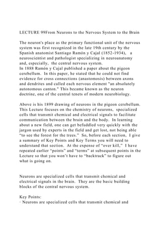
  • 1. LECTURE 99From Neurons to the Nervous System to the Brain The neuron's place as the primary functional unit of the nervous system was first recognized in the late 19th century by the Spanish anatomist Santiago Ramón y Cajal (1852-1934), a neuroscientist and pathologist specializing in neuroanatomy and, especially, the central nervous system. In 1888 Ramón y Cajal published a paper about the pigeon cerebellum. In this paper, he stated that he could not find evidence for cross connections (anastomosis) between axons and dendrites and called each nervous element "an absolutely autonomous canton." This became known as the neuron doctrine, one of the central tenets of modern neurobiology. Above is his 1899 drawing of neurons in the pigeon cerebellum. This Lecture focuses on the chemistry of neurons, specialized cells that transmit chemical and electrical signals to facilitate communication between the brain and the body. In learning about a new field, one can get befuddled very quickly with the jargon used by experts in the field and get lost, not being able “to see the forest for the trees.” So, before each section, I give a summary of Key Points and Key Terms you will need to understand that section. At the expense of “over kill,” I have repeated earlier “points” and “terms” at subsequent points in the Lecture so that you won’t have to “backtrack” to figure out what is going on. Neurons are specialized cells that transmit chemical and electrical signals in the brain. They are the basic building blocks of the central nervous system. Key Points: · Neurons are specialized cells that transmit chemical and
  • 2. electrical signals in the brain; they are the basic building blocks of the central nervous system. · The primary components of the neuron are the soma (cell body), the axon (a long slender projection that conducts electrical impulses away from the cell body), dendrites (tree- like structures that receive messages from other neurons), and synapses (specialized junctions between neurons). · Some axons are covered with myelin, a fatty material that acts as an insulator and conductor to speed up the process of communication. · Sensory neurons are neurons responsible for converting external stimuli from the environment into corresponding internal stimuli. · Motor neurons are neurons located in the central nervous system (CNS); they project their axons outside of the CNS to directly or indirectly control muscles. · Interneurons act as the “middle men” between sensory and motor neurons, which convert external stimuli to internal stimuli and control muscle movement, respectively. Key Terms: · glial cell: Non-neuronal cells that provide structure and support to neurons. · synapse: The junction between the terminal of a neuron and either another neuron or a muscle or gland cell, over which nerve impulses pass. · myelin: A white, fatty material composed of lipids and lipoproteins that surrounds the axons of nerves and facilitates swift communication. · nodes of Ranvier: Periodic gaps in the myelin sheath where the signal is recharged as it moves along the axon. The neuron is the basic building block of the brain and central nervous system. The brain is made up entirely of neurons and glial cells. Nearly 86 billion neurons work together within the nervous system to communicate with the rest of the body. Fun facts: The Milky Way galaxy has an estimated 100 billion
  • 3. stars. It is estimated that there are 10 trillion galaxies in the observable universe. Multiplying that by the Milky Way's estimated 100 billion stars results in a large number indeed: 1,000,000,000,000,000,000,000,000 stars. Given this astronomically-large value, the probability that there is (at least) one star with a planet “just the right distance” from that star is quite high. Whether there are large bodies of H2O on this planet is another matter but, if so, assuming the same distribution of elements as on planet Earth, the chemistry of Carbon can kick in and life on that planet is possible. Neurons are responsible for consciousness and thought to pain and hunger. You need almost as many neurons to function as there are stars in our Milky Way. Structures of a Neuron In addition to having all the normal components of a cell (nucleus, organelles, etc.), neurons also contain unique structures for receiving and sending the electrical signals that make neuronal communication possible. The structure of a neuron: The above image shows the basic structural components of an average neuron, including the dendrite, cell body, nucleus, Node of Ranvier, myelin sheath, Schwann cell, and axon terminal. Dendrite Dendrites are branch-like structures extending away from the cell body, and their job is to receive messages from other neurons and allow those messages to travel to the cell body. Although some neurons do not have any dendrites, other types of neurons have multiple dendrites. Dendrites can have small protrusions called dendritic spines, which further increase surface area for possible connections with other neurons.
  • 4. Cell Body Like other cells, each neuron has a cell body (or soma) that contains a nucleus, smooth and rough endoplasmic reticulum, Golgi apparatus, mitochondria, and other cellular components. Axon An axon is a tube-like structure that carries an electrical impulse from the cell body (or from another cell’s dendrites) to the structures at opposite end of the neuron—axon terminals, which can then pass the impulse to another neuron. The cell body contains a specialized structure, the axon hillock, which serves as a junction between the cell body and the axon. Synapse The synapse is the chemical junction between the axon terminals of one neuron and the dendrites of the next. It is a gap where specialized chemical interactions can occur, rather than an actual structure. Function of a Neuron The specialized structure and organization of neurons allows them to transmit signals in the form of electric impulses from the brain to the body and back. Individually, neurons can pass a signal all the way from their own dendrites to their own axon terminals. At a higher level, neurons are organized in long chains, allowing them to pass signals very quickly from one to the other. One neuron’s axon will connect chemically to another neuron’s dendrite at the synapse between them. Electrically charged chemicals flow from the first neuron’s axon to the second neuron’s dendrite, and that signal will then flow from the second neuron’s dendrite, down its axon, across a synapse, into a third neuron’s dendrites, and so on. This is the basic chain of neural signal transmission, which is how the brain sends signals to the muscles to make them move, and how sensory organs send signals to the brain. It is important that these signals can happen quickly, and they do. Think of how fast you drop a hot potato—before you even
  • 5. realize it is hot. This is because the sense organ (in this case, the skin) sends the signal “This is hot!” to neurons with very long axons that travel up the spine to the brain. If this didn’t happen quickly, people would burn themselves. Other Structures Dendrites, cell bodies, axons, and synapses are the basic parts of a neuron, but other important structures and materials surround neurons to make them more efficient. Myelin Sheath Some axons are covered with myelin, a fatty material that wraps around the axon to form the myelin sheath. This external coating functions as insulation to minimize dissipation of the electrical signal as it travels down the axon. Myelin’s presence on the axon greatly increases the speed of conduction of the electrical signal, because the fat prevents any electricity from “leaking out”. This insulation is important, as the axon from a human motor neuron can be as long as a meter—from the base of the spine to the toes. Periodic gaps in the myelin sheath are called nodes of Ranvier. At these nodes, the signal is “recharged” as it travels along the axon. Glial Cells The myelin sheath is not actually part of the neuron. Myelin is produced by glial cells (or simply glia, or “glue” in Greek), which are non-neuronal cells that provide support for the nervous system. Glia function to hold neurons in place (hence their Greek name), supply them with nutrients, provide insulation, and remove pathogens and dead neurons. In the central nervous system, the glial cells that form the myelin sheath are called oligodendrocytes; in the peripheral nervous system, they are called Schwann cells. Neuron in the central nervous system: This neuron diagram also shows the oligodendrocyte, myelin sheath, and nodes of Ranvier. Types of Neurons
  • 6. There are three major types of neurons: sensory neurons, motor neurons, and interneurons. All three have different functions, but the brain needs all of them to communicate effectively with the rest of the body (and vice versa). Sensory Neurons Sensory neurons are neurons responsible for converting external stimuli from the environment into corresponding internal stimuli. They are activated by sensory input, and send projections to other elements of the nervous system, ultimately conveying sensory information to the brain or spinal cord. Unlike the motor neurons of the central nervous system (CNS), whose inputs come from other neurons, sensory neurons are activated by physical stimuli (such as visible light, sound, heat, physical contact, etc.) or by chemical signals (such as smell and taste). Most sensory neurons are pseudounipolar, meaning they have an axon that branches into two extensions—one connected to dendrites that receive sensory information and another that transmits this information to the spinal cord. Motor Neurons Motor neurons are neurons located in the central nervous system, and they project their axons outside of the CNS to directly or indirectly control muscles. The interface between a motor neuron and muscle fiber is a specialized synapse called the neuromuscular junction. The structure of motor neurons is multipolar, meaning each cell contains a single axon and multiple dendrites. This is the most common type of neuron. Interneurons Interneurons are neither sensory nor motor; rather, they act as the “middle men” that form connections between the other two types. Located in the CNS, they operate locally, meaning their axons connect only with nearby sensory or motor neurons. Interneurons can save time and therefore prevent injury by sending messages to the spinal cord and back instead of all the way to the brain. Like motor neurons, they are multipolar in
  • 7. structure. Stages of the Action Potential Neural impulses occur when a stimulus depolarizes a cell membrane, prompting an action potential which sends an “all or nothing” signal. Key Points: · The neurons (or excitable nerve cells) of the nervous system conduct electrical impulses, or signals, that serve as communication between sensory receptors, muscles and glands, and the brain and spinal cord. · An action potential occurs when an electrical signal disrupts the original balance of Na+ and K+ within a cell membrane, briefly depolarizing the concentrations of each. · An electrical impulse travels along the axon via depolarized voltage-gated ion channels in the membrane, and can either “jump” along a myelinated area or travel continuously along an unmyelinated area. · While an action potential is being generated by a cell, no other action potential may be generated until the cell’s channels return to their resting state. · Action potentials generated by neural impulses are “all or nothing,” meaning the signal reaches the threshold for communication or it doesn’t. No signal is stronger or weaker than another. Key Terms: · polarity: The spatial differences in the shape, structure, and function of cells. Almost all cell types exhibit some sort of polarity, which enables them to carry out specialized functions. · action potential: A short-term change in the electrical potential that travels along a cell, such as a nerve or muscle fiber, and allows nerves to communicate. · neural impulse: The signal transmitted along a nerve fiber, either in response to a stimulus (such as touch, pain, or heat), or as an instruction from the brain (such as causing a muscle to contract).
  • 8. · resting potential: The nearly latent membrane potential of inactive cells. Neural Impulses in the Nervous System The central nervous system (CNS) goes through a three-step process when it functions: sensory input, neural processing, and motor output. The sensory input stage is when the neurons (or excitable nerve cells) of the sensory organs are excited electrically. Neural impulses from sensory receptors are sent to the brain and spinal cord for processing. After the brain has processed the information, neural impulses are then conducted from the brain and spinal cord to muscles and glands, which is the resulting motor output. A neuron affects other neurons by releasing a neurotransmitter that binds to chemical receptors. The effect upon the postsynaptic (receiving) neuron is determined not by the presynaptic (sending) neuron or by the neurotransmitter itself, but by the type of receptor that is activated. A neurotransmitter can be thought of as a key, and a receptor as a lock: the key unlocks a certain response in the postsynaptic neuron, communicating a particular signal. However, in order for a presynaptic neuron to release a neurotransmitter to the next neuron in the chain, it must go through a series of changes in electric potential. Stages of Neural Impulses “Resting potential ” is the name for the electrical state when a neuron is not actively being signaled. A neuron at resting potential has a membrane with established amounts of sodium (Na+) and potassium (K+) ions on either side, leaving the inside of the neuron negatively charged relative to the outside. The action potential is a rapid change in polarity that moves along the nerve fiber from neuron to neuron. In order for a neuron to move from resting potential to action potential—a short-term electrical change that allows an electrical signal to be passed from one neuron to another—the neuron must be stimulated by pressure, electricity, chemicals, or another form of stimuli. The level of stimulation that a neuron must receive
  • 9. to reach action potential is known as the threshold of excitation, and until it reaches that threshold, nothing will happen. Different neurons are sensitive to different stimuli, although most can register pain. The action potential has several stages. 1. Depolarization: A stimulus starts the depolarization of the membrane. Depolarization is caused when positively charged sodium ions rush into a nerve cell. As these positive ions rush in, the membrane of the stimulated cell reverses its polarity so that the outside of the membrane is negative relative to the inside. 2. Repolarization. Once the electric gradient has reached the threshold of excitement, the “downswing” of repolarization begins. The channels that let the positive sodium ion channels through close up, while channels that allow positive potassium ions open, resulting in the release of positively charged potassium ions from the neuron. This expulsion acts to restore the localized negative membrane potential of the cell, bringing it back to its normal voltage. 3. Refractory Phase. The refractory phase takes place over a short period of time after the depolarization stage. Shortly after the sodium gates open, they close and go into an inactive conformation. The sodium gates cannot be opened again until the membrane is repolarized to its normal resting potential. A sodium-potassium pump returns sodium ions to the outside and potassium ions to the inside. During the refractory phase this particular area of the nerve cell membrane cannot be depolarized. Therefore, the neuron cannot reach action potential during this “rest period.” Action potentials: A neuron must reach a certain threshold in order to begin the depolarization step of reaching the action potential. This process of depolarization, repolarization, and recovery moves along a nerve fiber from neuron to neuron like a very fast wave. While an action potential is in progress, another
  • 10. cannot be generated under the same conditions. In unmyelinated axons (axons that are not covered by a myelin sheath), this happens in a continuous fashion because there are voltage-gated channels throughout the membrane. In myelinated axons (axons covered by a myelin sheath), this process is described as saltatory because voltage-gated channels are only found at the nodes of Ranvier, and the electrical events seem to “jump” from one node to the next. Saltatory conduction is faster than continuous conduction. The diameter of the axon also makes a difference, as ions diffusing within the cell have less resistance in a wider space. Damage to the myelin sheath from disease can cause severe impairment of nerve-cell function. In addition, some poisons and drugs interfere with nerve impulses by blocking sodium channels in nerves. More on this point in the next Lecture. All-or-none Signals The amplitude of an action potential is independent of the amount of current that produced it. In other words, larger currents do not create larger action potentials. Therefore, action potentials are said to be all-or-none signals, since either they occur fully or they do not occur at all. The frequency of action potentials is correlated with the intensity of a stimulus. This is in contrast to receptor potentials, whose amplitudes are dependent on the intensity of a stimulus. Reuptake Reuptake refers to the reabsorption of a neurotransmitter by a presynaptic (sending) neuron after it has performed its function of transmitting a neural impulse. Reuptake is necessary for normal synaptic physiology because it allows for the recycling of neurotransmitters and regulates the neurotransmitter level in the synapse, thereby controlling how long a signal resulting from neurotransmitter release lasts. Mechanics of the Action Potential The synapse is the site at which a chemical or electrical exchange occurs between the presynaptic and postsynaptic cells.
  • 11. Key Points: · Receptors are pores that admit chemical or electrical signals into the postsynaptic cell. There are two main types of receptor: ligand-gated ion channels, which receive neurostransmitters, and g-protein coupled receptors, which do not. · There are two types of possible reactions at the synapse: a chemical reaction or an electrical reaction. · During a chemical reaction, neurotransmitters trigger the opening of ligand-gated ion channels on the membrane of the postsynaptic cell, resulting in a modification of the cell’s interior chemical composition and, in some cases, physical structure. · In an electrical reaction, the electrical charge of one cell is influenced by another. · Although electrical synapses yield faster reactions, chemical synapses result in stronger, more complex changes to the postsynaptic cell. Key Terms: · vesicle: A membrane-bound compartment found in a cell. · action potential: A short-term change in the electrical potential that travels along a cell, such as a nerve or muscle fiber, and allows nerves to communicate. · depolarization: The act of depriving of polarity, or the result of such action; reduction to an unpolarized condition. · membrane potential: The voltage across the cell membrane, with the inside relative to the outside. Synapses The synapse is the junction where neurons trade information. It is not a physical component of a cell but rather a name for the gap between two cells: the presynaptic cell (giving the signal) and the postsynaptic cell (receiving the signal). There are two types of possible reactions at the synapse—chemical or electrical. During a chemical reaction, a chemical called a neurotransmitter is released from one cell into another. In an electrical reaction, the electrical charge of one cell is influenced by the charge an adjacent cell.
  • 12. The electrical response of a neuron to multiple synaptic inputs: Synaptic responses summate in order to bring the postsynaptic neuron to the threshold of excitation, so it can fire an action potential (represented by the peak on the chart). All synapses have a few characteristics in common: · Presynaptic cell: a specialized area within the axon of the giving cell that transmits information to the dendrite of the receiving cell. · Synaptic cleft: the small space at the synapse that receives neurotransmitters. · G-protein coupled receptors: receptors that sense molecules outside the cell and thereby activate signals within it. · Ligand-gated ion channels: receptors that are opened or closed in response to the binding of a chemical messenger. · Postsynaptic cell: a specialized area within the dendrite of the receiving cell that contains receptors designed to process neurotransmitters. The Electrical Synapse The stages of an electrical reaction at a synapse are as follows: 1. Resting potential. The membrane of a neuron is normally at rest with established concentrations of sodium ions (Na+) and potassium ions (K+) on either side. The membrane potential (or, voltage across the membrane) at this state is -70 mV, with the inside being negative relative to the outside. 2. Depolarization. A stimulus begins the depolarization of the membrane. Depolarization, also referred to as the “upswing,” occurs when positively charged sodium ions (Na+) suddenly rush through open sodium gates into a nerve cell. If the membrane potential reaches -55 mV, it has reached the threshold of excitation. Additional sodium rushes in, and the membrane of the stimulated cell actually reverses its polarity so that the outside of the membrane is negative relative to the inside. The change in voltage stimulates the opening of additional sodium channels (called a voltage-gated ion channel), providing what is known as a positive feedbackloop. Eventually,
  • 13. the cell potential reaches +40 mV, or the action potential. 3. Repolarization. The “downswing” of repolarization is caused by the closing of sodium ion channels and the opening of potassium ion channels, resulting in the release of positively charged potassium ions (K+) from the nerve cell. This expulsion acts to restore the localized negative membrane potential of the cell. 4. Refractory Phase. The refractory phase is a short period of time after the repolarization stage. Shortly after the sodium gates open, they close and go into an inactive conformation where the cell’s membrane potential is actually even lower than its baseline -70 mV. The sodium gates cannot be opened again until the membrane has completely repolarized to its normal resting potential, -70 mV. The sodium-potassium pump returns sodium ions to the outside and potassium ions to the inside. During the refractory phase this particular area of the nerve cell membrane cannot be depolarized; the cell cannot be excited. The Chemical Synapse The process of a chemical reaction at the synapse has some important differences from an electrical reaction. Chemical synapses are much more complex than electrical synapses, which makes them slower, but also allows them to generate different results. Like electrical reactions, chemical reactions involve electrical modifications at the postsynaptic membrane, but chemical reactions also require chemical messengers, such as neurotransmitters, to operate. Neuron & chemical synapse: This image shows electric impulses traveling between neurons; the inset shows a chemical reaction occurring at the synapse. A basic chemical reaction at the synapse undergoes a few additional steps: 1. The action potential (which occurs as described above) travels along the membrane of the presynaptic cell until it reaches the synapse. The electrical depolarization of the membrane at the synapse causes channels to open that are
  • 14. selectively permeable, meaning they specifically only allow the entry of positive sodium ions (Na+). 2. The ions flow through the presynaptic membrane, rapidly increasing their concentration in the interior. 3. The high concentration activates a set of ion-sensitive proteins attached to vesicles, which are small membrane compartments that contain a neurotransmitter chemical. 4. These proteins change shape, causing the membranes of some “docked” vesicles to fuse with the membrane of the presynaptic cell. This opens the vesicles, which releases their neurotransmitter contents into the synaptic cleft, the narrow space between the membranes of the pre- and postsynaptic cells. 5. The neurotransmitter diffuses within the cleft. Some of it escapes, but the rest of it binds to chemical receptor molecules located on the membrane of the postsynaptic cell. 6. The binding of neurotransmitter causes the receptor molecule to be activated in some way. Several types of activation are possible, depending on what kind of neurotransmitter was released. In any case, this is the key step by which the synaptic process affects the behavior of the postsynaptic cell. 7. Due to thermal shaking, neurotransmitter molecules eventually break loose from the receptors and drift away. 8. The neurotransmitter is either reabsorbed by the presynaptic cell and repackaged for future release, or else it is broken down metabolically. Differences Between Electrical and Chemical Synapses · Electrical synapses are faster than chemical synapses because the receptors do not need to recognize chemical messengers. The synaptic delay for a chemical synapse is typically about 2 milliseconds, while the synaptic delay for an electrical synapse may be about 0.2 milliseconds. · Because electrical synapses do not involve neurotransmitters, electrical neurotransmission is less modifiable than chemical neurotransmission. · The response is always the same sign as the source. For example, depolarization of the presynaptic membrane will
  • 15. always induce a depolarization in the postsynaptic membrane, and vice versa for hyperpolarization. · The response in the postsynaptic neuron is generally smaller in amplitude than the source. The amount of attenuation of the signal is due to the membrane resistance of the presynaptic and postsynaptic neurons. · Long-term changes can be seen in electrical synapses. For example, changes in electrical synapses in the retina of your eyeare seen during light and dark adaptations of the retina. Neurotransmitters Neurotransmitters are chemicals that transmit signals from a neuron across a synapse to a target cell. Key Points: · Neurotransmitters dictate communication between cells by binding to specific receptors and depolarizing or hyperpolarizing the cell. · Inhibitory neurotransmitters cause hyperpolarization of the postsynaptic cell; excitatory neurotransmitters cause depolarization of the postsynaptic cell. · Too little of a neurotransmitter may cause the over accumulation of proteins, leading to disorders like Alzheimer’s disease. Too much of a neurotransmitter may block receptors required for proper brain function, leading to disorders like schizophrenia. · The three neurotransmitter systems in the brain are cholinergic, amino acids, and biogenic amines. Key Terms · reuptake: The reabsorption of a neurotransmitter by a neuron after the transmission of a neural impulse across a synapse. · vesicle: A membrane-bound compartment found in a cell. · action potential: A short-term change in the electrical
  • 16. potential that travels along a cell (such as a nerve or muscle fiber); the basis of neural communication. Neurotransmitters are chemicals that transmit signals from a neuron to a target cell across a synapse. When called upon to deliver messages, they are released from their synaptic vesicles on the presynaptic (giving) side of the synapse, diffuse across the synaptic cleft, and bind to receptors in the membrane on the postsynaptic (receiving) side. An action potential is necessary for neurotransmitters to be released, which means that neurons must reach a certain threshold of electric stimulation in order to complete the reaction. A neuron has a negative charge inside the cell membrane relative to the outside of the cell membrane; when stimulation occurs and the neuron reaches the threshold of excitement this polarity is reversed. This allows the signal to pass through the neuron. When the chemical message reaches the axon terminal, channels in the postsynaptic cell membrane open up to receive neurotransmitters from vesicles in the presynaptic cell. Inhibitory neurotransmitters cause hyperpolarization of the postsynaptic cell (that is, decreasing the voltage gradient of the cell, thus bringing it further away from an action potential), while excitatory neurotransmitters cause depolarization (bringing it closer to an action potential). Neurotransmitters match up with receptors like a key in a lock. A neurotransmitter binds to its receptor and will not bind to receptors for other neurotransmitters, making the binding a specific chemical event. There are several systems of neurotransmitters found at various synapses in the nervous system. The following groups refer to the specific chemicals, and within the groups are specific systems, some of which block other chemicals from entering the cell and some of which permit the entrance of chemicals that were blocked before. Cholinergic System The cholinergic system is a neurotransmitter system of its own,
  • 17. and is based on the neurotransmitter acetylcholine (ACh). This system is found in the autonomic nervous system, as well as distributed throughout the brain. The cholinergic system has two types of receptors: the nicotinic receptor and the acetylcholine receptor, which is known as the muscarinic receptor. Both of these receptors are named for chemicals that interact with the receptor in addition to the neurotransmitter acetylcholine. Nicotine, the chemical in tobacco, binds to the nicotinic receptor and activates it similarly to acetylcholine. Muscarine, a chemical product of certain mushrooms, binds to the muscarinic receptor. However, they can not bind to each others’ receptors. Amino Acids Another group of neurotransmitters are amino acids, including glutamate (Glu), GABA (gamma-aminobutyric acid, a derivative of glutamate), and glycine (Gly). These amino acids have an amino group and a carboxyl group in their chemical structures. Glutamate is one of the 20 amino acids used to make proteins. See Lecture 6. Each amino acid neurotransmitter is its own system, namely the glutamatergic, GABAergic, and glycinergic systems. They each have their own receptors and do not interact with each other. Amino acid neurotransmitters are eliminated from the synapse by reuptake. A pump in the cell membrane of the presynaptic element, or sometimes a neighboring glial cell, clears the amino acid from the synaptic cleft so that it can be recycled, repackaged in vesicles, and released again. The reuptake process: This illustration shows the process of reuptake, in which leftover neurotransmitters are returned to vesicles in the presynaptic cell. Biogenic Amines Another class of neurotransmitter is the biogenic amine, a group of neurotransmitters made enzymatically from amino acids. They have amino groups in them, but do not have carboxyl
  • 18. groups and are therefore no longer classified as amino acids. Neuropeptides A neuropeptide is a neurotransmitter molecule made up of chains of amino acids connected by peptide bonds, similar to proteins. However, proteins are long molecules while some neuropeptides are quite short. Neuropeptides are often released at synapses in combination with another neurotransmitter. Dopamine Dopamine is the best-known neurotransmitter of the catecholamine group. The brain includes several distinct dopamine systems, one of which plays a major role in reward- motivated behavior. Most types of reward increase the level of dopamine in the brain, and a variety of addictive drugs increase dopamine neuronal activity. Other brain dopamine systems are involved in motor control and in controlling the release of several other important hormones ( a regulatory substance produced in an organismand transported in tissue fluids such as blood or sap to stimulate specific cells or tissues into action ). More on this in the next Lecture. Effect on the Synapse The effect of a neurotransmitter on the postsynaptic element is entirely dependent on the receptor protein. If there is no receptor protein in the membrane of the postsynaptic element, then the neurotransmitter has no effect. The depolarizing (more likely to reach an action potential) or hyperpolarizing (less likely to reach an action potential) effect is also dependent on the receptor. When acetylcholine binds to the nicotinic receptor, the postsynaptic cell is depolarized. However, when acetylcholine binds to the muscarinic receptor, it might cause depolarization or hyperpolarization of the target cell. The amino acid neurotransmitters (glutamate, glycine, and GABA) are almost exclusively associated with just one effect. Glutamate is considered an excitatory amino acid because Glu receptors in the adult cause depolarization of the postsynaptic cell. Glycine and GABA are considered inhibitory amino acids,
  • 19. again because their receptors cause hyperpolarization, making the receiving cell less likely to reach an action potential. The Right Dose Sometimes too little or too much of a neurotransmitter may affect an organism’s behavior or health. The underlying cause of some neurodegenerative diseases, such as Parkinson’s disease, appears to be related to over accumulation of proteins, which under normal circumstances would be regulated by the presence of dopamine. On the other hand, when an excess of the neurotransmitter dopamine blocks glutamate receptors, disorders like Schizophrenia can occur. Neural Networks Neural networks consist of a series of interconnected neurons, and serve as the interface for neurons to communicate with each other. Key Points: · The connections between neurons form a highly complex network through which signals or impulses are communicated across the body. · The basic kinds of connections between neurons are chemical synapses and electrical gap junctions, through which either chemical or electrical impulses are communicated between neurons. · Neural networks are primarily made up of axons, which in some cases deliver information as far as two meters. · Networks formed by interconnected groups of neurons are capable of a wide variety of functions. In fact the range of capabilities possible for even small groups of neurons are beyond our current understanding. · Modern science views the function of the nervous system both in terms of stimulus -response chains and in terms of intrinsically generated activity patterns within neurons. · Cell assembly, or Hebbian theory, asserts that “cells that fire together wire together,” meaning neural networks can be created through associative experience and learning. Key Terms:
  • 20. · cell assembly: Also referred to as Hebbian theory; the concept that “cells that fire together wire together,” meaning neural networks can be created through associative experience and learning. · action potential: A short-term change in the electrical potential that travels along a cell such as a nerve or muscle fiber, and allows nerves to communicate. · plasticity: The ability to change and adapt over time. A neural network (or neural pathway) is the interface through which neurons communicate with one another. These networks consist of a series of interconnected neurons whose activation sends a signal or impulse across the body. Neural networks: A neural network (or neural pathway) is the complex interface through which neurons communicate with one another. See top of Lecture. Perhaps now you can begin to appreciate the path breaking research of Ramón y Cajal. As a child he was transferred many times from one school to another because of behavior that was declared poor, rebellious, and showing an “anti-authoritarian attitude.” An extreme example of his precociousness and rebelliousness at the age of eleven is his 1863 imprisonment for destroying his neighbor's yard gate with a homemade cannon. He and Camillo Golgi received the Nobel Prize in Physiology or Medicine in 1906. Ramón y Cajal was the first person of Spanish origin to win a Nobel Prize. SUMMARY of LECTURE 9
  • 21. The connections between neurons form a highly complex network. The basic kinds of connections between neurons are chemical synapses and electrical gap junctions, through which either chemical or electrical impulses are communicated between neurons. The method through which neurons interact with neighboring neurons usually consists of several axon terminals connecting through synapses to the dendrites on other neurons. If a stimulus creates a strong enough input signal in a nerve cell, the neuron sends an action potential and transmits this signal along its axon. The axon of a nerve cell is responsible for transmitting information over a relatively long distance, and so most neural pathways are made up of axons. Some axons are encased in a lipid-coated myelin sheath, making them appear a bright white; others that lack myelin sheaths (i.e., are unmyelinated) appear a darker beige color, which is generally called gray. The process of synaptic transmission in neurons: Neurons interact with other neurons by sending a signal, or impulse, along their axon and across a synapse to the dendrites of a neighboring neuron. Some neurons are responsible for conveying information over long distances. For example, motor neurons, which travel from the spinal cord to the muscle, can have axons up to a meter in length in humans. The longest axon in the human body is almost two meters long in tall individuals and runs from the big toe to the medulla oblongata of the brain stem. The Capacity of Neural Networks The basic neuronal function of sending signals to other cells includes the capability for neurons to exchange signals with each other. Networks formed by interconnected groups of neurons are capable of a wide variety of functions, including feature detection, pattern generation, and timing. In fact, it is difficult to assign limits to the types of information processing that can be carried out by neural networks. Given that
  • 22. individual neurons can generate complex temporal patterns of activity independently, the range of capabilities possible for even small groups of neurons are beyond current understanding. However, we do know that we have neural networks to thank for much of our higher cognitive functioning. Behaviorist Approach Historically, the predominant view of the function of the nervous system was as a stimulus-response associator. In this conception, neural processing begins with stimuli that activate sensory neurons, producing signals that propagate through chains of connections in the spinal cord and brain, giving rise eventually to activation of motor neurons and thereby to muscle contraction or other overt responses. Charles Sherrington, in his influential 1906 book The Integrative Action of the Nervous System, developed the concept of stimulus-response mechanisms in much more detail, and behaviorism, the school of thought that dominated psychology through the middle of the 20th century, attempted to explain every aspect of human behavior in stimulus-response terms. Hybrid Approach Experimental studies of electrophysiology, beginning in the early 20th century and reaching high productivity by the 1940s, showed that the nervous system contains many mechanisms for generating patterns of activity intrinsically—without requiring an external stimulus. Neurons were found to be capable of producing regular sequences of action potentials (“firing”) even in complete isolation. When intrinsically active neurons are connected to each other in complex circuits, the possibilities for generating intricate temporal patterns become far more extensive. A modern conception views the function of the nervous system partly in terms of stimulus-response chains, and partly in terms of intrinsically generated activity patterns; both types of activity interact with each other to generate the full repertoire of behavior. Hebbian Theory In 1949, neuroscientist Donald Hebb proposed that simultaneous
  • 23. activation of cells leads to pronounced increase in synaptic strength between those cells, a theory that is widely accepted today. Cell assembly, or Hebbian theory, asserts that “cells that fire together wire together,” meaning neural networks can be created through associative experience and learning. Since Hebb’s discovery, neuroscientists have continued to find evidence of plasticity and modification within neural networks. LECTURE 9. From Alcohol and Aspirin to Hallucinogens and Opioids La Nuit Etoilée (The Starry Night) is an oil on canvas by Dutch post-impressionist painter Vincent van Gogh (1853-1890). Painted in June 1889, it describes the view from the east-facing window of his asylum room at Saint-Rémy-de-Provence, just before sunrise, with the addition of an ideal village. He spent a long period in 1889-90 in a clinic because of his mental instability, before committing suicide. You do not have to be a chemist to wonder about the source of the swirls, spirals and other strange effects. Van Gogh's instability and suicide have been blamed on the liqueur-like drink absinthe, a fashionable French beverage in the half century up to the first world war. Absinthe, is a green liquid with an anise smell, made by distilling a mixture of alcohol, herbs (notably wormwood) and water. In the late 19th century, it became a national drink in France. Fashionable among the artistic community, it became cheap enough to be the spirit “beverage of choice” among the poor. Writers such as Baudelaire, Edgar Allan Poe and Verlaine relied upon it, and a whole range of artists (Degas, Gauguin, Manet, Picasso, Toulouse-Lautrec, and Van Gogh) are associated with it, often for including it in their paintings. Known as la fée verte (the green fairy), absinthe gave rise to l'heure verte, the
  • 24. time (5 pm) when drinkers in all walks of life went to a café for their absinthe, what we would now call a “Happy Hour”. L'Absinthe Artist Edgar Degas Year 1875–76 We begin by considering alcoholic beverages and their effect on the brain. How does alcohol affect the brain? Alcohol has a profound effect on the complex structures of the brain. It blocks chemical signals between brain cells (neurons), leading to the common intermediate symptoms of intoxication, including impulsive behavior, slurred speech, poor memory, and slowed reflexes. If heavy drinking continues over extended periods of time, the brain adopts to the blocked signals by responding more dramatically to certain brain chemicals, the neurotransmitters. After alcohol leaves the system, the brain continues over- activating the neurotransmitters, causing painful and potentially dangerous withdrawal symptoms that can damage brain cells. This damage is made more acute by “binge drinking” and sudden withdrawal.
  • 25. Alcohols damage to the brain can take several forms. The first is neurotoxicity, which occurs when neurons over react to neurotransmitters for too long. Too much exposure to a neurotransmitter can cause neurons to eventually “burn out.” Since neurons make up the pathways between different parts of the brain, when they begin “burning out,” it can cause noticeable slowing in the response of these pathways. People with alcohol dependence often experience “brain shrinkage,” which is reduced volume of both gray matter (cell bodies) and white matter (cell pathways) over time. There are some subtle differences in how brain damage occurs in men and women, but regardless of gender, loss of brain matter increases with age and amount of alcohol consumed. What are the observable effects of this damage? Since alcohol affects a large portion of the brain, many different kinds of cognitive impairment can occur as a result of heavy drinking, including problems with verbal fluency and verbal learning, processing speed, working memory, attention, problem solving, spatial processing and impulsivity. Parts of the brain relating to memory and “higher functions” ( for example, problem solving and impulse control) are more susceptible to damage than other parts of the brain, so problems in these areas tend to be worse than others. Adolescents are especially at risk for long-lasting or permanent damage and performance deficits, since their most-impacted areas of the brain are still in development.
  • 26. Without treatment, cognitive impairment grows worse, eventually developing into a lasting syndrome known as alcohol related dementia. This syndrome represents about 10% of all dementia cases (additionally, alcohol is estimated to contribute to roughly 29% of all other dementia cases). Cognitive deficits are made worse by malnutrition, especially a deficiency of vitamin B (a common deficiency in alcohol- dependent individuals). Malnutrition and heavy alcohol consumption can cause serious impairments in memory and language over time and can potentially result in permanent cognitive disorder called Wernicke-Korsakoff syndrome, which causes amnesia and can lead to coma if left untreated. In Lecture 5, we commented on the different physiological effects induced by methanol and ethanol. methanol and ethanolMethanol is a highly toxic alcohol with a smell and taste similar to ethanol. Small amounts (around 50 - 100 ml ) cause permanent blindness and severe neurological dysfunction leading to death. More than half of methanol-related morbidity and mortality is classified as accidental and therefore preventable. In addition, it can be suicidal by ingestion of a variety of commercial paint thinners, gasoline anti-freeze, windshield products, organic solvents, shellac varnish, washer fluid, photocopying fluids, perfumes, and in some eau de cologne. Occasionally, it is due to the fraudulent adulteration of wine or other alcoholic beverages. Its ingestion causes high anion gap metabolic acidosis from the production of formic and lactic acids and central nervous system disturbances ranging from inebriation and drowsiness to obtundation (A condition in which the senses have been dulled by trauma, mistreatment, or psychological stress.), seizure and coma.
  • 27. Aspirin The active ingredient in aspirin is acetylsalicylic acid, C9 H8 O4 . This organic acid was found in leaves from the willow tree, and has been used for its health effects for at least 2,400 years. Medicines made from willow and other salicylate-rich plants appear in clay tablets from ancient Sumer as well as the Ebers papyrus from ancient Egypt. Hippocrates (around 400 BC) referred to the use of salicylic tea to reduce fevers. Aspirin has been manufactured since 1899 by the German company Bayer. There are many generic varieties of aspirin. A generic drug manufacturer must prove that their product contains the same active ingredient(s) as the brand name product. Aspirin can be used to fight a host of health problems: cerebral thromboses (with less than one tablet a day); general pain or
  • 28. fever (two to six tablets a day); and diseases such as rheumatic fever, gout, and rheumatoid arthritis. The drug is also beneficial in helping to ward off heart attacks. In addition, biologists use aspirin to interfere with white blood cell action, and molecular biologists use the drug to activate genes. The wide range of effects that aspirin can produce made it difficult to pinpoint how it actually works, and it wasn't until the 1970s that biologists hypothesized that aspirin and related drugs (such as ibuprofen) work by inhibiting the synthesis of certain hormones that cause pain and inflammation. [Hormones are regulatory substances produced in an organism and transported in tissue fluids such as blood or sap to stimulate specific cells or tissues into action.] Since then, scientists have made further progress in understanding how aspirin works. They now know, for instance, that aspirin and its relatives actually prevent the growth of cells that cause inflammation. As a further example of how a change in a single functional group (See Lecture 5) can change the chemical, physical and physiological properties of a molecule, note that both aspirin and oil of wintergreen are synthesized from the same precursor, salicilic acid. See structure and reactions below. Autonomic nervous system drugs The autonomic nervous system controls the involuntary processes of the glands, large internal organs, cardiac muscle, and blood vessels. It is divided functionally and anatomically into the sympathetic and the parasympathetic systems, which are associated with the fight-or-flight response or with rest and energy conservation, respectively. Organization of the autonomic nervous system.Encyclopædia
  • 29. Britannica, Inc. Modern pharmacological understanding of the autonomic nervous system emerged from several key insights made in the early 20th century. The first of these came in 1914, when British physiologist Sir Henry Dale suggested that acetylcholine was the neurotransmitter at the synapse between preganglionic and postganglionic sympathetic neurons and also at the ends of postganglionic, parasympathetic nerves. Preganglionic neurons originate in the central nervous system, whereas postganglionic neurons lie outside the central nervous system. Dale showed that acetylcholine could produce many of the same effects as direct stimulation of parasympathetic nerves. Firm evidence that acetylcholine was in fact the neurotransmitter emerged in 1921, when German physiologist Otto Loewi discovered that stimulation of the autonomic nerves to the heart of a frog caused the release of a substance, later identified to be acetylcholine, which slowed the beat of a second heart perfused with fluid from the first. Similar direct evidence of the release of a sympathetic neurotransmitter, later shown to be norepinephrine (noradrenaline), was obtained by American physiologist Walter Cannon in 1921. Norepinephrine
  • 30. Both acetylcholine and norepinephrine act on more than one type of receptor. Dale found that two foreign substances, nicotine and muscarine, could each mimic some, but not all, of the parasympathetic effects of acetylcholine. The structure of muscarine is given below, that of nicotin later in this Lecture. Muscarine Nicotine stimulates skeletal muscle and sympathetic ganglia cells. Muscarine stimulates receptor sites located only at the junction between postganglionic parasympathetic neurons and the target organ. Muscarine slows the heart, increases the secretion of body fluids, and prepares the body for digestion. Dale therefore classified the many actions of acetylcholine into nicotinic effects and muscarinic effects. Drugs that influence
  • 31. the activity of acetylcholine, including atropine, scopolamine, and tubocuraine, are known as cholinergic drugs (see later text). A similar analysis of the sympathetic effects of norepinephrine, epinephrine, and related drugs was carried out by American pharmacologist Raymond Ahlquist, who suggested that these agents acted on two principal receptors. A receptor that is activated by the neurotransmitter released by an adrenergic neuron is said to be an adrenoceptor. Ahlquist called the two kinds of adrenoceptor alpha (α) and beta (β). This theory was confirmed when Sir James Black developed a new type of drug that was selective for the β-adrenoceptor. Adrenoline is a hormone secreted by the adrenal glands, especially in conditions of stress, increasing rates of blood circulation, breathing, and carbohydrate metabolism and preparing muscles for exertion.
  • 32. α-adrenoceptors and β-adrenoceptors are divided into subclasses: α1 ,α2 and β1, β2, β3. These receptor subtypes were recognized by their responses to specific agonists and antagonists, which provided important leads for the development of new drugs. For example, salbutamol was discovered as a specific β2-adrenoceptor agonist. It is used to treat asthma and is a great improvement over its predecessor, isoproterenol. Because the activity of isoproterenol is not specific, it acts on β1-adrenoceptors as well as β2-adrenoceptors, resulting in cardiac effects that are sometimes dangerous. Salbutamol and other agents that act on adrenoceptors, including albuterol, ephedrine, and imipramine, are known as adrenergic drugs. Central nervous system drugsSeveral major groups of drugs, notably anethetics and psychiatric drugs, affect the central nervous system. These agents often are administered in order to produce changes in physical sensation, behavior, or mental state. General anesthetics induce a temporary loss of consciousness, enabling surgeons to operate on a patient without the patient’s feeling pain. Local anesthetics induce a loss of sensation in just one area of the body by blocking conduction in nerves at and near the injection site.Drugs that influence the operation of neurotransmitter systems in the brain can profoundly influence and alter the behavior of patients with mental disorders. Psychiatric drugs that affect mood and behavior may be classified as antidepressants, antianxiety agents, antipsychotics or antimanics. What is a hallucinogen?
  • 33. A hallucinogen is a psychoactive agent that often causes hallucinations, perceptual anomalies, and other substantial subjective changes in thought, emotion, and consciousness that are not typically experienced to such degrees with other categories of drugs. Research suggests that classic hallucinogens work at least partially by temporarily disrupting communication between brain chemical systems throughout the brain and spinal cord. Some hallucinogens interfere with the action of the brain chemical serotonin, which regulates mood and sensory perception. Serotonin What are some common hallucinogens? Caffine Caffeine is a central nervous system (CNS) stimulant. It is the world's most widely consumed psychoactive drug. Unlike many other psychoactive substances, it is legal and unregulated in nearly all parts of the world. There are several known mechanisms to explain the effects of caffeine. The most prominent is that it reversibly blocks the action of adenosine on its receptors and consequently prevents the onset of drowsiness induced by adenosine. Caffeine also stimulates certain portions of the autonomic nervous system. Nicotine Nicotine is a stimulant (alkaloid) that is naturally produced in the nightshade family of plants. It is highly addictive. Nicotine acts as a receptor agonist at most nicotinic acetylcholine receptors, except at two nicotinic receptor subunits where it acts
  • 34. as a receptor antagonist. Marijuana (Cannabis) Marijuana is a psychoactive drug from the Cannabis plant used primarily for medical or recreational purposes. The main psychoactive component of cannabis is tetrahydrocannabinol, which is one of the 483 known compounds in the plant, including at least 65 other cannabinoids. Cocaine Cocaine, also known as coke, is a strong stimulant. Mental effects may include loss of contact with reality, an intense feeling of happiness or agitation. Physical symptoms may include a fast heart rate, sweating and large pupils. High doses can result in very high blood pressure or body temperature. Effects begin within seconds to minutes of use and last between five and ninety minutes. Cocaine has a small number of accepted medical uses such a numbing and decreasing bleeding during nasal surgery. Cocaine is addictive due to its effect on the reward pathway in the brain. Colombia, Peru and Bolivia are the most important cocaine- producing countries. What is an opioid?
  • 35. Opioids are substances that act on opioid receptors to produce morphine-like effects. Medically they are primarily used for pain relief, including anesthesia. Other medical uses include suppression of diarrhea, replacement therapy for opioid use disorder, reversing opioid overdose, suppressing cough, as well as for executions in the US. Extremely potent opioids such as carfentanil are approved only for veterinary use. Opioids are also frequently used non-medically for their euphoric effects or to prevent withdrawal. Side effects of opioids may include: itchiness, sedation, respiratory depression, constipation, and euphoria. Long-term use can cause tolerance, meaning that increased doses are required to achieve the same effect, and physical dependence, meaning that abruptly discontinuing the drug leads to unpleasant withdrawal symptoms. The euphoria attracts recreational use and frequent, escalating recreational use of opioids typically results in addiction. An overdose or concurrent use with other depressant drugs like benzodiazepine commonly results in death from respiratory depression. Opioids act by binding to opioid receptors, which are found principally in the central and peripheral nervous system and the gastrointestinal tract. These receptors mediate both the psychoactive and the somatic effects of opioids. Opioid drugs include partial agonists, like the anti-diarrhea drug loperamide and atagonists like naloxegol for opioid-induced constipation, which do not cross the blood-brain barrier but can displace other opioids from binding to those receptors. What are some examples of opioids?
  • 36. Morphine Morphine is found naturally in a number of plants and animals, including humans. It acts directly on the central nervous system (CNS) to decrease the feeling of pain. It can be taken for both acute pain and chronic pain. It is frequently used for pain from myocardial infarction and during labor. After injection, maximum effect is reached after about 20 minutes when given intravenously and after 60 minutes when given by mouth, while duration of effect is 3–7 hours. Potentially serious side effects include decreased respiratory effort and low blood pressure. Morphine is addictive and prone to abuse.
  • 37. Heroin Heroin is a highly addictive drug processed from morphine, a naturally occurring substance extracted from the seed pod of certain varieties of poppy plants.Common side effects include decreased breathing, dry mouth, drowsiness, impaired mental function, constipation, and addiction. Side effects of use by injection can include abscesses, infected heart valves and pneumonia.Opium (or “poppy tears”, scientific name: Lachryma papaveris) is dried latex obtained from the seed capsules of the opium poppy Papaver somniferum. Opium poppy seed pod exuding latex from a cu Approximately 12 percent of opium is made up of morphine, which is processed chemically to produce heroin and other synthetic opioids for medicinal use and for illegal drug trade. The latex also contains the closely related opiates codeine, and non-analfesic alkaloids such as papaverine and noscapine.The super sleuth, Sherlock Holmes (Robert Downey Jr. in the movies), occasionally used addictive drugs, especially in the absence of stimulating cases. He sometimes used morphine and other times cocaine. Both drugs were legal in 19th-century England. His sidekick, Dr. John Watson (Jude Law), strongly
  • 38. disapproved of his friend's cocaine habit, describing it as the detective's only vice, and was concerned about its effect on Holmes's mental health and intellect. Carfentanil Loperamide Naloxegol LECTURE 11 CANCER: DRUGS, IMMUNOCHEMISTRY and CHEMOCHEMISTRY A dividing breast cancer cell. Cancer is the name given to a collection of related diseases. In all types of cancer, some of the body’s cells begin to divide without stopping and spread into surrounding tissues. Cancer can start almost anywhere in the human body, which is made up of trillions of cells. Normally, human cells grow and divide to form new cells as the body needs them. When cells grow old or become damaged, they die, and new cells take their
  • 39. place. When cancer develops, this orderly process breaks down. As cells become more and more abnormal, old or damaged cells survive when they should die, and new cells form when they are not needed. These extra cells can divide without stopping and may form growths called tumors. Many cancers form solid tumors, which are masses of tissue. Cancers of the blood, such as leukemia, generally do not form solid tumors. Cancerous tumors are malignant, which means they can spread into, or invade, nearby tissues. In addition, as these tumors grow, some cancer cells can break off and travel to distant places in the body through the blood or the lymph system and form new tumors far from the original tumor. Unlike malignant tumors, benign tumors do not spread into, or invade, nearby tissues. Benign tumors can sometimes be quite large, however. When removed, they usually don’t grow back, whereas malignant tumors sometimes do. Unlike most benign tumors elsewhere in the body, benign brain tumors can be life threatening. What are the differences between cancer cells and normal cells? Cancer cells differ from normal cells in many ways that allow them to grow out of control and become invasive. One important difference is that cancer cells are less specialized than normal cells. That is, whereas normal cells mature into very distinct cell types with specific functions, cancer cells do not. This is one reason that, unlike normal cells, cancer cells continue to divide without stopping. In addition, cancer cells are able to ignore signals that normally tell cells to stop dividing or that begin a process known as programmed cell death, or apoptosis, which the body uses to get rid of unneeded cells. Cancer cells may be able to influence the normal cells, molecules, and blood vessels that surround and feed a tumor, an
  • 40. area known as the microenvironment. For instance, cancer cells can induce nearby normal cells to form blood vessels that supply tumors with oxygen and nutrients, which they need to grow. These blood vessels also remove waste products from tumors. Cancer cells are also often able to evade the immune system, a network of organs, tissues, and specialized cells that protects the body from infections and other conditions. Although the immune system normally removes damaged or abnormal cells from the body, some cancer cells are able to “hide” from the immune system. Tumors can also use the immune system to stay alive and grow. For example, with the help of certain immune system cells that normally prevent a runaway immune response, cancer cells can actually keep the immune system from killing cancer cells. How does cancer arise? Cancer is caused by changes to genes, the basic physical units of inheritance. Genes are arranged in chromosomes, long strands of tightly packed DNA. Cancer is a genetic disease—that is, it is caused by changes to genes that control the way our cells function, especially how they grow and divide. Genetic changes that cause cancer can be inherited from our parents. They can also arise during a person’s lifetime as a result of errors that occur as cells divide or because of damage to DNAcaused by certain environmental exposures. Cancer- causing environmental exposures include substances, such as the chemicals in tobacco smoke, and radiation, such as ultraviolet rays from the sun. Each person’s cancer is a unique combination of genetic changes. As the cancer continues to grow, additional changes will occur. Even within the same tumor, different cells may have different genetic changes.
  • 41. In general, cancer cells have more genetic changes, such as mutations in DNA, than normal cells. Some of these changes may have nothing to do with the cancer; they may be the result of the cancer, rather than its cause. Fundamentals of Cancer Cancer is a disease caused when cells divide uncontrollably and spread into surrounding tissues. Cancer is caused by changes to DNA. Most cancer-causing DNA changes occur in sections of DNA called genes. These changes are also called genetic changes. A DNA change can cause genes involved in normal cell growth to become oncogenes, which unlike normal genes, cannot be turned off, hence causing uncontrolled cell growth. Physicians who specialize in treating cancers are called Oncologists. In normal cells, tumor suppressor genes prevent cancer by slowing or stopping cell growth. DNA changes that inactivate tumor suppressor genes can lead to uncontrolled cell growth and cancer. Within a tumor, cancer cells are surrounded by a variety of immune cells, fibroblasts, molecules, and blood vessels—what’s known as the tumor microenvironment. Cancer cells can change the microenvironment, which in turn can affect how cancer grows and spreads. Cells in our immune system can detect and attack cancer cells. But, some cancer cells can avoid detection or thwart an attack. Some cancer treatments help the immune system better detect
  • 42. and kill cancer cells. Each person’s cancer has a unique combination of genetic changes. Specific genetic changes may make a person’s cancer more or less likely to respond to certain treatments. Genetic changes that cause cancer can be inherited or arise from certain environmental exposures. Genetic changes can also happen because of errors that occur as cells divide. Most often, cancer-causing genetic changes accumulate slowly as a person ages, leading to a higher risk of cancer later in life. Cancer cells can break away from the original tumor and travel through the blood or lymph system to distant locations in the body, where they exit the vessels to form additional tumors. This is called metastasis. What are "Drivers" of Cancer” ? The genetic changes that contribute to cancer tend to affect three main types of genes—proto-oncogenes, tumor suppressor genes, and DNA repair genes. These changes are sometimes called “drivers” of cancer. Proto-oncogenes are involved in normal cell growth and division. However, when these genes are altered in certain ways or are more active than normal, they may become cancer- causing genes (or oncogenes), allowing cells to grow and
  • 43. survive when they should not. Tumor suppressor genes are involved in controlling cell growth and division. Cells with certain alterations in tumor suppressor genes may divide in an uncontrolled manner. DNA repair genes are involved in fixing damaged DNA. Cells with mutations in these genes tend to develop additional mutations in other genes. Together, these mutations may cause the cells to become cancerous. Certain mutations commonly occur in many types of cancer. Consequntly, cancers are sometimes characterized by the types of genetic alterations that are believed to be driving them, not just by where they develop in the body and how the cancer cells look under the microscope.How does cancer spread? ENLARGE In metastasis, cancer cells break away from where the first formed (primary cancer), travel through the blood or lymph system, and form new tumors (metastatic tumors) in other parts of the body. The metastatic tumor is the same kind of cancer as the primary tumor. A cancer that has spread from the place where it first started to another place in the body is called metastatic cancer. The process by which cancer cells spread to other parts of the body is called metastasis. Metastatic cancer has the same name and the same type of cancer cells as the original, or primary, cancer. For example, breast cancer that spreads to and forms a metastatic tumor in the lung is metastatic breast cancer, not lung cancer. Under a microscope, metastatic cancer cells generally look the same as cells of the original cancer. Moreover, metastatic cancer cells and cells of the original cancer usually have some molecular features in common, such as the presence of specific chromosome changes. Treatment may help prolong the lives of some people with metastatic cancer. In general, though, the primary goal of
  • 44. treatments for metastatic cancer is to control the growth of the cancer or to relieve symptoms caused by it. Metastatic tumors can cause severe damage to how the body functions, and most people who die of cancer die of metastatic disease. Are there tissue changes that are not cancer? Not every change in the body’s tissues is cancer. Some tissue changes may develop into cancer if they are not treated, however. Here are some examples of tissue changes that are not cancer but, in some cases, are monitored: Hyperplasia occurs when cells within a tissue divide faster than normal and extra cells build up, or proliferate. However, the cells and the way the tissue is organized look normal under a microscope. Hyperplasia can be caused by several factors or conditions, including chronic irritation. Dysplasia is a more serious condition than hyperplasia. In dysplasia, there is also a buildup of extra cells. But the cells look abnormal and there are changes in how the tissue is organized. In general, the more abnormal the cells and tissue look, the greater the chance that cancer will form. Some types of dysplasia may need to be monitored or treated. An example of dysplasia is an abnormal mole (called a dysplastic nevus) that forms on the skin. A dysplastic nevus can turn into melanoma, although most do not. An even more serious condition is carcinoma in situ. Although it is sometimes called cancer, carcinoma in situ is not cancer because the abnormal cells do not spread beyond the original tissue. That is, they do not invade nearby tissue the way that cancer cells do. But, because some carcinomas in situ may become cancer, they are usually treated. Normal cells may become cancer cells. Before cancer cells form in tissues of the body, the cells go through abnormal changes called hyperplasia and dysplasia. In hyperplasia, there is an increase in the number of cells in an organ or tissue that appear normal under a microscope.
  • 45. Are there different types of cancer? There are more than 100 types of cancer. Types of cancer are usually named for the organs or tissues where the cancers form. For example, lung cancer starts in cells of the lung, and brain cancer starts in cells of the brain. Cancers also may be described by the type of cell that formed them, such as an epithelial cell or a squamous cell. Following are some categories of cancers that begin in specific types of cells. Carcinoma Carcinomas are the most common type of cancer. They are formed by epithelial cells, which are the cells that cover the inside and outside surfaces of the body. There are many types of epithelial cells, which often have a column-like shape when viewed under a microscope. Carcinomas that begin in different epithelial cell types have specific names: Adenocarcinoma is a cancer that forms in epithelial cells that produce fluids or mucus. Tissues with this type of epithelial cell are sometimes called glandular tissues. Most cancers of the breast, colon, and prostate are adenocarcinomas. Basal cell carcinoma is a cancer that begins in the lower or basal (base) layer of the epidermis, which is a person’s outer layer of skin. Squamous cell carcinoma is a cancer that forms in squamous cells, epithelial cells that lie just beneath the outer surface of the skin. Squamous cells also line many other organs, including the stomach, intestines, lungs, bladder, and kidneys. Squamous cells look flat, like fish scales, when viewed under a microscope. Squamous cell carcinomas are sometimes called epidermoid carcinomas. Transitional cell carcinoma is a cancer that forms in a type of epithelial tissue called transitional epithelium, or urothelium.
  • 46. This tissue, which is made up of many layers of epithelial cells that can get bigger and smaller, is found in the linings of the bladder, ureters, and part of the kidneys (renal pelvis), and a few other organs. Some cancers of the bladder, ureters, and kidneys are transitional cell carcinomas. Sarcoma ENLARGE Sarcomas are cancers that form in bone and soft tissues, including muscle, fat, blood vessels, lymph vessels, and fibrous tissue (such as tendons and ligaments). Osteosarcoma is the most common cancer of bone. The most common types of soft tissue sarcoma are leiomyosarcoma, Kaposi sarcoma, malignant fibrous histiocytoma, liposarcoma, and dermatofibrosarcoma protuberans. Leukemia Cancers that begin in the blood-forming tissue of the bone marrow are called leukemias. These cancers do not form solid tumors. Instead, large numbers of abnormal white blood cells (leukemia cells and leukemic blast cells) build up in the blood and bone marrow, crowding out normal blood cells. The low level of normal blood cells can make it harder for the body to get oxygen to its tissues, control bleeding, or fight infections. There are four common types of leukemia, which are grouped based on how quickly the disease gets worse (acute or chronic) and on the type of blood cell the cancer starts in (lymphoblastic or myeloid). Lymphoma Lymphoma is cancer that begins in lymphocytes (T cells or B cells). These are disease-fighting white blood cells that are part of the immune system. In lymphoma, abnormal lymphocytes build up in lymph nodes and lymph vessels, as well as in other organs of the body.
  • 47. There are two main types of lymphoma: Hodgkin lymphoma – People with this disease have abnormal lymphocytes that are called Reed-Sternberg cells. These cells usually form from B cells. Non-Hodgkin lymphoma – This is a large group of cancers that start in lymphocytes. The cancers can grow quickly or slowly and can form from B cells or T cells. Multiple Myeloma Multiple myeloma is cancer that begins in plasma cells, another type of immune cell. The abnormal plasma cells, called myeloma cells, build up in the bone marrow and form tumors in bones all through the body. Multiple myeloma is also called plasma cell myeloma and Kahler disease. Melanoma Melanoma is cancer that begins in cells that become melanocytes, specialized cells that make melanin (the pigment that gives skin its color). Most melanomas form on the skin, but melanomas can also form in other pigmented tissues, such as the eye. Brain and Spinal Cord Tumors There are different types of brain and spinal cord tumors. These tumors are named based on the type of cell in which they formed and where the tumor first formed in the central nervous system. For example, an astrocytic tumor begins in star-shaped brain cells called astrocytes, which help keep nerve cells healthy. Brain tumors can be benign (not cancer) or malignant (cancer). Are there other types of tumors?Germ Cell Tumors Germ cell tumors are a type of tumor that begins in the cells that give rise to sperm or eggs. These tumors can occur almost
  • 48. anywhere in the body and can be either benign or malignant.Neuroendocrine Tumors Neuroendocrine tumors form from cells that release hormones into the blood in response to a signal from the nervous system. These tumors, which may make higher-than-normal amounts of hormones, can cause many different symptoms. Neuroendocrine tumors may be benign or malignant.Carcinoid Tumors Carcinoid tumors are a type of neuroendocrine tumor. They are slow-growing tumors that are usually found in the gastrointestinal system (most often in the rectum and small intestine). Carcinoid tumors may spread to the liver or other sites in the body, and they may secrete substances such as serotonin or prostaglandins, causing carcinoid syndrome. ANTICANCER DRUGS Anticancer drugs are agents that demonstrate activity against malignant disease. They include hormones, natural products, antibodies, metabolites, alkylating agents as well as a variety of other chemicals that do not fall within these discrete classes but are capable of preventing the replication of cancer cells. Hormones are used primarily in the treatment of cancers of the breast and sex organs. These tissues require hormones such as estrogens, androgens or progestins for growth and development. By countering these hormones with an antagonizing hormone, the growth of that tissue is inhibited, as is the cancer growing in the area. For example, estrogens are required for female breast development and growth. Tamoxifen competes with endogenous estrogens for receptor sites in breast tissue where the estrogens normally exert their actions. The result is a decrease in the growth of breast tissue and of breast cancer tissue. Adrenocorticosteroids are also used for treating some types of cancer. These hormones are an example of a site-specific antineoplastic drug, but they work only on certain types of
  • 49. cancer. Understanding of the basic biology of cancer cells has led to drugs with entirely new targets. One agent, interleukin-2 , regulates the proliferation of tumor-killing lymphocytes. Interleukin-2 is used in the treatment of malignant melanoma and renal (kidney)carcinoma. Trans-retinoic acid can promote remission in patients with acute promyelocytic leukemia by inducing normal differentiation of the cancerous cells. A related compound, 13-cis-retinoic acid, prevents the development of secondary tumors in some individuals. A particularly exciting application of cancer biochemistry stems from the understanding of DNA translocation in chronic myelocytic leukemia. This translocation codes for a tyrosine kinase, an enzyme that phosphorylates other proteins and is essential for cell survival. Inhibition of the kinase by imatininib has been shown to be highly effective in treating patients who are resistant to standard therapies. Hydroxyuren inhibits the enzyme ribonucleotide reductase, an important element in DNA synthesis. It is used to reduce the high granulocyte count found in chronic myelocytic leukemia. Mitotane, a derivative of the insecticide DDT, causes necrosis of adrenal glands. A number of agents synthesized from plants are used in the treatment of cancer. Paclitaxel was first isolated from the bark of the western yew tree. It stops cell division by an action on the microtubules and has been tested for activity against ovarian and breast cancers. The camptothecins are a class of antineoplastic agents that target DNA replication. The first compound in this class was
  • 50. isolated from the Chinese camptotheca tree. Irinotecan and topotecan are used in the treatment of colorectal, ovarian, and small-cell lung cancer. Vinblastin and vincristine, derived from the periwinkle plant, along with etoposide, act primarily to stop spindle formation within the dividing cell during DNA replication and cell division. These drugs are important agents in the treatment of leukemias, lymphomas, and testicular cancer. Etoposide, a semisynthetic derivative of a toxin found in roots of the American mayapple, affects an enzyme and causes breakage of DNA strands. IMMUNOCHEMISTRY Immunochemistry is a branch of chemistry that involves the study of the molecular mechanisms underlying the function of the immune system, especially the nature of antibodies, antigens and their interactions. Various methods in immunochemistry have been developed and refined, and been used in scientific study, from virology to molecular evolution. One of the earliest examples of immunochemistry is the Wasserman test to detect syphilis. Svante Arrhenius, whose contribution to the understanding of ionic crystals (Lecture 4), was also one of the pioneers in this field. He published Immunochemistry in 1907 which described the application of the methods of physical chemistry to the study of the theory of toxins and antitoxins. An antibody (Ab), also known as an immunoglobulin (Ig),is a large, Y-shaped protein produced mainly by plasma cells that is used by the immune system to neutralize pathogens such as pathogenic bacteria and viruses The antibody recognizes a unique molecule of the pathogen,
  • 51. called an antigen, via the fragment antigen-binding (Fab) variable region. Each tip of the "Y" of an antibody contains a paratope (analogous to a lock) that is specific for one particular epitope (analogous to a key) on an antigen, allowing these two structures to bind together with precision. See figure below. Each antibody binds to a specific antigen; an interaction similar to a lock and key. Using this binding mechanism, an antibody can tag a microbe or an infected cell for attack by other parts of the immune system, or can neutralize its target directly (for example, by inhibiting a part of a microbe that is essential for its invasion and survival). Depending on the antigen, the binding may impede the biological process causing the disease or may activate macrophages to destroy the foreign substance. The ability of an antibody to communicate with the other components of the immune system is mediated via its Fc region (located at the base of the "Y"), which contains a conserved glycosylation site involved in these interactions. The production of antibodies is the main function of the humoral immune system. CHEMOTHERAPY Chemotherapy is a type of cancer treatment that uses one or more anti-cancer drugs as part of a standardized chemotherapy regimen. Chemotherapy may be given with a curative intent (which almost always involves combinations of drugs), or it may aim to prolong life or to reduce symptoms (palliative chemotherapy).
  • 52. The term chemotherapy has come to connote non-specific usage of agents to inhibit cell division (mitosis). Chemotherapy kills cells that are in the process of splitting into 2 new cells. Body tissues are made of billions of individual cells. Once we are fully grown, most of the body's cells don't divide and multiply much. They only divide if they need to repair damage. When cells divide, they split into 2 identical new cells. So where there was 1 cell, there are now 2. Then these divide to make 4, then 8 and so on. In cancer, the cells keep on dividing until there is a mass of cells. This mass of cells becomes a lump, called a tumor. Because cancer cells divide much more often than most normal cells, chemotherapy is much more likely to kill them. Some drugs kill dividing cells by damaging the part of the cell's control center that makes it divide. Other drugs interrupt the chemical processes involved in cell division. Chemotherapy damages cells as they divide. In the center of each living cell is a dark blob, called the nucleus. The nucleus is the control center of the cell. It contains chromosomes, which are made up of genes. These genes have to be copied exactly each time a cell divides into 2 to make new cells. Chemotherapy damages the genes inside the nucleus of cells. The term, chemotherapy, excludes more selective agents that block extracellular signals (signal transduction). The development of therapies with specific molecular or genetic targets, which inhibit growth-promoting signals from classic endocrine hormones (primarily estrogens for breast cancer and androgens for prostate cancer) are now called hormonal therapies. By contrast, other inhibitions of growth-signals like those associated with receptor tyrosine kinases (kinases are
  • 53. enzymes that catalyzes the transfer of a phosphate group from ATP to a specified molecule ) are referred to as targeted therapy.