Mg japan
•
0 likes•36 views

This document summarizes the diagnosis and prevention of chronic respiratory disease (CRD) caused by Mycoplasma gallisepticum infection in chickens in Japan. It describes how the disease spread throughout Japan in the 1960s following the development of the poultry industry. Diagnosis involves isolating M. gallisepticum through culture or detecting antibodies through agglutination or hemagglutination inhibition tests. Prevention efforts include using antibiotics like tylosin, implementing sanitary measures and eradication programs on farms, and developing clean breeding flocks free of the disease.

Recommended
More Related Content
What's hot
What's hot (20)
Similar to Mg japan
Similar to Mg japan (20)
More from Oumed Gerjis
Recently uploaded
Recently uploaded (20)
Mg japan
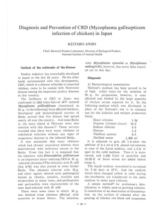
- 1. Diagnosis and Prevention of CRD (Mycoplasma gallisepticum infection of chicken) in Japan KEITARO ANDO Chief, Bacterial Product Laboratory, Division of Biological Product, National Institute of Animal Health Outlook of the outbreaks of the disease Poultry industry has remarkably developed in Japan in the last 10 years. On the other hand, accompanied with this development, CRD, which is a disease referable to imported chicken, came to be ranked with Newcastle disease among the important poultry diseases in this country. The incidence of CRD in Japan was confirmed in 1963 when Sato et al.8) isolated Mycoplasma gallisepticum (mentioned as M. g. in the following) from affected chickens. Serological and bacteriological surveys of flocks proved that this disease had spread nearly all over the country. And some flocks in the main island of Okinawa were also infected with this disease.s) These surveys revealed that there were many chickens of subclinical infection without any signs of respiratory distress in the infected flocks. It was noticeable that many chickens which had chronic respiratory distress were dual-infected with infectious coryza in the flocks. From this fact it is supposed that the infection with Haemophilus gallinarum is an important factor inducing CRD in M. g. -infected chickens.2)The infection with E. coli (0-2, 0-78) was also proved in some broiler flocks. T he chickens infected with M.g. and other agents showed such pathological lesions as rhinitis, sinusitis, trachitis and airsacculitis in many cases, and perihepatitis and pericarditis were characteristic of the ones dual-infected with E. coli. There were some cases in which M. g. was isolated from chickens affected with synovitis or breast blister. The infection with Mycoplasma synoviae or Mycoplasma meleagridis, however, has never been report- ed yet to this day. Diagnosis 1) Bacteriological examination Hofstad's medium has been proved to be of high utility value for the isolation of M. g. Its preparation, however, is com- plicated and limited by the large quantity of chicken serum required for it. So the following medium which was developed in place of the Hofstad's one is in common use for the isolation and antigen production lately. Heart infusion 1. Og Peptone (Animal tissue) 10. 0 Sodium chloride 5. O Glucose 1.0 Thallium acetate O. 5 Distilled water L 000 ml It is adjusted to g ive pH 7. 8 after the addition of O. 5 ml of 2 % phenol red solution in case of the liquid medium, and 1. 5 % of agar in the solid medium. T hen it is auto- claved, and 1, 000 U/ml of penicillin and 10-20 % of horse serum are added before using it. The liquid medium inoculated is incubated for 7-10 days at 37°C. And the medium which have changed yellow in color during the incubation are transferred to the plate medium to make pure cultures. A simple method9) for preliminary iden- tification is widely used on growing colonies. It constitutes of an observation of hemadsorp- tion on the surface of colonies after the pouring of chicken red blood cell suspension ...., 14 -
- 2. onto the plate medium. The final identifi- cation for M. g. is made by agglutination test and growth inhibition test. The isolation of M.g. from the affected chickens by the culture method mentioned above was high in percentage in the upper respiratory tract, especially in sinus or trachea, but it was extremely low in the case of subclinical infection. 2) Serological reaction A) Agglutination The authors prepared the antigen for agglutination using the strain S6 of M. g. obtained from University of California.1) It is a concentrated bacterial suspension (Mc- Farland No. 1 x 25), and contains crystal violet and Merthiolate at the rate of O. 01%, respectively. The whole blood plate agglutination is principally practiced, as in the case of Pullorum disease, in the field test. Practica- lly, in serological diagnosis, sample test has been adopted on flock basis. Flock tested, especially adult chicken flocks, often showed high percentage (50% or over) of reactor rate. The concentrated antigen is diluted to 1/12. 5 with saline solution for the serum tube agglutination. The tube method is applied to given final judgement for the flocks free from M.g. And it is more useful than the hemagglutination inhibition test (HI test) mentioned below in case quantitative data of antibody titer is needed. The ag- glutination titer of chickens 1 : 20 or over in most of the clinical cases, sometimes being 1: 640 or over, while they are 1 : 10 or lower in most of the healthy flocks. B) Hemagglutination inhibition test (HI test) The antigen is prepared as a mixture consisting of a concentrated bacterial suspen- sion of strain S6 and glycerol. The test method was improved by Kuniyasu.3) It was known that the HT antibody is contained in the 7S fraction of the serum, and appears a little later than the aggluti- nating antibody which is contained in the 19S and 7S fractions (Kuniyasu 1968). The purpose of the application of HI test is nearly the same as that of the serum agglutination method. But it is so compli- cated in technique that it is applicable only to special tentative flock. Prevention 1) Application of medicines Many kinds of antibiotics are in use to control chronic respiratory distress. The sensitivity test of M. g. to antibiotics was carried out in vitro by Matsui et al.4), obtaining the result that macrolide antibio- tics were the strongest in the antibacterial action, that is, tylosin had becteriostatic and bacteriocidal effects at the concentration of O. 01- 0. 1 /ml, and spiramycin was bacterios- tatic at O. 1 /ml. Resistant strain to antibio- tics other than streptomycin has never been found (see table). According to Azechi et al. (1967), who made experiments on the distribution of the tylosin applied in the body of chicken, it was 3 /ml in blood level 48 hours after the subcutaneous injection of 300 mg/kg dose, while it was proved to by only 1. 5/ml 24 hours after the oral administration of the same dose. These results show that tylosin remains in the blood for a considerably long time after subcutaneous injection. For the treatment and prevention of this disease, such antibiotics as tylosin, spiramy- cin, chlortetracycline and oxytetracycline are in use. These medicines are applied by injection or orally with drinking water and feed. They are generally injected at the dose of 25 mg/kg, added to drinking water to O. 02-0. 05%, and given at dose of 300 g/t or more by feeding. The effect of these antibiotics is fairly good, when treatment and prevention are practiced in artifically infected chickens. They, however, are unsatisfactory under natural conditions of poultry farms. Namely they has not a complete effect, only im- proving clinical conditions of affected chick- ens. There always remain carriers of M. g. in the flock.7) The dipping of hatching eggs in antibiotics is not in practical use at present. It, - 15 -
- 3. however, has been proved that the injection of tylosin has a germicidal effect into the eggs carrying M. g. without serious affect on hatchability (Isogai, 1967). 2) Eradication program Attempts are being made in many breeding farms to eradicate this disease in infected flocks for the production and supply of chickens free from M. g. It is practiced by rearing of the growing chicks under the strict sanitary condition in company with slaughtering of infected birds or flock, sero- logical examination and application of anti- biotics. The M. g. cleaning program made by Minamimoto et al.6) in the Niigata Prefectural Poultry Experimental Station is an example of the success in the eradication of this disease. This station was a layer farm where being suffered from recurrent consump- tion of chickens due to chronic respiratory distress, and the percentage of reactors was 25% in the year when the eradication program started. Agglutination test and frequent application of tylosin completely improved the condition, resulting in such a perfect state of health as there were no attack of the disease and no reactors in the progeny flock. Though the application of tylosin was considerably reduced, there occurred also no reactors in the flock of the second generation. Furthermore the production of chicken and eggs was improved. And they could be obtained a clean flock which was also free from other main infec- tious diseases than CRD. Vaccination (planed exposure) against this disease has never been practiced even for tentative trial in this country. Table 1. Minimum inhibitory concentration of antibiotics and nitrofurans against 8 strains of Mycoplasma gallisepticum (ug/ml * u/ml) Strain Drug S6 Tex C6 KP-3 KP-13 21TTc Sip-1 396S C30as Penicillin*G )100 )100 )100 )100 )100 )100 )100 )100 Streptomycin 10 10 )100 )100 )100 )100 )100 10 Dihydrostreptomycin 1 10 )100 )100 )100 )100 )100 1 Chlorainphenicol 10 10 10 10 10 10 10 10 Tiophenicol 10 10 10 10 10 10 10 10 l3acitracin )100 )100 )100 )100 )100 )100 )100 )100 Mikamycin A 10 1 1 1 1 1 1 1 Mikamycin B 100 100 100 100 100 100 100 100 Colistin* )100 )100 )100 )100 )100 )100 )100 100 Polymyxin B• )100 )100 )100 )100 )100 )100 )100 100 Noboviocin 10 10 10 10 10 10 10 10 Erythromycin 0.01 0.1 0.1 0.1 o. 1 0.01 0.01 0.01 Spiramycin 0.01 0.1 0.01 0.01 0.01 0.01 0.01 0.01 Tylosin (0.01 0.01 0.01 (0.01 (0.01 0.01 (0.01 (0.01 Tertiomycin 0.1 0.1 0.1 0.1 o.1 0.1 0.1 0.1 Oleandomycin 0.1 1 1 1 0.1 0.1 0.1 1 Leucomycin 0.1 0.1 0.1 0.1 0. 1 o. 1 0.1 0.1 Tetracycline 1 1 1 1 1 1 1 1 Oxytetracycline 10 1 10 1 1 1 1 1 ~hlortetracycline 1 1 10 10 1 10 10 10 Furazolidone 100 10 10 10 10 10 10 10 Nitrofurazone 100 10 10 10 10 10 10 100 Pana;i:on 1 1 1 1 1 1 1 1 - 16 -
- 4. ,. References 1) Ando, K., Matsui, IC, Sato, S., Yoshida, I., Kato, K. and Kuniyasu, C.: Evaluation of antigcni. city of the agglutination antigen for avian respiratory mycoplasmosis and availability of the antigen for field test. Nat. Inst. Anim. Hlth Quart. 5, 13-19(1965) · 2) Kato, K.: Infectious coryza of chickens. V. Influenceof Mycoplasma galliscpticum infection of chicken infected with Haemophilus galJJ. narum. Nat. Inst. Anim, Hlth Quart. 5,183 -189(1965) 3) Kuniyasu, C. and Ando, K.: Studies on the hemagglutination inhibition test for Mycolasma galliscpticum in chickens. Nat. Inst. Anim. ffith Quart. 6, 136-143(1966) 4) Matsui, K., Ando, K., Hayami, T. and Okubo T.: The invitro sensitivity of Mycoplasma gallisepticum to antibiotics and nitro(urans. English summary: Nat. Inst. Anim. Hlth. Quart. 7, 124(1967) 5) Machida, S., Ando, K. and Kawamura, H.: -a 17 - Serological survey on infectious respiratory diseases of poultry in Okinawa, Nat. Inst. Anim. Hlth Quart. 8, 55-56(1968). 6) llfinamimoto, S., Suzuki, K. , Tanaka, K. ,Otaki, K. and Murayama, J. : Eradication of Myco- plasma gallisepticum infccton in a chicken flock on a breeding farm. Nat. Inst. Anim. Hlth Quart. 8, 164-168(1968). 7) Okubo, T. ct al. : Prophylactic effects of an- tibiotics anp other drugs on natural iniection of Mycoplasma gallisepticum and Hacmophilus gallinarum in chickens. English summary: J. Jap. Vet. Med. Assoc. 19, 620-635(1966) 8) Sato, S., Matsui, K., Watase, H., Ando, K., Kawamura, H. and Tsubahara, H.: Isolation of Mycoplasma galJisepticum from chickens affected with chronic respiratory distress in Japan. Nat. Inst. Anim. Hlth Quart. 4, 68-76 (1964). 9) Sato. S., Matsui, K., and Yoshida, Y.: chick- en red blood cell adsorption test for detection of colonies of llfycoplasma. gallisepticum devel- oped on agar media. Nat. Inst. Anim. mth Quart. 5, 45-46(1965).